Download presentation
Presentation is loading. Please wait.

1
COMMON SUTURING TECHNIQUES
Professor Magdy Amin RIAD Professor of Otolaryngology. Ain shames University Senior Lecturer in Otolaryngology University of Dundee

2
LIGATURES A suture tied around a vessel to occlude the lumen is called a ligature or tie. It may be used to effect hemostasis or to close off a structure to prevent leakage. There are two primary types of ligatures.

3
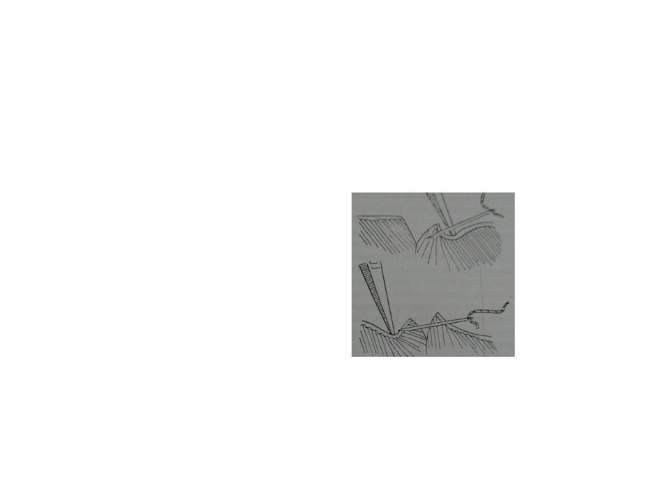
Free tie or freehand ligatures
single strands of suture material used to ligate a vessel, duct, or other structure. After a hemostat or other similar type of surgical clamp has been placed on the end of the structure, the suture strand is tied around the vessel under the tip of the hemostat. The hemostat is removed after the first throw and the surgeon tightens the knot using fingertips. Additional throws are added as needed to square and secure the knot.

4
Stick tie, suture ligature, or transfixion suture
Is a strand of suture material attached to a needle to ligate a vessel, duct, or other structure. This technique is used on deep structures where placement of hemostat is difficult or on vessels of large diameter. The needle is passed through the structure or adjacent tissue first to anchor the suture, then tied around the structure. Additional throws are used as needed to secure the knot.

5
THE PRIMARY SUTURE LINE CONTINUOUS SUTURES
running stitches: continuous sutures are a series of stitches taken with one strand of material. The strand may be tied itself at each end, or looped, with both cut ends of the strand tied together. placed rapidly. tension distributed evenly apply firm tension, rather than tight tension, to avoid tissue strangulation. Excessive tension and instrument damage should be avoided to prevent suture breakage which could disrupt the entire line of a continuous suture.

6
Continuous suturing leaves less foreign body mass in the wound.
In the presence of infection, it may be desirable to use a monofilament suture material because it has no interstices which can harbor microorganisms. This is especially critical as a continuous suture line can transmit infection along the entire length of the strand.

25
Interrupted sutures Number of strands to close the wound.
Each strand is tied and cut after insertion. This provides a more secure closure, because if one suture breaks, the remaining sutures will hold the wound. Interrupted sutures may be used if a wound is infected, because microorganisms may be less likely to travel along a series of interrupted stitches.

30
DEEP SUTURES placed completely under the epidermal skin layer.
They may be placed as continuous or interrupted sutures Not removed postoperatively.

31
BURIED SUTURES placed so that the knot protrudes to the inside, under the layer to be closed. This technique is useful when using sutures on airway Burying sutures in deeper layers prevent stitch sinuses.

32
PURSE-STRING SUTURES Continuous sutures placed around a lumen and tightened like a drawstring to invert the opening.

33
SUBCUTICULAR SUTURES Continuous or interrupted sutures
placed in the dermis, beneath the epithelial layer. Continuous subcuticular sutures are placed in a line parallel to the wound. This technique involves taking short, lateral stitches the full length of the wound.

34
SUBCUTICULAR SUTURES After the suture has been drawn taut, the distal end is anchored in the same manner as the proximal end. This may involve tying or any of a variety of anchoring devices. Subcuticular suturing may be performed with absorbable suture which does not require removal. Or with monofilament non absorbable suture that is later removed by simply removing the anchoring device at one end and pulling the opposite end.

35
THE SECONDARY SUTURE LINE
Called retention, stay, or tension sutures. To reinforce and support the primary suture line. Eliminate dead space, and prevent fluid accumulation. To support wounds for healing by second intention. For secondary closure following wound disruption when healing by third intention.

36
THE SECONDARY SUTURE LINE
If secondary sutures are used in cases of non healing, they should be placed in opposite fashion from the primary sutures i.e., interrupted if the primary sutures were continuous. Retention sutures are placed approximately 2 inches from each edge of the wound. Retention sutures utilize Non absorbable suture material. They should therefore be removed as soon as the danger of wound bursting is over, usually 2 to 6 weeks, with an average of 3 weeks.

38
STITCH PLACEMENT

39
FASCIA Fascia regains approximately 40% of its original strength in 2 months. It may take up to a year or longer to regain maximum strength. Full original strength is never regained. suture line. Because of the slow healing time non absorbable suture may be used. interrupted simple or figure-of eight suture.

40
MUSCLE Muscle does not tolerate suturing well.
muscles may be either cut, split (separated), or retracted, Where avoid interfering with the blood supply and nerve function by making a muscle-splitting incision or retracting the entire muscle toward its nerve supply. During closure, muscles handled in this manner do not need to be sutured. The fascia is sutured rather than the muscle. Interrupted sutures or "figure of eight“, VICRYL sutures are usually used or a Monofilament PROLENE
, or retracted, Where avoid interfering with the blood supply and nerve function by making a muscle-splitting incision or retracting the entire muscle toward its nerve supply. During closure, muscles handled in this manner do not need to be sutured. The fascia is sutured rather than the muscle. Interrupted sutures or figure of eight , VICRYL sutures are usually used or a Monofilament PROLENE.")
41
SUBCUTANEOUS FAT Neither fat nor muscle tolerate suturing well.
Has little tensile strength due to its composition, which is mostly water. Place at least a few sutures in a thick layer of subcutaneous fat to prevent dead space, especially in obese patients. Absorbable sutures are usually selected for the subcutaneous layer. VICRYL suture is especially suited for use in fatty,avascular tissue because it is absorbed by hydrolysis.

42
SUBCUTICULAR TISSUE To minimize scarring, suturing the subcuticular layer of tough connective tissue will hold the skin edges in close approximation. In a single-layer subcuticular closure, less evidence of scar gaping or expansion may be seen after a period of 6 to 9 months than is evident with simple skin closure. Continuous short lateral stitches beneath the epithelial layer of skin. Either absorbable or non absorbable sutures may be used.

43
SUBCUTICULAR TISSUE To produce only a hair-line scar on the face, the skin can be held in very close approximation with skin closure tapes in addition to subcuticular sutures. Tapes may be left on the wound for an extended period of time Chromic surgical gut and polymeric materials, such as MONOCRYL suture, are acceptable for placement within the dermis. They are capable of maintaining sufficient tensile strength through the collagen synthesis stage of healing which lasts approximately 6 weeks. The sutures must not be placed too close to the epidermal surface to reduce extrusion. MONOCRYL suture is particularly well-suited for this closure because, as a monofilament, it does not harbor infection and, as a synthetic absorbable suture, tissue reaction is minimized. After this layer is closed, the skin edges may then be approximated.

44
SKIN Skin is composed of the epithelium and the underlying dermis.
It is so tough that a very sharp needle is essential for every stitch to minimize tissue trauma. Skin wounds regain tensile strength slowly. If a non absorbable suture material is used, it is typically removed between 3 and 10 days postoperatively, when the wound has only regained approximately 5% to 10% of its strength. This is possible because most of the stress placed upon the healing wound is absorbed by the fascia, which the surgeon relies upon to hold the wound closed.

45
Suturing technique for skin closure may be either continuous or interrupted.
Skin edges should be everted. Interrupted technique is usually preferred. stitch abscess. The interstices of multifilament sutures may provide a haven for microorganisms. Therefore, monofilament non absorbable sutures may be preferred for skin closure. Monofilament sutures also induce significantly less tissue reaction than multifilament sutures. For cosmetic reasons, nylon or polypropylene monofilament sutures may be preferred.

46
And before contamination is converted into infection.
THE RAILROAD TRACK SCAR CONFIGURATION The key to success is early suture removal before epithelialization of the suture tract occurs And before contamination is converted into infection.

47
The oral cavity and pharynx
Absorbable sutures

48
The esophagus The esophagus is a difficult organ to suture.
It lacks a serosal layer. The mucosa heals slowly. The thick muscular layer does not hold sutures well. If multifilament sutures are used, penetration through the mucosa into the lumen should be avoided to prevent infection.

49
VESSELS Excessive tissue reaction to suture material may lead to decreased luminal diameter or to thrombus. More inert synthetics including nylon and polypropylene are the materials of choice. Interrupted monofilament sutures PROLENE sutures are used formicrovascular anastomoses.

50
SUTURES FOR BONE In repairing facial fractures, monofilament surgical steel has proven The suture material must remain in place for a long period of time—perhaps months—until the fibrous tissue is laid down and remodeled. Steel sutures immobilize the fracture line and keep the tissues in good apposition.

51
CLOSING CONTAMINATED OR INFECTED WOUNDS
Contamination exists when microorganisms are present, but in insufficient numbers to overcome the body's natural defenses. Infection exists when the level of contamination exceeds the tissue's ability to defend against the invading microorganisms. Generally, contamination becomes infection when it reaches approximately 1 million bacteria per gram of tissue in an immunologically normal host.

52
CLOSING CONTAMINATED OR INFECTED WOUNDS
Inflammation without discharge and/or the presence of culture positive serous fluid indicate possible infection. Presence of purulent discharge indicates positive infection. Contaminated wounds can become infected when hematomas, necrotic tissue, devascularized tissue, or large amounts of devitalized tissue especially in fascia, muscle, and bone are present. Microorganisms multiply rapidly under these conditions, where they are safe from cells that provide local tissue defenses.

53
CLOSING CONTAMINATED OR INFECTED WOUNDS
In general, contaminated wounds should not be closed but should be left open to heal by secondary intention because of the risk of infection. Foreign bodies, including sutures, perpetuate localized infection. Nonabsorbable monofilament nylon sutures are commonly used in anticipation of delayed closure of dirty and infected wounds. The sutures are laid in but not tied. Instead, the loose suture ends are held in place with PROXI-STRIP skin closures (sterile tape).
.")
54
CLOSING CONTAMINATED OR INFECTED WOUNDS
The wound should be packed to maintain a moist environment. When the infection has subsided, the surgeon can easily reopen the wound, remove the packing and any tissue debris, and then close using the previously inserted monofilament nylon suture.

55
SUTURE FOR DRAINS one or two non absorbable sutures.
Purse string suture Roman garter

56
The dura mater It tears with ease and cannot withstand too much tension. Drain some of the cerebrospinal fluid to decrease volume, easing the tension on the dura before closing. If it is too damaged to close, a graft must be inserted and sutured in place. NUROLON sutures or coated VICRYL sutures because they tie easily, offer greater strength than surgical silk, and cause less tissue reaction. PROLENE sutures for potentially infected wounds, or repair dural tears.

Similar presentations