Download presentation
Presentation is loading. Please wait.

1
The Integumentary System A high quality saran wrap

2
Functions PROTECTION from: – Mechanical damage (damage from the external environment/damage caused by movement and physical stress) – Chemical damage (damage from chemicals harmful to the skin) – Bacterial damage (damage from harmful bacteria trying to enter the body) – UV radiation (melanin protects us from the harmful rays of the sun) – Thermal damage (damage from heat/cold) – Desiccation (drying out – the skin helps us maintain our aqueous internal environment)
 – Chemical damage (damage from chemicals harmful to the skin) – Bacterial damage (damage from harmful bacteria trying to enter the body) – UV radiation (melanin protects us from the harmful rays of the sun) – Thermal damage (damage from heat/cold) – Desiccation (drying out – the skin helps us maintain our aqueous internal environment)")
3
Functions Aids in heat loss or heat retention. (helps us maintain our body’s temperature within its homeostatic range) Aids in excretion of urea and uric acid (waste that is excreted in sweat) Synthesizes Vitamin D (modified cholesterol molecules in the skin are converted into Vitamin D when exposed to sunlight)
 Aids in excretion of urea and uric acid (waste that is excreted in sweat) Synthesizes Vitamin D (modified cholesterol molecules in the skin are converted into Vitamin D when exposed to sunlight).")
4
Two Layers Epidermis (this is what you see when you look at someone!) -Outer covering. Composed of epithelial tissue. (epithelial tissue is avascular – it does not have its own blood supply) Dermis -Lies deep to the epidermis. -Composed mostly of dense connective tissue. (dense connective tissue is highly vascularized – it provides the oxygen and nutrients carried in the blood to the epidermis)
 Dermis -Lies deep to the epidermis. -Composed mostly of dense connective tissue. (dense connective tissue is highly vascularized – it provides the oxygen and nutrients carried in the blood to the epidermis).")
6
The Epidermis Composed solely of epithelial cells -Stratified Squamous Epithelial cells Why can a man shave and not bleed despite ripping off several layers of cells? This is because epithelial tissue is avascular – a person will not bleed when shaving until they have cut all the way through the epidermis and struck the dermis! How do epidermal cells receive nourishment? They receive nourishment provided by the blood that flows to the dermis.

7
The Epidermis Most of the epidermal cells are KERATINOCYTES. -Produce keratin, a tough fibrous protein (insoluble); the more keratin in a cell, the “tougher” the cell is!
; the more keratin in a cell, the tougher the cell is!.")
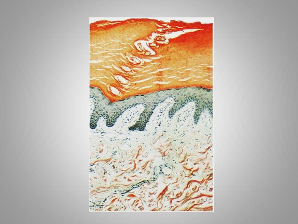
8
Epidermis Composed of five layers: – Stratum basale – Stratum spinosum – Stratum granulosum – Stratum lucidum – Stratum corneum Beagles Should Grow Less Corn

9
Stratum Basale Closest to dermis. This is the deepest layer of the epidermis Constantly dividing. Division of cells in the basale layer make it possible for our skin to “recycle” itself, as new cells push up to become more superficial layers. Contains Melanocytes – Melanocytes are cells that produce melanin, a pigment that gives skin its color. Phagocytosis of melanin by basale cells allows melanin to form a superficial shield for the DNA in our cells to protect it from the sun’s harmful rays.

10
Stratum Spinosum Lies superficial to stratum basale. Cells become flatter. Increasingly filled with keratin.

11
Stratum Granulosum Extremely flat. Full of keratin. Last layer of living cells.

12
Stratum Lucidum Clear layer of dead cells. Found only in hairless, extra-thick skin. Examples? Skin of the palms and soles of the feet The cells in the stratum lucidum are dead because of an extremely high keratin content and the distance between them and the vascular dermis.

13
Stratum Corneum Most superficial layer. 20-30 cell layers thick. Dead cells. Most keratinized layer of cells Replaced every 25-45 days.

14
The Dermis The hide. Composed of dense fibrous connective tissue. Two major regions: – Papillary layer – Reticular layer What happens when the Dermis separates from the Epidermis? Typically the dermis will separate from the epidermis when the skin experiences friction (an example of this would be changing directions quickly during basketball) A pocket of air will form, exposing our touch receptors in the papillary layer. This is a blister.
 A pocket of air will form, exposing our touch receptors in the papillary layer. This is a blister..")
15
Papillary Layer Dermal papillae form finger-like projections. Indent the overlying epidermis…this is why we have fingerprints. Contain capillary loops (these are blood vessels that provide nutrients to epidermis Contain pain and touch receptors as well (these are called meissner’s corpuscles)
.")
16
Reticular Layer Deepest layer of cutaneous membrane (the cutaneous membrane refers to the dermis and the epidermis combined). Contains skin appendages (hair, nails, glands). Composed of collagen and elastin (it is the loss of collagen and elastin in the reticular layer later in life as well as the loss of adipose tissue beneath the skin that results in “wrinkles”)
. Composed of collagen and elastin (it is the loss of collagen and elastin in the reticular layer later in life as well as the loss of adipose tissue beneath the skin that results in wrinkles ).")
17
Reticular Layer Cont’d Contains blood vessels, sweat and oil glands, and deep pressure receptors (pacinian corpuscles) Phagocytes protect against harmful bacteria (this is the last layer of protection against harmful bacteria before it can enter the body) Capillaries maintain body temperature – Swell with blood to release heat at surface (this lowers core body temperature, even though it makes our skin feel “flushed”) – Constrict for bypass to retain heat (this keeps core body temperatures high, even though it makes our extremities feel cold)
 Phagocytes protect against harmful bacteria (this is the last layer of protection against harmful bacteria before it can enter the body) Capillaries maintain body temperature – Swell with blood to release heat at surface (this lowers core body temperature, even though it makes our skin feel flushed ) – Constrict for bypass to retain heat (this keeps core body temperatures high, even though it makes our extremities feel cold)")
18
Hypodermis Hypo = below “Below dermis” NOT PART OF SKIN Composed of adipose tissue Functions: – Anchor skin – Shock absorber – Insulation

19
Summary Reticular Layer Cutaneous Membrane Epidermis Dermis Stratum Basale Stratum Spinosum Stratum Granulosum Stratum Lucidum Stratum Corneum Papillary Layer

20
Accessory organs of the skin

21
Types of Appendages Nails Hair Cutaneous Glands -Sebaceous glands -Sweat glands -Eccrine glands -Apocrine glands

22
Nails: Protection for ends of fingers/toes Heavily keratinized (these cells are dead). nail plate- the nail itself; this is the hard, shiny surface that we see nail bed- overlies skin surface. If you pull back the tip of your finger and see where the nail connects to the skin, you are looking at the nail bed Lunula (white, half moon circle at the base of the nail)- cells under lunula divide quickly, keratinize, and become the nail plate
- cells under lunula divide quickly, keratinize, and become the nail plate.")
23
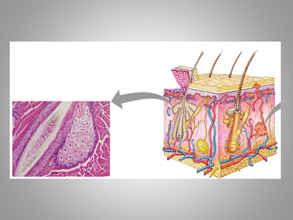
Hair: insulate, protect, conceal

24
Hair Grows from epidermal cells at base of hair follicle (tubelike depression) These cells are nourished from dermal blood vessels Epidermal cells divide push cells toward surface cells die/Keratinize form shaft of hair
 These cells are nourished from dermal blood vessels Epidermal cells divide push cells toward surface cells die/Keratinize form shaft of hair")
25
Arrector Pili Smooth muscle Cause hair to “stand on end” goosebumps

26
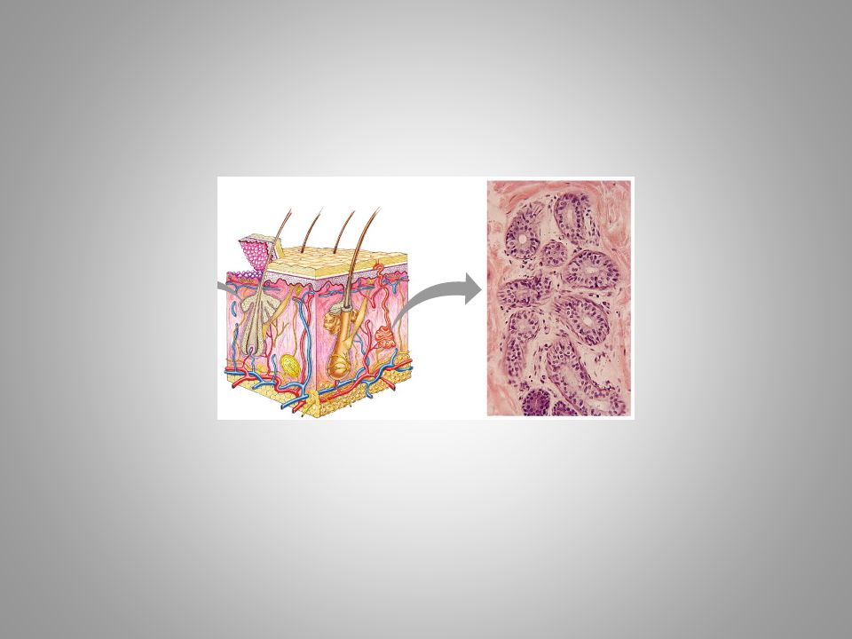
Sebaceous Glands: keeps hair & skin soft, pliable, waterproof Everywhere except palms of hands and soles of feet. Secrete sebum: oily mixture of fatty material & cellular debris Associated w/ hair follicles Acne: bacteria will accumulate in a pore, irritating the pore and causing it to swell shut. The newly formed cavity will fill with sebum, and result in a “pimple” that needs to be popped.

28
Eccrine Sweat Glands: Respond to body temperature Forehead, neck, back, etc. Sweat leaves through pores Sweat = water + some salts (electrolytes) + metabolic wastes (urea) Sweat is one of the skin’s methods of lowering body temperature
 + metabolic wastes (urea) Sweat is one of the skin’s methods of lowering body temperature.")
30
Apocrine Glands Found in the axillary and genital areas Activate during puberty – Tend to excrete when emotionally upset/frightened/in pain

31
In the following picture, label: Epidermis Dermis Hypodermis Hair follicle Arrector pili Sebaceous gland Eccrine sweat gland

Similar presentations


 Largest organ of the body (15% of body weight) Skin thickness variable, normally 1-2 mm Protection –chemical barrier (waterproof)>")
>")


