Download presentation
Presentation is loading. Please wait.

1
Geometric Factors Focal Spot Object Film Object b a h c Film B A H C
a b c h ---- = --- = --- = --- A B C H

2
Magnification Defined
Focal Spot size of image size of object Object Film (image)
")
3
Using Similar Triangles
size of image Magnification = size of object Focal Spot h H Object Film (image) focus to film distance H Magnification = = focus to object distance h
 focus to film distance H Magnification = = --- focus to object distance h.")
4
Using Similar Triangles
size of image Magnification = size of object Focal Spot h focus to film distance H magnification = = focus to object distance h H Object Film (image) SO focus to film dist. size of image = size of object X focus to object dist
 SO. focus to film dist. size of image = size of object X focus to object dist.")
5
Optimizing Image Quality
* focus to film distance H magnification = = focus to object distance h Focal Spot h H Object Film (image) Minimize magnification Minimize object-film distance Maximize focal-film distance
 Minimize magnification. Minimize object-film distance. Maximize focal-film distance.")
6
Relative Position Distortion
Distortion Types X-Ray Tube Film Image Shape Distortion X-Ray Tube Film Image Relative Position Distortion minimal distortion when object near central beam & close to film

7
Penumbra Latin for “almost shadow” region of partial illumination
also called edge gradient region of partial illumination caused by finite size of focal spot smears edges on film zone of unsharpness called geometric unsharpness penumbra edge gradient Line source focal spot Film Image

8
True Magnification m = geometric mag M = true mag Function of ratio of focal spot to object size (f / d) true & geometric magnification equal only when object very large compared to focal spot f a d b M=m + (m-1) X (f / d)
 X (f / d)")
9
Penumbra Calculation Minimizing Penumbra
Minimize object-film distance (OID) Maximize source-object distance (SOD) Makes focal spot appear smaller Minimize focal spot size F Line source focal spot SOD Object SID OID P = F x SOD OID P
 Maximize source-object distance (SOD) Makes focal spot appear smaller. Minimize focal spot size. F. Line source focal spot. SOD. Object. SID. OID P = F x SOD. OID. P.")
10
Magnification m = geometric mag M = true mag M = m = (a+b) / a
d m = (a+b) / a M=m + (m-1) X (f / d) Finite sized focal spot M = m = (a+b) / a Infinitely small focal spot Geom = True m = geometric mag M = true mag a b
 / a. M=m + (m-1) X (f / d) Finite sized focal spot. M = m = (a+b) / a. Infinitely small focal spot. Geom = True. m = geometric mag M = true mag. a. b.")
11
For general radiography purposes the geometric unsharpeness dominates the other components
Therefore the unsharpeness will increase with increasing magnification. To keep magnification small (close to m=1) requires the image receptor to be as close as possible to the patient and the focus patient distance to be large. Typical conditions are: a 1mm d1 1 m d2 10 cm
 requires the image receptor to be as close as possible to the patient and the focus patient distance to be large. Typical conditions are: a 1mm. d1 1 m. d2 10 cm.")
12
Motion Unsharpness Caused by motion during exposure of Effect
patient tube film Effect similar to penumbra Minimize by immobilizing patient short exposure times

13
Absorption Unsharpness
Cause gradual change in x-ray absorption across an object’s edge or boundary thickness of absorber presented to beam changes Effect produces poorly defined margin of solid objects X-Ray Tube X-Ray Tube X-Ray Tube

14
Inverse Square Law intensity of light falling on flat surface from point source is inversely proportional to square of distance from point source if distance 2X, intensity drops by 4X Assumptions point source no attenuation Cause increase in exposure area with distance Intensity a 1/d2 d

15
Trade-off Geometry vs. Intensity
maximize SID to minimize geometric unsharpness but doubling SID increases mAs by X4 increased tube loading longer exposure time possible motion going from 36 to 40 inch SID requires 23% mAs increase F SOD SID OID P

16
Magnification Types Geometric Magnification True Magnification
Focal Spot Object Film h H Geometric Magnification assumes point source calculated from similar triangles True Magnification takes into account finite size of focal spot focal spot is area (not point) source
 source.")
17
Automatic Artifact Occurs whenever we image a 3D object in 2D
Work-around Multiple views ? ?

18
Film Construction Radiographic Film has two basic parts. Base Emulsion
Most film has two layers of emulsion so it is referred to as Double Emulsion Film

19
Emulsion The emulsion is the heart of the film. The x-rays or light from the intensifying screens interact with the emulsion and transfer information to the film The emulsion consists or a very homogeneous mixture of gelatin and silver halide crystals about 3 to 5 µm thick.

20
Silver Halide Crystals
98% Silver Bromide 2% Silver Iodide Tabular shape used most commonly for general radiography. About 1µm thick for screen film exposure.

21
Silver Halide Crystals
The differences in speed, contrast and resolution depend upon the process by which the silver halide crystals are manufactured and by the mixture of these crystals into the gelatin. Size and concentration of crystals have a primary influence on speed.

22
Producing the Latent Image
The resulting silver grain is formed. Silver halide that is not irradiated remain inactive. The irradiated and non-irradiated silver halide produces the latent image.

23
Types of X-ray Film Two main types:
Screen film used with intensifying screens. Single emulsion- emulsion on one side of base. Double emulsion used with two screens. Direct exposure film or non-screen film. Special purpose: Duplication, Cine, Dental

24
Film Dosimeters OD Dose (log)
")
26
Optical density X-ray film is a negative recorder – increased light (or x-ray) exposure causes the developed film to become darker Degree of darkness is quantified by the OD, measured with a densitometer Transmittance and OD defined as:

27
OD examples T OD Comment 1.0000 Perfectly clear (does not exist)
Perfectly clear (does not exist) 0.7760 0.11 Unexposed film (base + fog) 0.1000 1 Medium gray 0.0100 2 Dark 0.0010 3 Very dark; requires hot lamp 3.6 Maximum OD used in medical radiography
 Unexposed film (base + fog) Medium gray Dark Very dark; requires hot lamp Maximum OD used in medical radiography.")
29
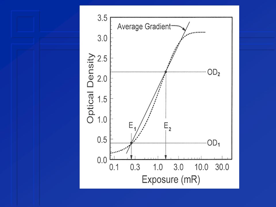
Contrast Contrast of a radiographic film is related to the slope of the H&D curve: Regions of higher slope have higher contrast Regions of reduced slope (e.g., the toe and shoulder) have lower contrast A single number, which defines the overall contrast of a given type of radiographic film, is the average gradient
 have lower contrast. A single number, which defines the overall contrast of a given type of radiographic film, is the average gradient.")
31
Average gradient OD1 = 0.25 + base + fog OD2 = 2.0 + base + fog
Average gradients for radiographic film range from 2.5 to 3.5

33
Scattered radiation For virtually all radiographic procedures except mammography, most photon interactions in soft tissue produce scattered x-ray photons Detection of scattered photons causes film darkening but does not add information content to the image

35
Effect of collimation As the field of view is reduced, the scatter is reduced An easy way to reduce the amount of x-ray scatter is by collimating the x-ray field to include only the anatomy of interest and no more

36
Antiscatter grid An antiscatter grid is placed between the patient and the screen-film cassette The grid uses geometry to reduce the amount of scattered reaching the detector

38
Antiscatter grid (cont.)
Antiscatter grid is composed of a series of small slits, aligned with the focal spot, that are separated by highly attenuating septa Primary x-rays have a higher chance of passing through the slits unattenuated by the adjacent septa Septa (grid bars) are usually made of lead; openings (interspaces) between the bars can be made of carbon fiber, aluminum, or even paper
 are usually made of lead; openings (interspaces) between the bars can be made of carbon fiber, aluminum, or even paper.")
41
Bar Phantom Setup Who is this?

42
The Rise & Fall of Joe Camel

43
Focal Spot Size Varies with Technique
Electron beam focuses more poorly at high mA or low kV Blooming increase of focal spot size with increasing mA more of a problem at low kV’s more blooming perpendicular to cathode-anode axis kilovoltage effects size decreases slightly with increasing kVp size always measured & specified at particular technique

44
Off-Axis Variation focal spot measurements normally made on central ray apparent focal spot size changes in anode-cathode direction smaller toward anode side larger toward cathode side less effect in cross-axis direction

45
Focal Spot Size Trade-off Focal Spot Size most critical for
heat vs. resolving power exposure time vs. resolving power Focal Spot Size most critical for magnification mammography

46
Modulation Transfer Function (MTF)
How well information reproduced (fraction of contrast retained) at various input spatial frequencies
 at various input spatial frequencies.")
47
Modulation Transfer Function (MTF)
Fraction of contrast reproduced as a function of frequency Freq. = line pairs / cm 1 MTF 50% Recorded Contrast (reduced by blur) Contrast provided to film frequency
 Contrast provided. to film. frequency.")
48
MTF If MTF = 1 all contrast reproduced at this frequency
Contrast provided to film Recorded Contrast

49
MTF If MTF = 0.5 half of contrast reproduced at this frequency
Contrast provided to film Recorded Contrast

50
MTF If MTF = 0 no contrast reproduced at this frequency
Contrast provided to film Recorded Contrast

51
Modulation Transfer Function (MTF)
value between 0 and 1 MTF = 1 indicates all information reproduced at this frequency MTF = 0 indicates no information reproduced at this frequency

52
Component MTF Each component of imaging system has its own MTF
each component retains a fraction of contrast as function of frequency System MTF is product of MTF’s for each component.

53
Modulation Transfer Function (MTF)
Since MTF is between 0 and 1, composite MTF <= MTF of poorest component 1/2 * 1/3 * 1/4 * 1/5 = ? ? < 1/5

54
Film MTF Resolves 10-20 line pairs per mm
MTF ~ 1 for clinical applications

55
Focal Spot MTF Function of magnification
Deteriorates with increased magnification all clinical imaging involves some magnification The larger the focal spot, the more deterioration of MTF with increased magnification

56
Imaging Screen MTF improves with magnification Why?
magnification of an object of given frequency reduces frequency seen by screen

57
Focal Spot & Screen MTF focal spot MTF degrades with magnification
screen MTF improves with magnification film MTF unaffected with magnification best system resolution detail screens minimize magnification BUT detail screens decrease speed increase exposure time more potential for motion

58
Imaging Physics Wrap Up

59
Object Motion independent of magnification depends on
exposure time velocity If motion < 0.1 mm usually doesn’t contribute to unsharpness If motion > 1 mm, it generally dominates

60
Quantum Mottle statistical fluctuation in # of x-ray photons used by imaging system to form image quantum mottle independent of geometric (radiographic) magnification for a given density the same # of photons must strike a given area of screen/film
 magnification. for a given density the same # of photons must strike a given area of screen/film.")
61
Quantum Mottle quantum mottle directly affected by photographic (optical) magnification less photons per unit area of image

62
Quantum Mottle influences perception of low-contrast objects with poorly defined borders not well addressed by MTF measures sharp high contrast borders geometric (radiographic) magnification may improve visibility of low contrast objects quantum mottle noise does not increase
 magnification may improve visibility of low contrast objects. quantum mottle noise does not increase.")
63
Skin Exposure & Magnification
Patient closer to x-ray tube Increases exposure grids not used with magnification work because of air gap Decreases exposure No bucky factor loss (3-6) Grid Film
 Grid. Film.")
64
Skin Exposure & Magnification
Increase is less than calculated by inverse square law for 2X mag skin not twice as close routine radiography also involves some magnification smaller volume of patient exposed high speed screen/film sometimes used for magnification Grid Film

65
Ilse, we’ll always have physics.
You Must Remember This Ilse, we’ll always have physics. The End

Similar presentations