Download presentation
Presentation is loading. Please wait.

1
The Neurological Exam Made Simple

2
Syndrome Diagnosis (Anatomical Localization)
1. Pattern (syndrome) Recognition 2. Nine (Anatomic) syndrome patterns
 Recognition. 2. Nine (Anatomic) syndrome patterns.")
3
Nine Syndrome Patterns
Cortical Sub-Cortical Brainstem Cerebellum Spinal cord Nerve root Peripheral nerve Neuromuscular junction Muscle

4
1. Muscle – Proximal symmetric weakness without sensory loss
History Lower Ext – difficulty rising from sitting position Upper Ext – difficulty lifting grocery bags, small children etc., Normal sensation – may have myalgia or cramps

5
1. Muscle (cont’) Exam Proximal symmetric weakness without sensory loss Muscles – normal size, no atrophy or fasciculations -- Tone and DTRs are normal to slightly decreased

6
Proximal Weakness

7
2. Neuromuscular Junction Resembles muscle: proximal variable weakness
History Fatigability (waxing and waning weakness) Patient fatigues with prolonged activity (myasthenia gravis) Patient strength improves with activity (myasthenia syndrome)
 Patient fatigues with prolonged activity (myasthenia gravis) Patient strength improves with activity (myasthenia syndrome)")
8
2. Neuromuscular Junction (cont’)
Exam – resembles muscle (proximal weakness) Fatigability of proximal muscles without sensory loss Looses strength after exercise (eg., ptosis after sustained upward gaze) Muscles normal size, no atrophy or fasciculations Normal tone and DTRs
 Fatigability of proximal muscles without sensory loss. Looses strength after exercise (eg., ptosis after sustained upward gaze) Muscles normal size, no atrophy or fasciculations. Normal tone and DTRs.")
9
Variable Weakness

10
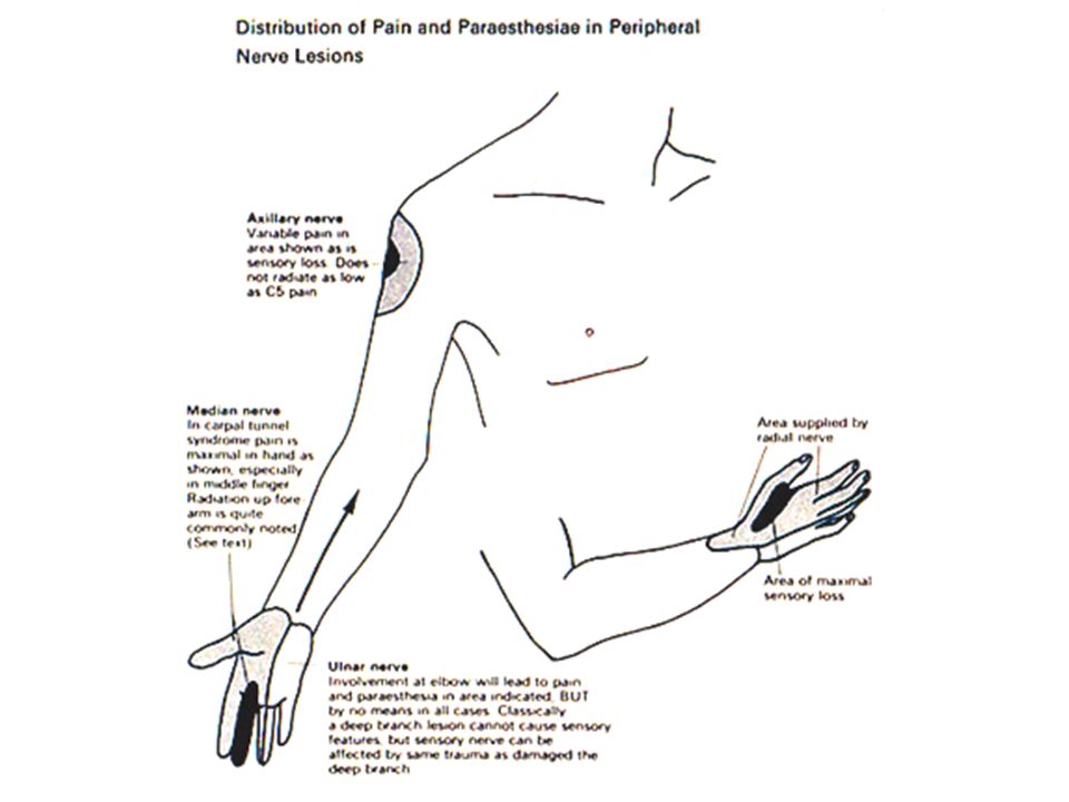
3. Peripheral Nerve Distal Weakness
History Lower ext – trips, drags feet, wears out toes of shoes Upper ext – drops objects, problems with grip Asymmetric weakness – localized to involved nerve (compression syndromes) Symmetric weakness – secondary to metabolic changes (eg., diabetes, renal etc) Muscle atrophy, twitching or quivering (fasciculations) Sensory changes - paresthesias
 Symmetric weakness – secondary to metabolic changes (eg., diabetes, renal etc) Muscle atrophy, twitching or quivering (fasciculations) Sensory changes - paresthesias.")
11
Clinical Findings in Upper and Lower Motor Neuron Defects
Upper motor neuron defect Spastic weakness No significant muscle atrophy No fasciculations and fibrillations Hyperreflexia Babinski’s reflex may be present Lower motor neuron defect Flaccid weakness Significant atrophy Fasciculations and fibrillations Hyporeflexia No Babinski’s reflex

12
3. Peripheral Nerve (cont’)
Exam Distal often asymmetric weakness Atrophy and Fasciculations Sensory loss Muscle tone normal or slightly decreased DTRs decreased Autonomic changes Trophic changes – smooth shinny skin Vasomotor changes – swelling or temperature dysregulation, loss of hair or nails

16
Nerve Hypertrophy

18
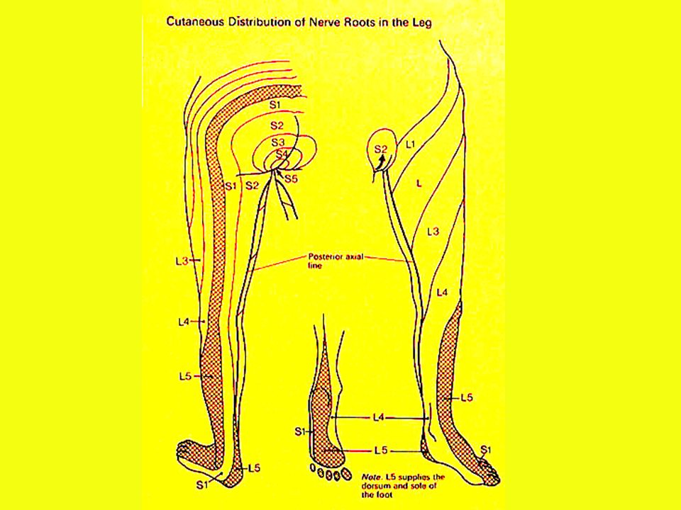
4. Nerve Root *Pain is the hallmark
History – sharp, stabbing, hot, electric, shooting or radiating pain Resembles peripheral nerve but weakness may be proximal or distal depending on the involved nerve root Lower ext L5 – S1 is most common; distal Upper ext C5-C6 is most common: proximal

20
4. Nerve Root (cont’) Exam Distal often asymmetric weakness
Atrophy and fasciculations Tone normal or decreased DTR decreased or absent in involved muscles Sensory loss (dermatomal) Maneuvers that stretch the nerve root increase pain ( eg., valsalva, SLR etc.,)
 Maneuvers that stretch the nerve root increase pain ( eg., valsalva, SLR etc.,)")
23
5. Spinal Cord - Triad of Symptoms
1. Sensory level - Pathognomonic 2. Distal symmetric, spastic weakness (UMN) mimics peripheral nerve 3. Bladder and bowel dysfunction due to autonomic fibers in spinal cord
 mimics peripheral nerve 3. Bladder and bowel dysfunction due to autonomic fibers in spinal cord")
24
5. Spinal Cord (cont’) History
Lower ext. weakness – drags toes or trips Upper ext. weakness – drops objects or problem with grip Symmetric – both legs or both arms and legs equally Sensory complaint – belt, band, girdle or tightness around trunk or abdomen Sphincter dysfunction – retention or incontinence of bladder more common than bowel

25
5. Spinal Cord (cont’) Sensory level (tested with pinprick) Exam
Weakness more common in legs than arms Urinary retention or incontinence Superficial reflexes decreased (anal wink, bulbocavernosus and cremasteric) UMN damage - distal > proximal weakness (weakness of extensor and (anti-gravity muscles greater than flexors)
 UMN damage - distal > proximal weakness (weakness of extensor and (anti-gravity muscles greater than flexors)")
26
Commissural syndrome

27
Sensory loss with sacral sparing due to the intramedullary lesion shown on the left, involving lateral spinothalamic tracts bilaterally.

28
Brown-Sequard Syndrome

29
6. Brainstem – Ipsilateral cranial nerve and contralateral long tract signs (essentially the spinal cord with embedded cranial nerves) History Long tracts (hemiparesis or hemisensory loss) Cranial nerves (the 6 Ds) Diplopia Dysarthria Dysphagia Dizziness Deafness Decreases strength or sensation over the face (crossed signs may be bilateral)
 Cranial nerves (the 6 Ds) Diplopia. Dysarthria. Dysphagia. Dizziness. Deafness. Decreases strength or sensation over the face (crossed signs may be bilateral)")
30
Posterior fossa

31
Major nervous system connections

32
6. Brainstem (cont’) Exam
Cranial nerves Ipsilateral -ptosis, pupillary abnormality, extraocular paralysis, diplopia, nystagmus, decreased corneal and blink reflexes, facial weakness or numbness, deafness, vertigo, dysarthria, dysphagia, weakness or deviation of the palate, decreased gag reflex, weakness of neck, shoulders or tongue Long tracts – Contralateral distal extensor (UMN) hemiparesis, increased DTRs, spasticity, Babinski, loss of some and possibly all modalities
 hemiparesis, increased DTRs, spasticity, Babinski, loss of some and possibly all modalities.")
33
Distribution of pain and temperature sensation loss characteristic of lesions at the posterior fossa level.

34
Sensory Pathways

35
7. Cerebellum - In-coordination, clumsiness, intention tremor
7. Cerebellum - In-coordination, clumsiness, intention tremor *(smooths and refines voluntary movements) History Clumsiness in lower ext. –staggers, drunken walk Clumsiness in upper ext. – difficulty with targeting movements (such as lighting cigarettes, keys in car ignition) and intention tremor Brainstem symptoms are common with cerebellar disease and vice versa
 History. Clumsiness in lower ext. –staggers, drunken walk. Clumsiness in upper ext. – difficulty with targeting movements (such as lighting cigarettes, keys in car ignition) and intention tremor. Brainstem symptoms are common with cerebellar disease and vice versa.")
36
7. Cerebellum (cont’) Exam
Lower Ext. - Gait (staggering, wide based, ataxic, difficulty with tandem walking, Heel-shin, or tracing patterns on floor with toe Upper ext. – Intention tremor, difficulty targeting movements (such as finger-nose, heel shin) difficulty with rapid alternating movements (dysdiadochokinesis)
 difficulty with rapid alternating movements (dysdiadochokinesis)")
37
8. Sub-cortical verses 9. Cortical
History – generally diagnosed by Specific cortical defects Pattern of motor and sensory defects The type of sensory defects Presence of visual field defects

38
Sub-cortical v Cortical (cont’)
1.Specific Cortical Defects Language (dominant hemisphere) Speech – aphasia Writing – agraphia Reading – alexia Comprehension (eg., apraxia) Visual-spatial (Non-dominant hemisphere) Denial or neglect of physical signs and symptoms (agnosia)
 Speech – aphasia. Writing – agraphia. Reading – alexia. Comprehension (eg., apraxia) Visual-spatial (Non-dominant hemisphere) Denial or neglect of physical signs and symptoms (agnosia)")
39
Sub-cortical v Cortical (cont’)
2. Patterns of motor & sensory defects (homunculus) Cortical lesions - complete paralysis or sensory loss of face and arm (spares legs) Subcortical lesions – complete paralysis or sensory loss of face, arm, trunk and legs
 Cortical lesions - complete paralysis or sensory loss of face and arm (spares legs) Subcortical lesions – complete paralysis or sensory loss of face, arm, trunk and legs.")
40
Sub-cortical v Cortical (cont’)
3. Type of sensory defect *(most primary sensory modalities reach consciousness in the thalamus and do not require the cortex for their perception) Cortical lesions – patients can still feel pain, touch, vibration and position but have impaired higher sensory processing, ie., graphesthesia or astereognosis) Subcortical defect – patient complains of significant numbness
 Cortical lesions – patients can still feel pain, touch, vibration and position but have impaired higher sensory processing, ie., graphesthesia or astereognosis) Subcortical defect – patient complains of significant numbness.")
41
Sub-Cortical v Cortical (cont’)
4. Visual field defects *(fibers run subcortically) Cortical – no visual field defect unless occipital lobe involved (cortical blindness-Anton’s syndrome) Sub-cortical has visual field defects
 Cortical – no visual field defect unless occipital lobe involved (cortical blindness-Anton’s syndrome) Sub-cortical has visual field defects.")
42
Sub-cortical v Cortical (cont’) Exam
1. Cortical – aphasia, visual-spatial dysfunction or seizures 2. Motor – UMN weakness Cortical - Face and arm Sub-cortical - Face, arm, trunk and leg

43
Suprathalamic syndrome

44
Thalamic syndrome

45
Sub-cortical v Cortical (cont’) Exam
3. Sensory Cortical – impaired higher sensory processing, (eg.,graphesthesia or astereognosis) with relatively normal sensation Sub-cortical – decrease primary sensory modalities, (eg., pinprick and touch etc.,) 4. Visual Cortical – no defect unless occipital lobe Sub-cortical – visual field defects
 with relatively normal sensation. Sub-cortical – decrease primary sensory modalities, (eg., pinprick and touch etc.,) 4. Visual. Cortical – no defect unless occipital lobe. Sub-cortical – visual field defects.")
46
Visual Field Defects

47
Cortical centers related to vision and ocular movement

48
Differential Diagnosis
Axis levels Atrophy Fascic- ulations Babinski Hyper- reflexia Pain, severe Sensory Loss Weakness Cortex + Brainstem Cerebellum Cord Root Nerve NMJ Muscle Another important point is to realize is that sensory loss and weakness is pervasive throughout the levels. That is why you need to be able to distinguish the patterns innervated by nerve, nerve root, spinal tracts, and cerebral hemispheres. This is where you earn your stripes and the excitement of pinpointing an anatomic level hence the pathology affecting the level….or more precisely ……………….YOU HAVE JUST MADE THE DIAGNOSIS. Lets not get ahead of ourselves because you will need more information in the slides which follows in order to accomplish this. The process in the neurologic diagnosis is an elegant exercise for your brain and will serve you well no matter what field of medicine you enter…..remember you have to have the right diagnosis before any treatment can be considered.

Similar presentations


 (MND) are a group of neurological disorders that selectively affect motor neurons.>")


 Is it a stroke? (2) What part of the brain is affected? (3) What caused this stroke? Is it a haemorrhage or an infarct? Can.>")


