Download presentation
Presentation is loading. Please wait.

2
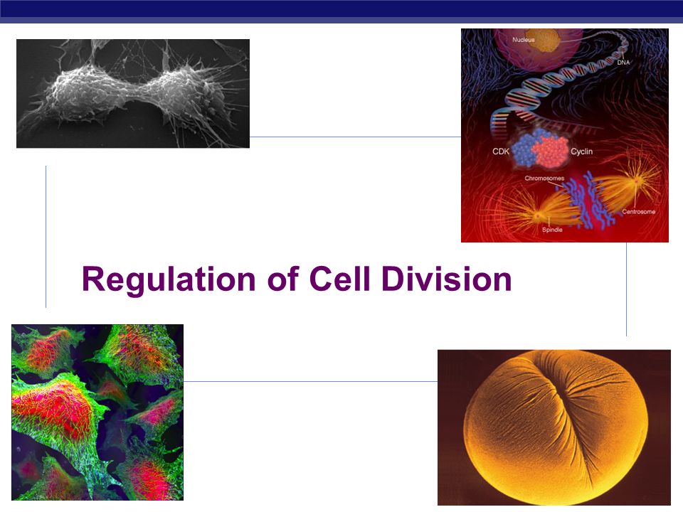
AP Biology 2008-2009 Regulation of Cell Division

3
AP Biology Coordination of cell division A multicellular organism needs to coordinate cell division across different tissues & organs critical for normal growth, development & maintenance coordinate timing of cell division coordinate rates of cell division not all cells can have the same cell cycle

4
AP Biology G2G2 S G1G1 M metaphase prophase anaphase telophase interphase (G 1, S, G 2 phases) mitosis (M) cytokinesis (C) C Frequency of cell division varies by cell type embryo cell cycle < 20 minute skin cells divide frequently throughout life 12-24 hours cycle New skin every 4-6 weeks liver cells retain ability to divide, but keep it in reserve divide once every year or two mature nerve cells & muscle cells do not divide at all after maturity permanently in G 0 Frequency of cell division
 mitosis (M) cytokinesis (C) C Frequency of cell division varies by cell type embryo cell cycle < 20 minute skin cells divide frequently throughout life hours cycle New skin every 4-6 weeks liver cells retain ability to divide, but keep it in reserve divide once every year or two mature nerve cells & muscle cells do not divide at all after maturity permanently in G 0 Frequency of cell division")
5
AP Biology Overview of Cell Cycle Control Two irreversible points in cell cycle replication of genetic material separation of sister chromatids Checkpoints process is assessed & possibly halted centromere sister chromatids single-stranded chromosomes double-stranded chromosomes

6
AP Biology Checkpoint control system Checkpoints cell cycle controlled by STOP & GO chemical signals at critical points signals indicate if key cellular processes have been completed correctly

7
AP Biology Checkpoint control system 3 major checkpoints: G 1 /S can DNA synthesis begin? G 2 /M has DNA synthesis been completed correctly? commitment to mitosis spindle checkpoint are all chromosomes attached to spindle? can sister chromatids separate correctly?

8
AP Biology G 1 /S checkpoint G 1 /S checkpoint is most critical primary decision point “restriction point” if cell receives “GO” signal, it divides internal signals: cell growth (size), cell nutrition external signals: “growth factors” if cell does not receive signal, it exits cycle & switches to G 0 phase non-dividing, working state
, cell nutrition external signals: growth factors if cell does not receive signal, it exits cycle & switches to G 0 phase non-dividing, working state")
9
AP Biology G 0 phase G 0 phase non-dividing, differentiated state most human cells in G 0 phase liver cells in G 0, but can be “called back” to cell cycle by external cues nerve & muscle cells highly specialized arrested in G 0 & can never divide

10
AP Biology How do cells know when to divide? cell communication signals chemical signals in cytoplasm give cue signals usually mean proteins activators inhibitors Activation of cell division experimental evidence: Can you explain this?

11
AP Biology “Go-ahead” signals Protein signals that promote cell growth & division internal signals “promoting factors” external signals “growth factors” Primary mechanism of control phosphorylation kinase enzymes either activates or inactivates cell signals

12
AP Biology Cell cycle s ignals Cell cycle controls cyclins regulatory proteins levels cycle in the cell Cdks cyclin-dependent kinases phosphorylates cellular proteins activates or inactivates proteins Cdk-cyclin complex MPF- “maturation promoting factor” triggers passage through different stages of cell cycle activated Cdk inactivated Cdk

13
AP Biology Cyclin levels rise sharply in interphase, fall abruptly during mitosis Peaks in the activity of cyclin-Cdk complex, MPF, correspond to M phase

14
AP Biology Cdk / G 1 cyclin Cdk / G 2 cyclin (MPF) G2G2 S G1G1 C M G 2 / M checkpoint G 1 / S checkpoint APC Active Inactive Active Inactive Active mitosis cytokinesis MPF = Maturation Promoting Factor APC = Anaphase Promoting Complex Replication completed DNA integrity Chromosomes attached at metaphase plate Spindle checkpoint Growth factors Nutritional state of cell Size of cell
 G2G2 S G1G1 C M G 2 / M checkpoint G 1 / S checkpoint APC Active Inactive Active Inactive Active mitosis cytokinesis MPF = Maturation Promoting Factor APC = Anaphase Promoting Complex Replication completed DNA integrity Chromosomes attached at metaphase plate Spindle checkpoint Growth factors Nutritional state of cell Size of cell")
15
AP Biology External signals Growth factors coordination between cells protein signals released by body cells that stimulate other cells to divide density-dependent inhibition crowded cells stop dividing each cell binds a bit of growth factor not enough activator left to trigger division in any one cell anchorage dependence to divide cells must be attached to a substrate “touch sensor” receptors

16
AP Biology Example of a Growth Factor Platelet Derived Growth Factor (PDGF) made by platelets in blood clots binding of PDGF to cell receptors stimulates cell division in connective tissue heal wounds Don’t forget to mention erythropoietin! (EPO)
.")
17
AP Biology Learning Check Summarize the internal external methods used to control cell division What happens if these check points fail??

18
Loss of Normal Growth Control Cancer cell division Fourth or later mutation Third mutation Second mutation First mutation Uncontrolled growth Cell Suicide or Apoptosis Cell damage— no repair Normal cell division

19
AP Biology Cancer & Cell Growth Cancer is essentially a failure of cell division control unrestrained, uncontrolled cell growth What control is lost? lose checkpoint stops Tumor supressor gene p53 plays a key role in G 1 /S restriction point p53 protein halts cell division if it detects damaged DNA options: stimulates repair enzymes to fix DNA forces cell into G 0 resting stage keeps cell in G 1 arrest causes apoptosis of damaged cell ALL cancers have to shut down p53 activity p53 is the Cell Cycle Enforcer

20
AP Biology DNA damage is caused by heat, radiation, or chemicals. p53 allows cells with repaired DNA to divide. Step 1 DNA damage is caused by heat, radiation, or chemicals. Step 1 Step 2 Damaged cells continue to divide. If other damage accumulates, the cell can turn cancerous. Step 3 p53 triggers the destruction of cells damaged beyond repair. ABNORMAL p53 NORMAL p53 abnormal p53 protein cancer cell Step 3 The p53 protein fails to stop cell division and repair DNA. Cell divides without repair to damaged DNA. Cell division stops, and p53 triggers enzymes to repair damaged region. Step 2 DNA repair enzyme p53 protein p53 protein p53 — master regulator gene

21
Example of Normal Growth Cell migration Dermis Dividing cells in basal layer Dead cells shed from outer surface Epidermis

22
The Beginning of Cancerous Growth Underlying tissue During the development of skin cancer, the normal balance between cell division and cell loss is disrupted

23
Tumors (Neoplasms) Underlying tissue Tumors increase in size because new cells are being produced in greater numbers than needed.
 Underlying tissue Tumors increase in size because new cells are being produced in greater numbers than needed.")
24
Invasion and Metastasis 3 Cancer cells reinvade and grow at new location 1 Cancer cells invade surrounding tissues and blood vessels 2 Cancer cells are transported by the circulatory system to distant sites

25
AP Biology Growth Factors and Cancer Growth factors can create cancers proto-oncogenes normally activates cell division growth factor genes become oncogenes (cancer-causing) when mutated if switched “ON” can cause cancer example: RAS (activates cyclins) tumor-suppressor genes normally inhibits cell division if switched “OFF” can cause cancer example: p53
 when mutated if switched ON can cause cancer example: RAS (activates cyclins) tumor-suppressor genes normally inhibits cell division if switched OFF can cause cancer example: p53")
26
AP Biology Development of Cancer Cancer develops only after a cell experiences ~6 key mutations (“hits”) unlimited growth turn on growth promoter genes ignore checkpoints turn off tumor suppressor genes (p53) escape apoptosis turn off suicide genes immortality = unlimited divisions turn on chromosome maintenance genes promotes blood vessel growth turn on blood vessel growth genes overcome anchor & density dependence turn off touch-sensor gene It ’ s like an out-of-control car with many systems failing!
 unlimited growth turn on growth promoter genes ignore checkpoints turn off tumor suppressor genes (p53) escape apoptosis turn off suicide genes immortality = unlimited divisions turn on chromosome maintenance genes promotes blood vessel growth turn on blood vessel growth genes overcome anchor & density dependence turn off touch-sensor gene It ’ s like an out-of-control car with many systems failing!")
27
AP Biology What causes these “hits”? Mutations in cells can be triggered by UV radiation chemical exposure radiation exposure heat cigarette smoke pollution age genetics

28
AP Biology Tumors Mass of abnormal cells Benign tumor abnormal cells remain at original site as a lump p53 has halted cell divisions most do not cause serious problems & can be removed by surgery Malignant tumor cells leave original site lose attachment to nearby cells carried by blood & lymph system to other tissues start more tumors = metastasis impair functions of organs throughout body

29
Malignant versus Benign Tumors Malignant (cancer) cells invade neighboring tissues, enter blood vessels, and metastasize to different sites Time Benign (not cancer) tumor cells grow only locally and cannot spread by invasion or metastasis
 cells invade neighboring tissues, enter blood vessels, and metastasize to different sites Time Benign (not cancer) tumor cells grow only locally and cannot spread by invasion or metastasis")
30
Why Cancer Is Potentially Dangerous Melanoma cells travel through bloodstream Melanoma (initial tumor) Brain Liver Cancer cells in the liver would be called metastatic melanoma, not liver cancer. Metastases share the name of the original (“primary”) tumor. 1 3 2
 tumor")
31
AP Biology Traditional treatments for cancers Treatments target rapidly dividing cells high-energy radiation kills rapidly dividing cells chemotherapy stop DNA replication stop mitosis & cytokinesis stop blood vessel growth

32
AP Biology 2008-2009 Any Questions??

Similar presentations


 Coordination of cell division A multicellular organism needs to coordinate cell division across different tissues.>")


