Download presentation
Presentation is loading. Please wait.

1
IB Fertilization & Pregnancy

2
IB Assessment Statement
Describe the process of fertilization, including the acrosome reaction, penetration of the egg membrane by a sperm and the cortical reaction.

3
Fertilization Fertilisation occurs in the oviducts (fallopian tubes)
pH of vagina is acidic and pH of semen is basic. Thus they neutralize each other.

4
Fertilization Contraction of the uterus and oviducts help move the sperm into the oviducts. One or more sperm will reach the oocyte in the oviduct.

5
Fertilisation The oocyte is surrounded by a coat that consists of a glycoprotein called a zona pellucida . The zona pellucida must be crossed by the sperm.

6
Fertilisation Contact between the zona pellucida and proteins in the sperm cells membrane trigger a the acrosome reaction.

7
Fertilisation The acrosome vesicle fuses with the sperm plasma membrane and releases enzymes that digest a path through the zona pellucida.

8
Fertilisation Hydrolytic enzyme that are located in the sperm’s head, called acrosomes. These acrosomes enzymes digest a pathway for the sperm to enter the oocyte. This process is called capacitation.

9
Fertilisation The membrane of the sperm cell and the ovum fuse together. At the same time this results in a release of Ca2+ from the endoplasmic reticulum.

10
Fertilisation The cortical vesicle fuse with the plasma membrane of egg cell releasing enzymes that destroy the sperm binding proteins on the zona pellucida. This prevents polyspermy. (more than one sperm from entering

11
Fertilisation The release of Ca2+ also activate meiosis and prepare the oocyte cell for completion Meiosis II. Oocyte undergoes meiosis II.

12
Fertilisation The head of the sperms contain its nucleus.
The sperms nucleus fuses with the oocyte nucleus for the final stage of fertilisation.

13
Fertilisation The new diploid nucleus undergoes mitosis
The division of cytoplasm occurs forming the first two cells of the embryo.

14
Fertilization & Pregnancy
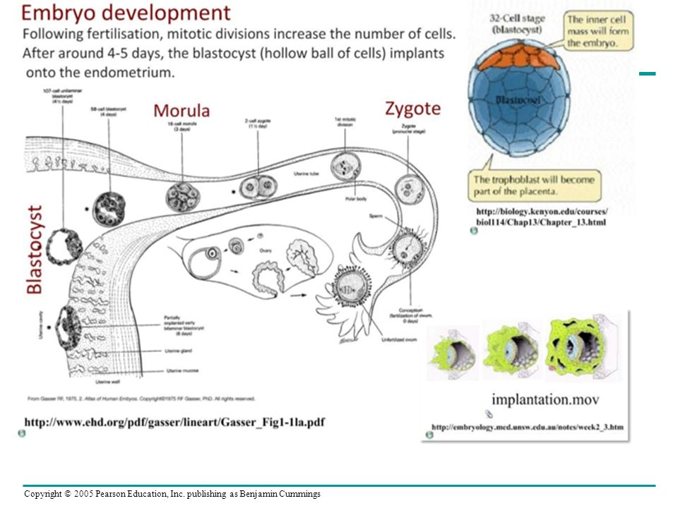
Fertilization and Implantation If an egg is fertilized, a zygote forms and begins to undergo cell division (mitosis) as it travels to the uterus.
 as it travels to the uterus.")
15
Early development of Zygote
Fertilization occurs in the oviduct

17
IB LEARNING OBJECTIVE Outline early embryo development up to the implantation of the blastocyst.

18
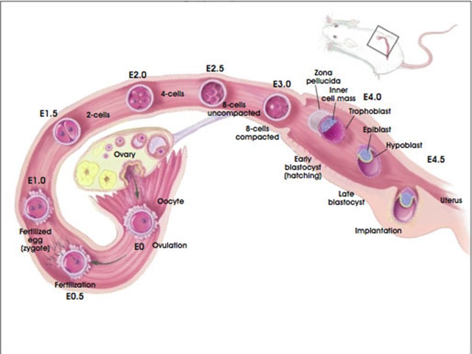
Early development of Zygote
After fertilization, the zygote undergoes cleavage and develops into a blastocyst before implantation in the endometrium

19
Early development of Zygote
Zygote is transplant to the uterus by ciliary (small moving hair-like proteins) action in the oviduct The fertilized egg undergoes cleavage. Cleavage is the mitotic division of the zygote into a mass of daughter cells.
 action in the oviduct. The fertilized egg undergoes cleavage. Cleavage is the mitotic division of the zygote into a mass of daughter cells.")
20
Early development of Zygote
When the embryo reaches the uterus is has undergone both cleavage and has formed a blastocysts. A blastocyst is a tiny solid ball of cells.

22
Early development of Zygote
In humans, by 7 days the blastocysts consists of about 100 cells It becomes imbedded in the endometrium, a process called implantation.

23
Early development of Zygote
The inner cell mass of the blastocyst eventually becomes the fetus. Once implanted the embryo starts to receive nutrients from the endometrium of the uterus wall.

26
IB LEARNING QBJECTIVE State that the fetus is supported and protected by the amniotic sac and amniotic fluid.

27
Early development of Zygote
Pregnancy, or gestation, is the condition of carrying one or more embryos in the uterus

28
Gestation – zygote to embryo to fetus in human
Gestation – the period of development in the mother’s body, lasting from conception to birth. First 2 months of gestation the developing baby is referred as an embryo. The embryo is protected in amniotic fluid and amniotic sac.

29
Early Development Amniotic sac A fluid-filled amniotic sac, which cushions and protects the developing embryo. Placenta Umbilical cord Uterus Amnion Fetus

30
Early Development –Amniotic Fluid
Amniotic sac The fetus floats in the amniotic fluid. This fluid acts as a shock absorber. Placenta Umbilical cord Uterus Amnion Fetus

31
IB LEARNING OBJECTIVE State that materials are exchanged between the maternal and fetal blood in the placenta.

32
During its first 2 to 4 weeks, the embryo obtains nutrients directly from the endometrium
Meanwhile, the outer layer of the blastocyst mingles with the endometrium and eventually forms the placenta Blood from the embryo travels to the placenta through arteries of the umbilical cord and returns via the umbilical vein

33
The outer layer of the embryo gives rise to the placenta & the maternal endometrium
It is a disc shaped structure that allows for an exchange of material between fetus and mother

34
The outer layer of the embryo gives rise to the placenta & the maternal endometrium
It is a disc shaped structure that allows for an exchange of material between fetus and mother Maternal arteries Maternal veins Placenta Maternal portion of placenta Umbilical cord Chorionic villus containing fetal capillaries Fetal portion of placenta (chorion) Maternal blood pools Uterus Umbilical arteries Fetal arteriole Fetal venule Umbilical vein Umbilical cord
 Maternal blood. pools. Uterus. Umbilical arteries. Fetal arteriole. Fetal venule. Umbilical vein. Umbilical cord.")
35
The placenta

36
The Placenta A disc shaped structure composed of maternal endometrial and fetal membrane. Exchange in the placenta – is by diffusion and active transport, and involve: Respiratory gases – Oxygen and carbon dioxide Water Excretory products (urea) Antibodies ( immunity from diseases)
 Antibodies ( immunity from diseases)")
37
Maternal, foetal exchanges across placenta
The foetus develops and grows using materials obtained by exchange across the placental wall from mother to child. Excretory products are exchanged in the opposite direction from child to mother.

38
IB Learning Objective Explain how the structure and functions of the placenta, including its hormonal role in secretion of estrogen and progesterone, maintain pregnancy.

39
Structure and function of placenta
The female blood supply which supplies the foetus with oxygen and nutrient. It will also remove waste from the foetal blood and excrete this through the maternal systems.

42
Structure and function of placenta
a. Umbilical cord connects the fetus to the placenta b. There are two umbilical arteries that carry the deoxygenated fetal blood to the placenta. a) Umbilical cord connects the foetus to the placenta b) There are two umbilical arteries that carry the deoxygenated blood to the placenta. c) The single umbilical vein returns the blood to the rest of the foetal circulation. d) The placenta is normally about 190mm wide and 20 mm deep. The human placenta is more deeply integrated into the maternal tissue than any other animal. e) The myometrium is composed on smooth muscle that produces the contraction in labor. f) The endometrium which is maintained through out pregnancy by progesterone. Initially from the corpus luteum and later from the placenta itself. g) The female blood supply which supplies the foetus with oxygen and nutrient. It will also remove waste from the foetal blood and excrete this through the maternal systems. h) Open ended blood arterioles and capillaries that produce the inter-villous 'blood lakes'. i) Inter-villus spaces filled with maternal blood. These surround the placental villi and allow for very efficient exchange. j) Placental-Villi with large surface area for the exchange of nutrient and waste.As previously mentioned there are few membranes between the maternal blood and the foetal blood an association that is closer than an other mammal
 Umbilical cord connects the foetus to the placenta. b) There are two umbilical arteries that carry the deoxygenated blood to the placenta. c) The single umbilical vein returns the blood to the rest of the foetal circulation. d) The placenta is normally about 190mm wide and 20 mm deep. The human placenta is more deeply integrated into the maternal tissue than any other animal. e) The myometrium is composed on smooth muscle that produces the contraction in labor. f) The endometrium which is maintained through out pregnancy by progesterone. Initially from the corpus luteum and later from the placenta itself. g) The female blood supply which supplies the foetus with oxygen and nutrient. It will also remove waste from the foetal blood and excrete this through the maternal systems. h) Open ended blood arterioles and capillaries that produce the inter-villous blood lakes . i) Inter-villus spaces filled with maternal blood. These surround the placental villi and allow for very efficient exchange. j) Placental-Villi with large surface area for the exchange of nutrient and waste.As previously mentioned there are few membranes between the maternal blood and the foetal blood an association that is closer than an other mammal.")
43
Structure and function of placenta
c. The single umbilical vein returns the oxygenated blood to the rest of the foetal circulation. d. The placenta is normally about 190mm wide and 20 mm deep. The human placenta is more deeply integrated into the maternal tissue than any other animal. a) Umbilical cord connects the foetus to the placenta b) There are two umbilical arteries that carry the deoxygenated blood to the placenta. c) The single umbilical vein returns the blood to the rest of the foetal circulation. d) The placenta is normally about 190mm wide and 20 mm deep. The human placenta is more deeply integrated into the maternal tissue than any other animal. e) The myometrium is composed on smooth muscle that produces the contraction in labor. f) The endometrium which is maintained through out pregnancy by progesterone. Initially from the corpus luteum and later from the placenta itself. g) The female blood supply which supplies the foetus with oxygen and nutrient. It will also remove waste from the foetal blood and excrete this through the maternal systems. h) Open ended blood arterioles and capillaries that produce the inter-villous 'blood lakes'. i) Inter-villus spaces filled with maternal blood. These surround the placental villi and allow for very efficient exchange. j) Placental-Villi with large surface area for the exchange of nutrient and waste.As previously mentioned there are few membranes between the maternal blood and the foetal blood an association that is closer than an other mammal
 Umbilical cord connects the foetus to the placenta. b) There are two umbilical arteries that carry the deoxygenated blood to the placenta. c) The single umbilical vein returns the blood to the rest of the foetal circulation. d) The placenta is normally about 190mm wide and 20 mm deep. The human placenta is more deeply integrated into the maternal tissue than any other animal. e) The myometrium is composed on smooth muscle that produces the contraction in labor. f) The endometrium which is maintained through out pregnancy by progesterone. Initially from the corpus luteum and later from the placenta itself. g) The female blood supply which supplies the foetus with oxygen and nutrient. It will also remove waste from the foetal blood and excrete this through the maternal systems. h) Open ended blood arterioles and capillaries that produce the inter-villous blood lakes . i) Inter-villus spaces filled with maternal blood. These surround the placental villi and allow for very efficient exchange. j) Placental-Villi with large surface area for the exchange of nutrient and waste.As previously mentioned there are few membranes between the maternal blood and the foetal blood an association that is closer than an other mammal.")
44
Structure and function of placenta
e) The myometrium is composed on smooth muscle in the uterus that produces the contraction in labor. f. The endometrium which is maintained through out pregnancy by progesterone. Initially from the corpus luteum and later from the placenta itself. a) Umbilical cord connects the foetus to the placenta b) There are two umbilical arteries that carry the deoxygenated blood to the placenta. c) The single umbilical vein returns the blood to the rest of the foetal circulation. d) The placenta is normally about 190mm wide and 20 mm deep. The human placenta is more deeply integrated into the maternal tissue than any other animal. e) The myometrium is composed on smooth muscle that produces the contraction in labor. f) The endometrium which is maintained through out pregnancy by progesterone. Initially from the corpus luteum and later from the placenta itself. g) The female blood supply which supplies the foetus with oxygen and nutrient. It will also remove waste from the foetal blood and excrete this through the maternal systems. h) Open ended blood arterioles and capillaries that produce the inter-villous 'blood lakes'. i) Inter-villus spaces filled with maternal blood. These surround the placental villi and allow for very efficient exchange. j) Placental-Villi with large surface area for the exchange of nutrient and waste.As previously mentioned there are few membranes between the maternal blood and the foetal blood an association that is closer than an other mammal
 The myometrium is composed on smooth muscle in the uterus that produces the contraction in labor. f. The endometrium which is maintained through out pregnancy by progesterone. Initially from the corpus luteum and later from the placenta itself. a) Umbilical cord connects the foetus to the placenta. b) There are two umbilical arteries that carry the deoxygenated blood to the placenta. c) The single umbilical vein returns the blood to the rest of the foetal circulation. d) The placenta is normally about 190mm wide and 20 mm deep. The human placenta is more deeply integrated into the maternal tissue than any other animal. e) The myometrium is composed on smooth muscle that produces the contraction in labor. f) The endometrium which is maintained through out pregnancy by progesterone. Initially from the corpus luteum and later from the placenta itself. g) The female blood supply which supplies the foetus with oxygen and nutrient. It will also remove waste from the foetal blood and excrete this through the maternal systems. h) Open ended blood arterioles and capillaries that produce the inter-villous blood lakes . i) Inter-villus spaces filled with maternal blood. These surround the placental villi and allow for very efficient exchange. j) Placental-Villi with large surface area for the exchange of nutrient and waste.As previously mentioned there are few membranes between the maternal blood and the foetal blood an association that is closer than an other mammal.")
45
Structure and function of placenta
g) The female blood supply which supplies the foetus with oxygen and nutrient. It will also remove waste from the foetal blood and excrete this through the maternal systems. h) Open ended blood arterioles and capillaries that produce the inter-villous 'blood lakes’. i) Inter-villus spaces filled with maternal blood. These surround the placental villi and allow for very efficient exchange. a) Umbilical cord connects the foetus to the placenta b) There are two umbilical arteries that carry the deoxygenated blood to the placenta. c) The single umbilical vein returns the blood to the rest of the foetal circulation. d) The placenta is normally about 190mm wide and 20 mm deep. The human placenta is more deeply integrated into the maternal tissue than any other animal. e) The myometrium is composed on smooth muscle that produces the contraction in labor. f) The endometrium which is maintained through out pregnancy by progesterone. Initially from the corpus luteum and later from the placenta itself. g) The female blood supply which supplies the foetus with oxygen and nutrient. It will also remove waste from the foetal blood and excrete this through the maternal systems. h) Open ended blood arterioles and capillaries that produce the inter-villous 'blood lakes'. i) Inter-villus spaces filled with maternal blood. These surround the placental villi and allow for very efficient exchange. j) Placental-Villi with large surface area for the exchange of nutrient and waste.As previously mentioned there are few membranes between the maternal blood and the foetal blood an association that is closer than an other mammal
 The female blood supply which supplies the foetus with oxygen and nutrient. It will also remove waste from the foetal blood and excrete this through the maternal systems. h) Open ended blood arterioles and capillaries that produce the inter-villous blood lakes’. i) Inter-villus spaces filled with maternal blood. These surround the placental villi and allow for very efficient exchange. a) Umbilical cord connects the foetus to the placenta. b) There are two umbilical arteries that carry the deoxygenated blood to the placenta. c) The single umbilical vein returns the blood to the rest of the foetal circulation. d) The placenta is normally about 190mm wide and 20 mm deep. The human placenta is more deeply integrated into the maternal tissue than any other animal. e) The myometrium is composed on smooth muscle that produces the contraction in labor. f) The endometrium which is maintained through out pregnancy by progesterone. Initially from the corpus luteum and later from the placenta itself. g) The female blood supply which supplies the foetus with oxygen and nutrient. It will also remove waste from the foetal blood and excrete this through the maternal systems. h) Open ended blood arterioles and capillaries that produce the inter-villous blood lakes . i) Inter-villus spaces filled with maternal blood. These surround the placental villi and allow for very efficient exchange. j) Placental-Villi with large surface area for the exchange of nutrient and waste.As previously mentioned there are few membranes between the maternal blood and the foetal blood an association that is closer than an other mammal.")
46
Structure and function of placenta
Open ended blood arterioles and capillaries that produce the inter-villous 'blood lakes'. .

47
Structure and function of placenta
Inter-villus spaces filled with maternal blood. These surround the placental villi and allow for very efficient exchange.

48
Structure and function of placenta
g) Placental-Villi with large surface area for the exchange of nutrient and waste. a) Umbilical cord connects the foetus to the placenta b) There are two umbilical arteries that carry the deoxygenated blood to the placenta. c) The single umbilical vein returns the blood to the rest of the foetal circulation. d) The placenta is normally about 190mm wide and 20 mm deep. The human placenta is more deeply integrated into the maternal tissue than any other animal. e) The myometrium is composed on smooth muscle that produces the contraction in labor. f) The endometrium which is maintained through out pregnancy by progesterone. Initially from the corpus luteum and later from the placenta itself. g) The female blood supply which supplies the foetus with oxygen and nutrient. It will also remove waste from the foetal blood and excrete this through the maternal systems. h) Open ended blood arterioles and capillaries that produce the inter-villous 'blood lakes'. i) Inter-villus spaces filled with maternal blood. These surround the placental villi and allow for very efficient exchange. j) Placental-Villi with large surface area for the exchange of nutrient and waste.As previously mentioned there are few membranes between the maternal blood and the foetal blood an association that is closer than an other mammal
 Placental-Villi with large surface area for the exchange of nutrient and waste. a) Umbilical cord connects the foetus to the placenta. b) There are two umbilical arteries that carry the deoxygenated blood to the placenta. c) The single umbilical vein returns the blood to the rest of the foetal circulation. d) The placenta is normally about 190mm wide and 20 mm deep. The human placenta is more deeply integrated into the maternal tissue than any other animal. e) The myometrium is composed on smooth muscle that produces the contraction in labor. f) The endometrium which is maintained through out pregnancy by progesterone. Initially from the corpus luteum and later from the placenta itself. g) The female blood supply which supplies the foetus with oxygen and nutrient. It will also remove waste from the foetal blood and excrete this through the maternal systems. h) Open ended blood arterioles and capillaries that produce the inter-villous blood lakes . i) Inter-villus spaces filled with maternal blood. These surround the placental villi and allow for very efficient exchange. j) Placental-Villi with large surface area for the exchange of nutrient and waste.As previously mentioned there are few membranes between the maternal blood and the foetal blood an association that is closer than an other mammal.")
49
Structure and function of placenta
Placental-Villi with large surface area for the exchange of nutrient and waste.

50
Structures that allow the Exchange of Material Across the Placenta

51
IB Learning Objective Explain the oxygen dissociation curves of adult hemoglobin, fetal hemoglobin and myoglobin.

52
Fetal Haemoglobin This means that the fetal haemoglobin will combine more readily oxygen than the mother’s haemoglobin at the same partial pressure.

53
Fetal Haemoglobin Why is this the case?
If the mother’s haemoglobin had a stronger affinity for oxygen than the fetus’s haemoglobin, then the oxygen would pass from the fetus to the mother.

55
IB Learning Objective Outline the role of HCG in early pregnancy. .

56
The placenta as an endocrine gland
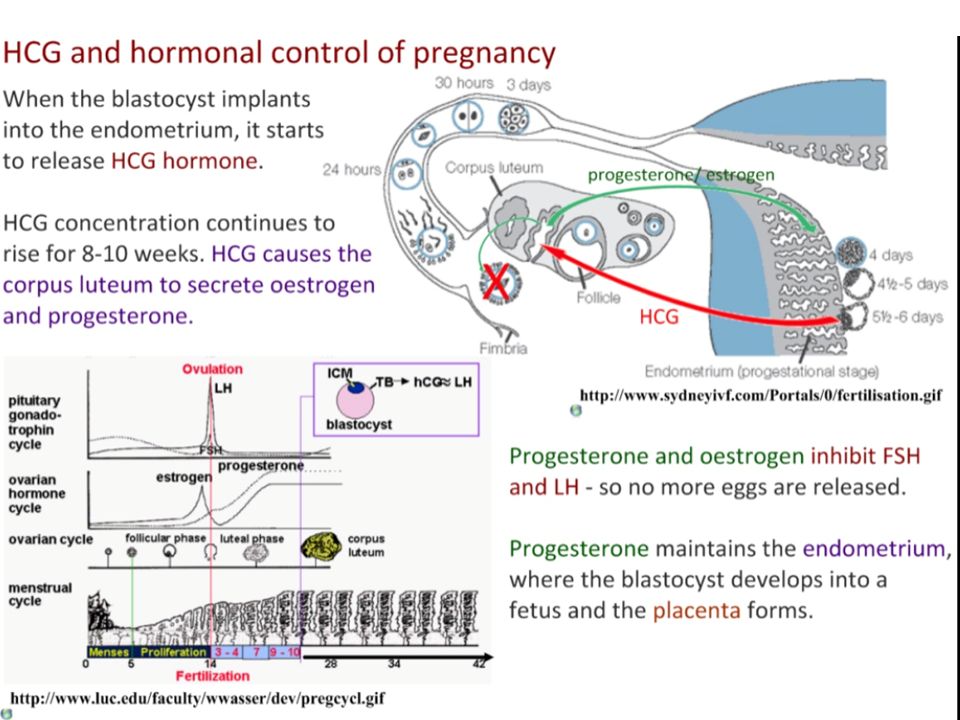
The placenta secretes the hormone, human chorionic gonadotrophin (HCG) HCG appears in the urine 3 days after conception.
 HCG appears in the urine 3 days after conception.")
57
The placenta as an endocrine gland
HCG is first secreted by the blastocyst cell of the embryo. HCG maintains the Corpus luteum ( a temporary gland) that secretes oestrogen and progesterone.
 that secretes oestrogen and progesterone.")
58
HCG HORMONE & Pregnancy
The HCG targets the ovary and the corpus luteum. The corpus luteum secretes progesterone and oestrogen at high levels .

59
HCG HORMONE & Pregnancy
The oestrogen and progesterone continue to inhibit FSH and LH secretion from the pituitary. The progesterone's prevent the breakdown of the endometrium and so the embryo can continue its development into a foetus

60
IB Learning Objective Outline the process of birth and its hormonal control, including the changes in progesterone and oxytocin levels and positive feedback

61
Hormone control & the process of giving Birth
Before birth, the level of progesterone declines sharply This decline causes contraction of the muscles of the uterine lining.

62
Hormone control & the process of giving Birth
At the same time, the pituitary gland releases the hormone oxytocin. This hormone relaxes the elastic fibers of the bones to the pelvic girdle, and dilates the cervix. Uterine contractions separate the placenta from the endometrium,

63
Positive Feedback between oxytocin & Uterine contraction
In this system the stimuli to the brain increases the oxytocin production In turn the oxytocin stimulate uterine contraction Uterine contractions further stimulates the pituitary of the mother to release more oxytocin

64
Positive Feedback between oxytocin & Uterine contraction
The strength and frequency of the uterine contractions is further increased. In turn this further stimulates more oxytocin production The process builds with stronger and stronger contractions

65
Positive Feedback between oxytocin & Uterine contraction
Finally the child passes though the cervix and vagina to be born Contractions continue for a further period until the placenta is delivered (after birth).
.")
66
Estrogen Oxytocin from ovaries from fetus and mother’s
LE 46-18 Estrogen Oxytocin from ovaries from fetus and mother’s posterior pituitary Induces oxytocin receptors on uterus Positive feedback Stimulates uterus to contract Stimulates placenta to make Progesterone Stimulate more contractions of uterus

70
LE 46-19 Placenta Umbilical cord Uterus Placenta (detaching) Uterus
Cervix Umbilical cord Dilation of the cervix Expulsion: delivery of the infant Delivery of the placenta

71
LE 46-21 Head Head Body Body

72
Trimesters

Similar presentations


>")


