Download presentation
Presentation is loading. Please wait.

1
NUR-224

2
Explain cardiac anatomy/physiology and the conduction system of the heart. Incorporate assessment of cardiac risk factors into the health history and physical assessment of the patient with cardiovascular disease. Discuss clinical indications, patient preparation and other elated nursing implications fro common test and procedures used to assess and diagnose cardiovascular diseases.

3
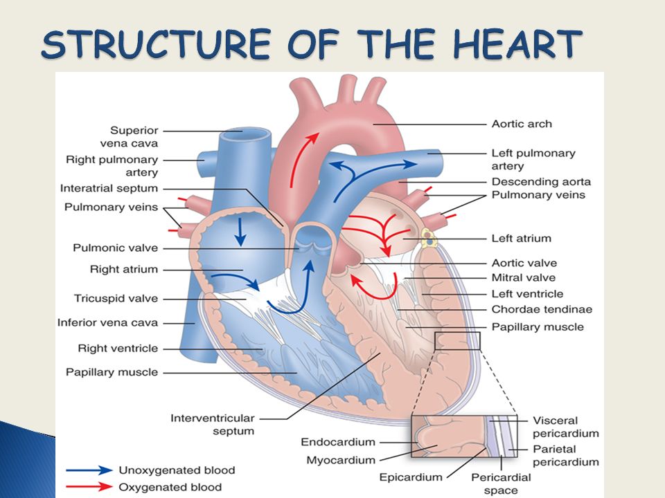
Three layers Endocardium Myocardium Epicardium Four chambers Heart valves

4
Surrounded by pericardium Pericardial fluid 10-30 mL Divided by septum Left ventricular wall 2-3 x as thick as right ventricle Atrial wall thinner than ventricles

5
Inferior and superior vena cava send deoxygenated blood to right atrium Blood passes through tricuspid valve to right ventricle blood passes from right ventricle through pulmonic valve via pulmonary artery to lungs Blood from lungs enters left atrium via pulmonary veins Passes through mitral valve to left ventricle Blood ejected to body through aortic valve aorta peripheral system

8
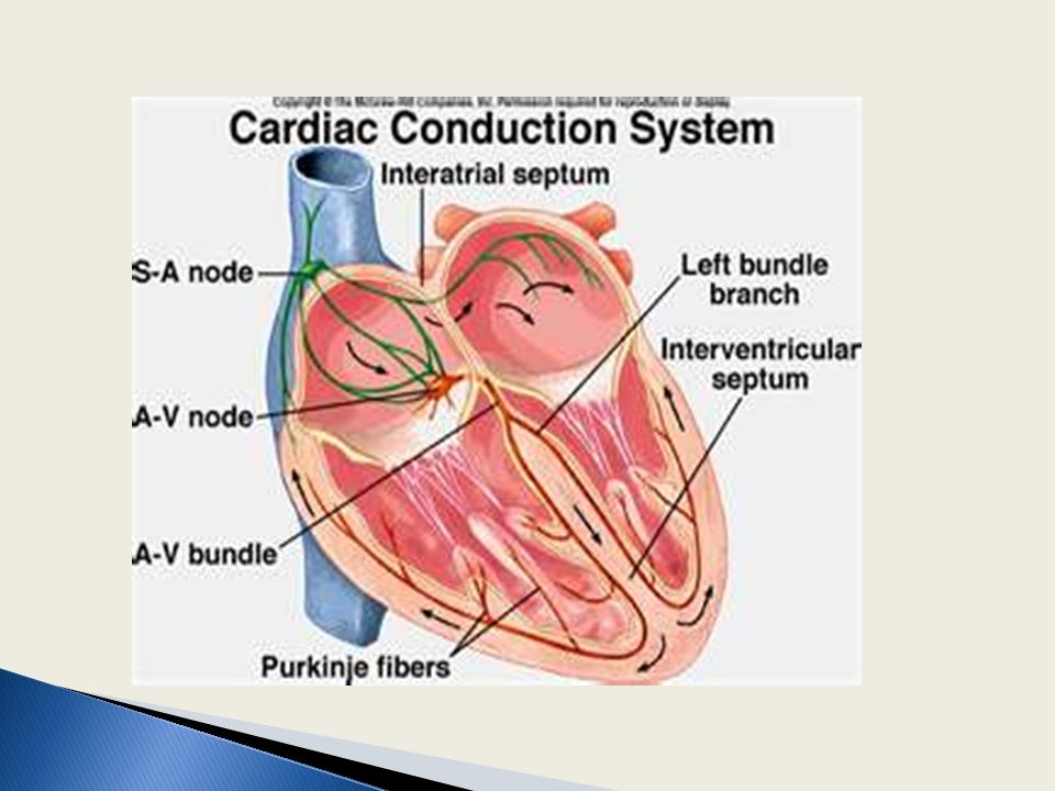
Depolarization (contraction of heart) Sinoatrial node – pacemaker of heart Contraction of atria AV node Bundle of His Right and left bundle branches Purkinje fibers
 Sinoatrial node – pacemaker of heart Contraction of atria AV node Bundle of His Right and left bundle branches Purkinje fibers")
10
Systole Contraction of myocardium Ejection of blood from ventricles Diastole Relaxation of myocardium Filling of coronary arteries Atrium is emptying into the ventricles

11
Number of times the ventricles contract each minute 60-100 Regulated by: Autonomic Nervous System Sympathetic Parasympathetic

12
Amount of blood pumped by each ventricle during a given period Amount of blood ejected from ventricle with each beat (stroke volume) x heart rate CO = SV x HR 4 – 7 L/min
 x heart rate CO = SV x HR 4 – 7 L/min")
13
Stroke volume: amount of blood ejected with each heartbeat Cardiac output: amount of blood pumped by ventricle in liters per minute Preload: degree of cardiac muscle fiber tension at end of diastole (prior to contraction) Afterload: resistance that ventricles must overcome to eject the blood Contractility: ability of cardiac muscle to shorten in response to electrical impulse
 Afterload: resistance that ventricles must overcome to eject the blood Contractility: ability of cardiac muscle to shorten in response to electrical impulse")
14
Health history Family/genetic history

15
Chest pain Dyspnea Peripheral edema, weight gain Palpitations Fatigue Dizziness, syncope, changes in level of consciousness

17
Medications Nutrition Elimination Activity, exercise Sleep, rest Self-concept Roles, relationships Sexuality Risk factors

18
Inspection Palpation Percussion Auscultation

19
Normal skin color Capillary refill < 3 seconds Thorax symmetrical No jugular vein distention with patient at 45° Absence of clubbing

20
PMI palpable at 5th ICS mid-clavicular line No thrills, heaves Slight pulsation of abdominal aorta in epigastric region Carotid and extremity pulses equal bilaterally No pedal edema

21
Normal heart sounds S1 and S2 heart sounds heard Apical-radial rate equal and regular No murmurs or extra heart sounds No S3 or S4 Pericardial friction rub

23
Extremities Lungs Abdomen

24
Laboratory test: Diagnose the cause of cardiac-related signs/symptoms Determine baseline values before initiating therapeutic interventions Ensure therapeutic levels of medication are maintained Evaluate the patient’s response to the therapeutic regimen Identify abnormalities

25
Cholesterol - normal level <200mg/dL Major sources – diet, liver Low density lipoproteins LDLs <160 High-density lipoproteins HDLs Triglycerides <200

26
CXR/Fluoroscopy Electrocardiography Cardiac stress testing Echocardiography

27
Coronary arteries dilate to 4x their normal in response to increased metabolic demands for oxygen. Coronary arteries affected by atherosclerosis dilate less, compromising blood flow to the myocardium ischemia Noninvasive test Abnormalities in CV function are more likely to be detected during times of increased stress.

28
Determine : presence of CAD cause of chest pain functional capacity of the heart after MI/ heart surgery effective of antianginal/antiarrhythmic dysrhythmias/ physical exercise

29
Pre-Test Physical and Baseline ECG Signed consent Patient teaching Report cardiac symptoms during test NPO 4 hours pre-test Withhold meds Emergency and resuscitation equipment need to be at site of test at all times

30
Testing procedure Exercise equipment Increase HR to target rate for age and gender OR c/o chest pain or fatigue Speed or incline increased every 2-3 minutes to increase stress on patient ECG and BP monitored throughout the test Rest for 15 minutes post test while being monitored

31
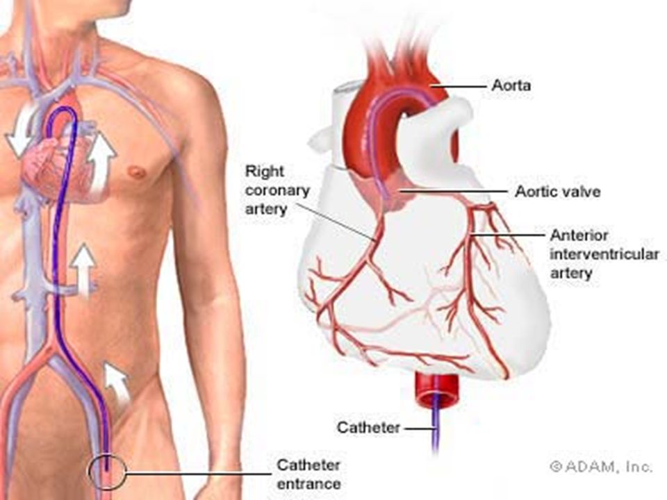
Invasive procedure study used to measure cardiac chamber pressures, assess patency of coronary arteries Requires ECG, emergency equipment must be available Assessment prior to test: allergies, blood work Assessment of patient postprocedure: circulation, potential for bleeding, potential for dysrhythmias Activity restrictions Patient education pre/postprocedure

33
Preparation √ allergies to shellfish Signed consent form D/C anticoagulants, ASA, salicylates, herbals affecting coagulants Contraindicated; patients with bleeding disorders Elderly, dehydrated Severe renal failure Patient Teaching Palpitations as catheter enters left ventricle Heat/hot flash as contrast medium injected Sensation of need to cough as medium injected into right side of heart

34
During Procedure nausea pain at insertion site STAT Intervention chest pain dysrhythmias changes in peripheral pulses neuro assessment Post Procedure VS & Neuro checks insertion site pressure dressing bleeding/hematoma Assessment extremities - s/s ischemia r/t clots bed rest 4-6 hrs post procedure

35
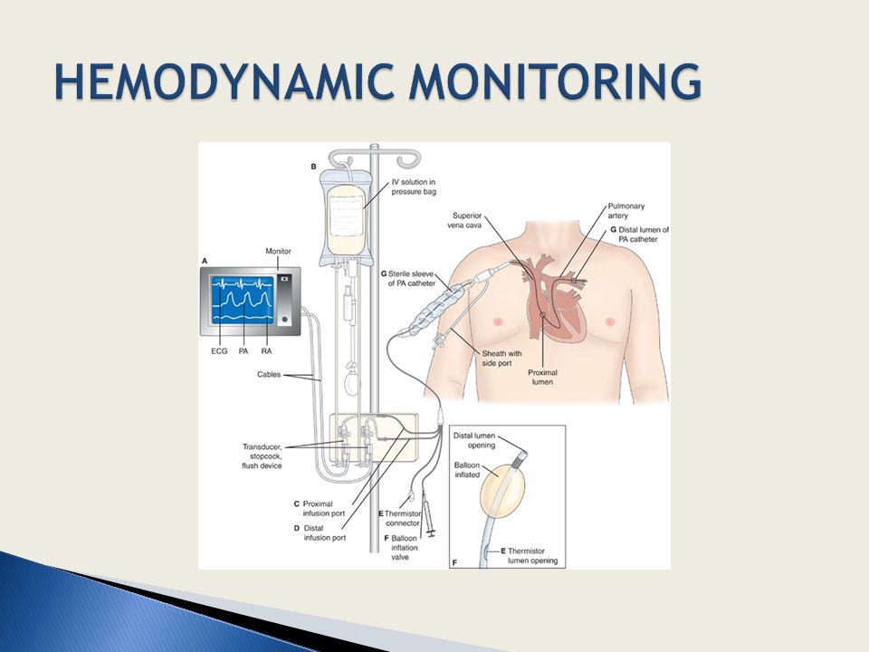
CVP Pulmonary artery pressure Intra-arterial BP monitoring

Similar presentations