Download presentation
Presentation is loading. Please wait.

1
Male Genital Surgical Conditions
Dr. S. Nishan Silva (MBBS)
")
2
Anatomy

3
Congenital Anomalies : Hypospadias and Epispadias
Malformations of urethral groove and/or urethral canal: Abnormal opening on ventral surface: hypospadias (1/300 live male births) Abnormal opening on dorsal surface: epispadias (less common than hypospadias) Associated with: Failure of normal descent of testes Other malformations of urinary tract Clinical sequelae: urinary tract obstructions, infections, sterility
 Abnormal opening on dorsal surface: epispadias (less common than hypospadias) Associated with: Failure of normal descent of testes. Other malformations of urinary tract. Clinical sequelae: urinary tract obstructions, infections, sterility.")
4
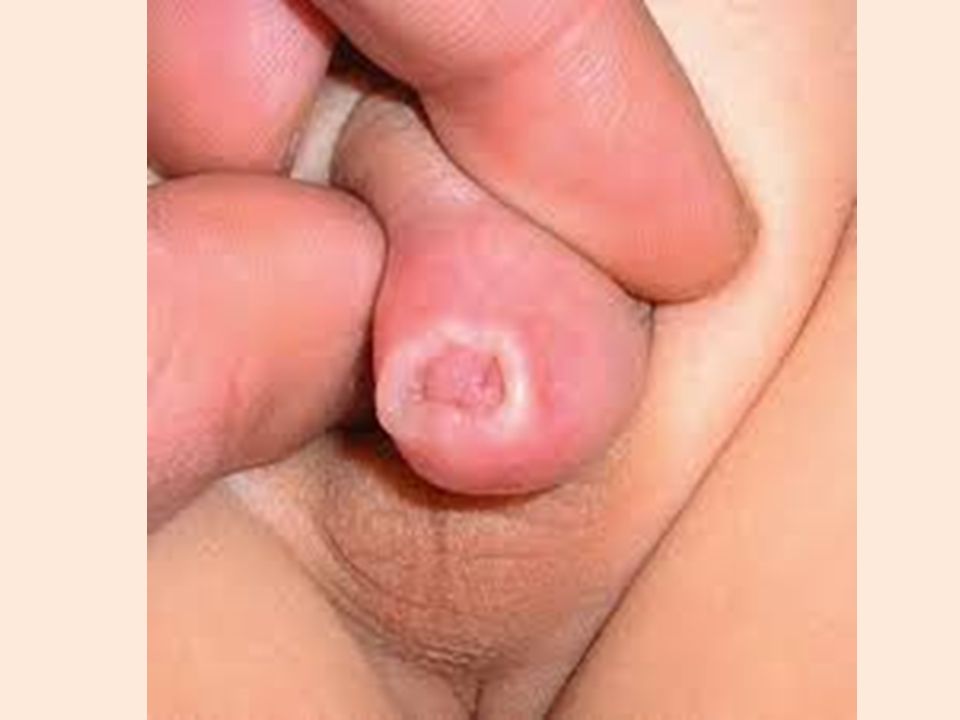
Phimosis Phimosis is the inability to retract the prepuce (foreskin) of penis over the shaft due to a narrow opening. Phimosis can be congenital or acquired:- In acquired phimosis there is chronic inflammation of the tip of the penis and prepuce (fore skin) or there are adhesions between glans & prepuce or due to malignancy. In congenital causes it is present since birth. Phimosis is usually caused by thickening and repeated inflammation of the foreskin.
 of penis over the shaft due to a narrow opening. Phimosis can be congenital or acquired:- In acquired phimosis there is chronic inflammation of the tip of the penis and prepuce (fore skin) or there are adhesions between glans & prepuce or due to malignancy. In congenital causes it is present since birth. Phimosis is usually caused by thickening and repeated inflammation of the foreskin.")
7
Symptoms of Phimosis ? Inability to retract foreskin. Straining during urination. Thin stream of urine. Recurrent urinary infections. Pus from penis - due to belanophosthitis. How can we diagnose Phimosis ? From history & examination On Examination: Pin hole opening of foreskin Difficulty to push back the foreskin over the shaft of the penis. Balooning of foreskin - A bulge in the tip of penis as urine accumulates under the foreskin.

8
How can Phimosis be treated ? Circumcision
If untreated complications of phimosis can occur: Infected foreskin leads to infection of glans also. Paraphimosis Back pressure due to obstruction of flow of urine. Meatal Stenosis - narrowing of penile opening. Sometimes a cancerous ulcer on glans can cause the adhesion to take place.

12
ParaPhimosis Paraphimosis occurs when the foreskin has been retracted and narrows below the glans, constricting the lymphatic drainage and causing the glans to swell. If not corrected, blood flow in the penis becomes impeded by the increasingly constricting band of foreskin, which causes further swelling of the glans. Because lack of oxygen from the reduced blood flow can cause tissue death (necrosis) paraphimosis is considered a medical emergency and requires immediate treatment.
 paraphimosis is considered a medical emergency and requires immediate treatment.")
15
Causes: Bacterial infection (e.g., balanoposthitis) Catheterization (i.e., if the foreskin is not returned to its original position after a urethral catheter is inserted, the glans may become swollen, which can initiate paraphimosis) Poor hygiene Swelling-producing injury Vigorous sexual intercourse Symptoms and Signs : Inability to urinate (urinary retention) Penile pain Swollen glans (the shaft of the penis is not swollen) Redness, Black tissue on the glans (indicates necrosis Band of retracted foreskin tissue beneath the glans Tenderness
 Poor hygiene. Swelling-producing injury. Vigorous sexual intercourse. Symptoms and Signs : Inability to urinate (urinary retention) Penile pain. Swollen glans (the shaft of the penis is not swollen) Redness, Black tissue on the glans (indicates necrosis. Band of retracted foreskin tissue beneath the glans. Tenderness.")
16
Diagnosis Paraphimosis is diagnosed during physical examination. Treatment Injection of hyaluronidase with lidocane followed by gentel pressure. This usually results in reduction Failure incision of he constricting band Circumcision to prevent reoccurrence

17
Epididymo-orchitis This is primarily an infection of the epididymis, but some oedema & inflammatory changes spread into the testis There maybe an associated urinary tract infection. Types: Acute Under 40 years old chalmydia trachomatis & gonorrhea In old pt enterococci, E.coli Chronic Follow recurrent acute attacks TB

18
Hisory: Sever pain (comes quickly-hrs- ,can be relieved by scrotal support) & swelling in one side of the scrotum Malaise, fever, sweating &loss of appetite Symptoms of urinary tract infection O/E: Swelling confined to one side of the scrotum Scrotal skin red &shiny, four days later become bronze in color Scrotal skin hot Not-tender but the testis& epididymis are very tender Surface of epididymis smooth Swelling is fluctuant (secondary hydrocele)
")
19
Investigation: CBC ,Leukocytosis MSU U/S & doplar Treatment: Bed rest Analgesia Scrotal support Broad spectrum Ab (ciprofloxacin) **The swelling may take as long as 2 months to resolve
 **The swelling may take as long as 2 months to resolve.")
20
Case 1: Patient T.R. What is the Differential Diagnosis?
HPI: 21 yo man presents with 3 hours of intense, constant testicular pain Began several hours after college track meet Associated nausea and vomiting PMH: None Meds: Glucosamine, condroitin, creatine supplements Alls: PCN FH: Non-contributory SH: Sexually active, multiple partners

21
Differential Diagnosis
Testicular Torsion Appendiceal Torsion Epididymitis Trauma Inguinal Hernia Henoch-Schonlein Purpura Mumps Fournier’s Gangrene Referred Pain

22
Case 1: Patient T.R. Exam:

23
Case 1: Patient T.R. What is the Next Step?
Exam: Right testicle higher than left Long axis oriented horizontally Significant swelling No cremasteric reflex on either side No relief of pain on elevation

24
Case 1: Patient T.R. Next Step
If Diagnosis Certain (Torsion): To the OR. Outcomes directly related to length of time from onset Irreversible ischemia at mean of 12 hours If Diagnosis Less Obvious Doppler Ultrasound Test 82% sensitive, 99% specific for torsion (loss of flow)
: To the OR. Outcomes directly related to length of time from onset. Irreversible ischemia at mean of 12 hours. If Diagnosis Less Obvious. Doppler Ultrasound. Test 82% sensitive, 99% specific for torsion (loss of flow)")
25
Case 1: Patient T.R. Operation: testicular detorsion and fixation
Unilateral or bilateral? Why?

26
Case 1: Patient T.R. Operation testicular detorsion and fixation
Unilateral or bilateral? Why? ANSWER: Bilateral-- Torsion associated with absence/insufficeincy of gubernaculum. Often bilateral. What if surgery not an option?

27
Testicular torsion This is twisting of the testis with interference to the arterial blood supply. the actual torsion is usually of the spermatic cord Possible mechanism; it is associated with: Imperfectly descended testis High investment of tunica vaginalis with a horizontal lie of testis Epididymis& testis are separated by a mesorchium, & twisting occurs at the mesorchium. The incidence is highest between 10 & 20 years.

28
Testicular Torsion 40% over 21 Associated with physical activity/sleep
Exam Absent cremasteric Doppler Surgical Emergency

30
Symptoms: pain in the scrotum &groin: Sever Sudden onset
Radiating to the lower abdomen Associated with vomiting May follow strain, lifting, exercise, or masturbation Signs: Swollen testis Tender Drawn up to the groin

31
Torsion of Testis

32
Treatment: Explore testis as soon as possible (untwisting should be carried out within 6 hrs of symptoms). Check that it is not irreversibly infarcted. Fix it to the scrotal septum. The other testis should be fixed at the same operation, since it is likely to have abnormal position. However If the testis is infarcted, it should be removed

33
Testicular torsion

34
Management – Orchiopexy

35
Management – Orchidectomy

36
Scrotal Swellings

37
Scrotal swllings Cystic Hydrocele Epididymal cyst/ spermatocele Hematocele Solid Tumor (benign/ Malignant) Neither varicocele Hernia
 Neither. varicocele. Hernia.")
38
Hydrocele Secondary Develop slowly develop rapidly Large small
A collection of serous fluid in the tunica vaginalis Types: Congenital: occurs in infants due to patent processus vaginalis peritoneal fluid can enter the scrotum Primary. (idiopathic) Develop slowly Large Hard & tense No defined cause Over 40s Secondary develop rapidly small lax secondary to inflammation, trauma or tumor of underling testes younger age group(20-40)
 Develop slowly. Large. Hard & tense. No defined cause. Over 40s. Secondary. develop rapidly. small. lax. secondary to inflammation, trauma or tumor of underling testes. younger age group(20-40)")
39
Congenital hydrocele: processus vaginalis is patent & connects to the peritoneal cavity. In children <3yrs Infentile hydrocele: the tunica and processus vaginalis are distended to the superficial inguinal ring. There is no conection. Occurs in all ages Hydrocele of the cord: swelling near the spermatic cord. D/D hernia, lipoma of the cord

40
Symptoms: Scrotal swelling Pain & discomfort if its secondary Frequent &painful micturation if secondary to epididymo-orchitis Malaise & weight loss if secondary to tumor with distant metastases Don’t affect fertility

41
O/E: often bilateral Can “get above it”
Testes cannot be felt separately Transilluminates Fluctuant Fluid thrill Dull to percussion Not campressible or pulsatial Can’t be reduced Normal skin color & temp Not tender if primary (may be tender if secondary) Size can be reach up to 10-20cm in diameter Surface smooth
 Size can be reach up to 10-20cm in diameter. Surface smooth.")
42
U/S of hydrocele Done to exclude testicular tumor or epididymitits

43
Treatment If congenital hydrocele persists beyond the age of 1year, surgical treatment is indicated. This involves the division and ligation of the processus. In an adult with primary hydrocele Surgery Opening the tunica vaginalis longitudinally Emptying hydrocele Everting the sac Suturing it behind the cord thus obliterating the potential space Aspiration reccurance In elderly patient who are not fit for surgery Secondary hydrocele treat the underlying cause

44
Lesions of tunica vaginalis, cont.
spermatocele Hematocele: post-trauma or torsion Spermatocele: cystic accumulation of semen in dilated efferent ducts of testis Varicocele: dilated veins of pampiniform plexus within spermatic cord; may cause infertility testis rope-like mass of dilated veins

45
Epididymal cyst Fluid-filled swellings connected with the epididymis.
If cyst contains clear fluid ,it is called epididymal cyst . However, if the fluid is grey opaque &contains few spermatozoa, it is called spermatocele (after aspiration) Symptoms: Over age of 40 years Scrotal swelling (as if having a 3rd testis) Painless Often multiple, bilateral Enlarge slowly Doesn’t affect fertility (maybe after surgical removal)
 Symptoms: Over age of 40 years. Scrotal swelling (as if having a 3rd testis) Painless. Often multiple, bilateral. Enlarge slowly. Doesn’t affect fertility (maybe after surgical removal)")
46
O/E: Frequently bilateral Lies above & slightly behind the testes, the cord is felt above it Cysts are not tender Elongated, measures from few millimeters to 5-10cm diameter Smooth surface Testis can be felt separately Can “get above it Fluctuant, fluid thrill, dull to percussion Can’t be reduced Transilluminates if contains clear fluid i.e Epididymal cyst (spermatocele; sometime depend on density of the fluid)
")
47
U/S Must be done to confirm your diagnosis & R/O testicular tumore
spermatocele

48
Treatment: None if asymptomatic But if large & interfere with walking: Aspiration may help Excision for large cysts; this may affect fertility of the testis

49
Testicular tumors Commonest malignancy in men < 35
Rare in men of African ancestry and before puberty Peaks in the early twenties 90% arise from germ cells &are either seminomas(30-40 years) or teratomas(20-30 years) 10% are lymphomas, sertoli cell tumors or leydig cell tumors One in 10 testicular tumors occurs in association with maldescent of the testis. Prognosis is good particularly if there was no lymph node involvement
 or teratomas(20-30 years) 10% are lymphomas, sertoli cell tumors or leydig cell tumors. One in 10 testicular tumors occurs in association with maldescent of the testis. Prognosis is good particularly if there was no lymph node involvement.")
51
Symptoms: Painless swelling of the testis, (sometime dull aching, dragging pain )(80%) Heaviness in the scrotum Maybe history of trauma delays diagnosis General malaise, wasting ,loss of appetite Abdominal pain if lymph nodes are enlarged Swelling of legs caused by lymphatic or venous obstruction Infertility Secondary hydrocele

52
Signs: can “get above it” Testes can not be felt separately Not translucent Not fluctuant Harder than normal testis Dull to percussion hydrocele If skin is affected, it maybe warm & discolored Usually not tender Irregular, different sizes Surface usually smooth (sometime irregular or nodular) Examine the para-aortic & supraclavicular lymph nodes for metastasis The liver maybe enlarged & there maybe sign of pulmonary secondaries (collapse, consolidation or a pleural effusion).
 Examine the para-aortic & supraclavicular lymph nodes for metastasis. The liver maybe enlarged & there maybe sign of pulmonary secondaries (collapse, consolidation or a pleural effusion).")
53
Investigation: US testis CXR mets Tumor markers :AFP (yolk-sac cell), βHCG (trophoblastic cells). CT scan abdomen and chest to identify lymph nodes and pulmonary mets

54
Explore testis through an inguinal incision Orchidectomy
Treatment: Explore testis through an inguinal incision Orchidectomy Further treatments depends on the type and stage Staging Treatment of seminoma Treatment of non-seminomatous germ cell tumor stageI confined to the testis DXT to abdominal nod Observation or RPLND stageII retroperitolneal LN involvement IIa: nodes <2cm IIb: nodes 2-5cm IIc: nodes >5cm DXT to abdominal nodes “ Chemotherapy Chemo &RPLD of residual dx stageIII nodal dx above the diaphragm DXT to abdominal wall & thoracic nodes or chemo Chemo stageIV visceral mets DXT=deep x-ray therapy, RPLND=retroperitoneal lymph node dissection

55
Varicocele It is a bunch of dilated& tortuous veins of the pampiniform plexus i.e. (varicose vines in the spermatic cord). More common on the left side 25% of normal men have small symptomless varicoceles. Causes of varicocele Incompetent valve btw the renal and testicular veins Nephrectomy Lt. Renal neoplasm Lymphadenopathy

56
Symptoms: Varicose veins in the scrotum on standing. Disappear on lying down Heavy or dragging sensation in scrotum Aching pain Bilateral varicoceles may case subfertility O/E: The pt must be examined standing, not to miss the diagnosis Vein often visible They are also palpable & fell like a “bag of worms” Affected testis may be smaller & more soft

57
U/s PIC

58
Treatment: In Asymptomatic pt ,no treatment is required
Scrotal support for aching &discomfort If symptoms fail to settle or there is evidence of subfertility; there are two options for treatment: Embolization &obliteration under radiological control (majority) Surgery is via an inguinal approach, all testicular veins bar on being ligated at the deep inguinal ring. Microsurgery is used in most cases. Has less recurrence rate and better success. Embolization is preferred in case of recurrence
 Surgery is via an inguinal approach, all testicular veins bar on being ligated at the deep inguinal ring. Microsurgery is used in most cases. Has less recurrence rate and better success. Embolization is preferred in case of recurrence.")
59
Indirect inguinal hernia
A peritoneal sac protrudes through the deep inguinal ring, passes down the inguinal canal, &may extend as far as the upper pole of the testis. The defect is congenital & is due to persistent processus vaginalis Symptoms: Often none (scrotal swelling that can be pushed back by the pt. Aching dragging sensation in the groin Some pt relate the development to an episode of straining or lifting

60
Signs: Can’t “get above it” There is a cough impulse Reducible Treatment: Herniotomy& Herniorrhaphy (excision of the sac &repair of the defect) in adult By: Lichtenstein repair (tension free mesh repair) Shouldice repair
 in adult By: Lichtenstein repair (tension free mesh repair) Shouldice repair.")
61
History Agetumors (20-40). Rare before puberty H/o trauma
Torsion usually in teens and children Hydrocele in an infantcommunicating H/o trauma Painepididymo-orchitis, varicocele, torsion Infertility Constitutional sympmalignancy PSHvaricocele SOH marital status & extramarital relation epididymo-orchitis

62
investigations CBC WBC MSU for culture and sensitivity
Tumor markers if indicated U/S ± doplar CT if indicated tumor

63
Vasectomy – Male Sterilization

64
LOCAL ANAESTHESIA

65
3 Finger technique for vas fixation

66
Holding with ringed clamp

67
Piercing the skin with dissecting forceps

68
Delivery of vas

69
Vas occlusion Ligature Cautery Haemoclips

70
Dressing

Similar presentations