Download presentation
Presentation is loading. Please wait.

1
Bicuspid Aortic Valve 06/01/2007

2
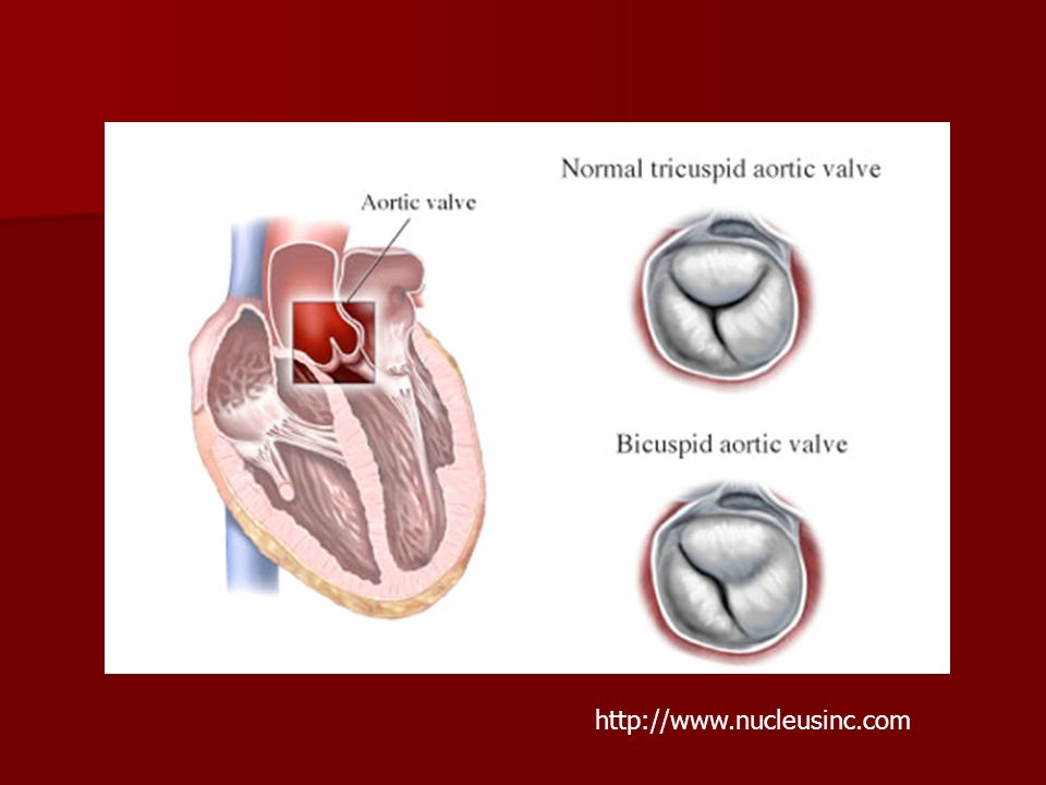
Bicuspid aortic valve Definition: Definition: –Two functional aortic valve leaflets with two complete commissures AKA “Bicommissural aortic valve”

3
Bicuspid aortic valve Felt to represent a complex continuum Felt to represent a complex continuum –Unicommissural, Bicommissural, Tricuspid, and Quadricuspid Not simply a fusion of two normal cusps Not simply a fusion of two normal cusps

4
Bicuspid aortic valve Three criteria: Three criteria: 1.) Unequally sized cusps 1.) Unequally sized cusps –Larger leaflet is the "conjoined" leaflet –Larger leaflet is the "conjoined" leaflet
 Unequally sized cusps 1.) Unequally sized cusps –Larger leaflet is the conjoined leaflet –Larger leaflet is the conjoined leaflet")
5
http://www.nucleusinc.com
6
Bicuspid Aortic Valve 2.) Presence of a central ridge (raphe) 2.) Presence of a central ridge (raphe) –Usually in the center of the conjoined leaflet
 Presence of a central ridge (raphe) 2.) Presence of a central ridge (raphe) –Usually in the center of the conjoined leaflet")
7
Bicuspid Aortic Valve 3.) Smooth cusp margins 3.) Smooth cusp margins –Excludes tricuspid valves which fused due to inflammatory processes (eg, rheumatic fever) Irregularity and scarring within the raphe.
 Smooth cusp margins 3.) Smooth cusp margins –Excludes tricuspid valves which fused due to inflammatory processes (eg, rheumatic fever) Irregularity and scarring within the raphe.")
8
Morphology Orientation: Orientation: –Anterior-posterior Left leaflet Right (conjoined) leaflet –Left-right Anterior (conjoined) leaflet Posterior leaflet
 leaflet –Left-right Anterior (conjoined) leaflet Posterior leaflet")
9
Physiology Normally functioning bicuspid valve Normally functioning bicuspid valve –Abnormal folding and creasing –Restricted motion –Turbulent flow Prolonged stress leads to valve damage Prolonged stress leads to valve damage

10
Statistics Estimated overall incidence of 1-2% Estimated overall incidence of 1-2% M:F at least 2:1 M:F at least 2:1 Familial clustering suggests AD with variable penetrance Familial clustering suggests AD with variable penetrance No race or geographical predilection No race or geographical predilection

11
Associated Anomalies Left dominant coronary artery Left dominant coronary artery –Up to 50% 10% with tricuspid valve Short left main coronary artery Short left main coronary artery –Less than 5mm

12
Associated Anomalies Coarctation of the aorta Coarctation of the aorta Interrupted aortic arch Interrupted aortic arch –>50% have bicuspid aortic valve

13
Associated Anomalies Turner's Syndrome Turner's Syndrome –Up to 30% William's Syndrome William's Syndrome –Up to 10%

14
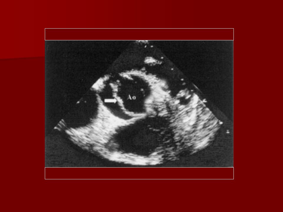
Imaging Echocardiography Echocardiography –Modality of choice Long-axis shows systolic doming due to limited valve opening Short-axis allows examination of the commisures, leaflet morphology, and mobility.

16
Radiographs Usually normal in pediatric population Usually normal in pediatric population Aortic root enlargement Aortic root enlargement Left ventricular enlargement Left ventricular enlargement May see calcified raphe or leaflets May see calcified raphe or leaflets

17
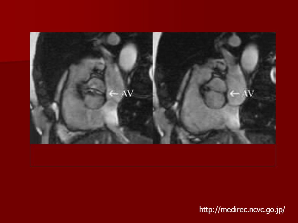
CT/MRI Allows for evaluation of the aorta and coronary arteries Allows for evaluation of the aorta and coronary arteries Functional information Functional information

19
http://medirec.ncvc.go.jp/
21
Pathology Aortic Stenosis Aortic Stenosis Aortic Insufficiency Aortic Insufficiency Bacterial Endocarditis Bacterial Endocarditis Aortic Dissection Aortic Dissection

22
Aortic Stenosis Poorly functioning valves may have incomplete systolic opening Poorly functioning valves may have incomplete systolic opening Responsible for 80-95 % of aortic valve disease detected in infancy Responsible for 80-95 % of aortic valve disease detected in infancy –May cause rapid deterioration –Progression over years is more common

23
Aortic Stenosis Bicuspid valve may be prone to accelerated aging Bicuspid valve may be prone to accelerated aging –Sclerosis begins in the second decade of life –Estimated that 50% of adults with severe AS have bicuspid valves.

24
Aortic Stenosis Cusps oriented in the AP direction demonstrate more rapid progression Cusps oriented in the AP direction demonstrate more rapid progression Presence of risk factors can also expedite the process Presence of risk factors can also expedite the process –High LDL, high lipoprotein (A) and smoking
 and smoking")
25
Aortic Insufficiency Isolated AI Isolated AI AI with aortic root dilatation AI with aortic root dilatation

26
Aortic Insufficiency Isolated AI Isolated AI –Prolapse of redundant larger cusp Rarely severe

27
Aortic Insufficiency Disruption of the elastic tissue within the upper aortic ring/sinotubular junction Disruption of the elastic tissue within the upper aortic ring/sinotubular junction –May occur due to inherent abnormality –May be due to coarctation of the aorta or bacterial endocarditis Often severe with high mortality

28
Bacterial Endocarditis Estimated 10-30% of patients with bicuspid aortic valve Estimated 10-30% of patients with bicuspid aortic valve –25% of cases of endocarditis occur on bicuspid valves –Tetralogy of Fallot, VSD, and MVP are the other lesions associated with SBE Prophylactic antibiotics for dental/surgical procedures Prophylactic antibiotics for dental/surgical procedures

29
Bacterial Endocarditis Responsible for half of cases of severe AI in patients with bicuspid valve Responsible for half of cases of severe AI in patients with bicuspid valve –Many due to cusp perforation Unexplained systemic emboli should raise suspicion Unexplained systemic emboli should raise suspicion

30
Aortic Dissection Approximately 5% of patients Approximately 5% of patients –Etiology is unclear Abnormal response to hemodynamic stress –Cystic medial necrosis similar to Marfan’s –Dysfunctional microfibrillar proteins, endothelial nitric oxide synthetase, etc.

31
Medical Management Lifestyle Lifestyle –Exercise, heart healthy diet, no smoking Cholesterol and hypertensive medication Cholesterol and hypertensive medication

32
Medical Management Surveillance echocardiography Surveillance echocardiography Early surgical referral Early surgical referral First-degree relative screening First-degree relative screening

33
Surgical Treatment Severe valvular dysfunction or aortic root dilatation Severe valvular dysfunction or aortic root dilatation Symptomatic patients Symptomatic patients Evidence of abnormal LV dimensions and function Evidence of abnormal LV dimensions and function

34
Surgical Treatment Pediatric cases Pediatric cases –Balloon Valvuloplasty Without calcified valves Isolated Aortic Insufficiency Isolated Aortic Insufficiency –Valve Repair Valve replacement Valve replacement –With or without aortic root replacement Prosthetic/Bioprosthetic/Homograft Ross procedure

35
References Fedak PWM, Verma S, David TE, Leask RL, Weisel RD, Butany J. Clinical and pathophysiological implications of a bicuspid aortic valve. Circulation. 2002; 106: 900–904 Fedak PWM, Verma S, David TE, Leask RL, Weisel RD, Butany J. Clinical and pathophysiological implications of a bicuspid aortic valve. Circulation. 2002; 106: 900–904 Ward, C. Clinical significance of the bicuspid aortic valve. Heart 2000 83: 81-85 Ward, C. Clinical significance of the bicuspid aortic valve. Heart 2000 83: 81-85 Pediatric Cardiac Surgery, Mavroudis et al., 3rd edition, Mosby, St. Louis. Pediatric Cardiac Surgery, Mavroudis et al., 3rd edition, Mosby, St. Louis. Aboulhosn, J, Child, JS. Left ventricular outflow obstruction: subaortic stenosis, bicuspid aortic valve, supravalvar aortic stenosis, and coarctation of the aorta. Circulation. 2006 Nov 28;114(22):2412-22. Aboulhosn, J, Child, JS. Left ventricular outflow obstruction: subaortic stenosis, bicuspid aortic valve, supravalvar aortic stenosis, and coarctation of the aorta. Circulation. 2006 Nov 28;114(22):2412-22. http://www.emedicine.com/ped/topic2486.htm http://www.emedicine.com/ped/topic2486.htm http://www.emedicine.com/ped/topic2486.htm
: Aboulhosn, J, Child, JS. Left ventricular outflow obstruction: subaortic stenosis, bicuspid aortic valve, supravalvar aortic stenosis, and coarctation of the aorta. Circulation Nov 28;114(22):")
Similar presentations


 Assistant Professor of Medicine Medical Unit-4 LUMHS, Jamshoro.>")


