Download presentation
Presentation is loading. Please wait.

1
FP 7 Collaborative Project Novel MR-compatible PET detectors for simultaneous PET/MRI imaging FP7-HEALTH-2009-single-stage -Scope -Issues -Actions -Deadline(s)
")
3
FP7 PET-MRI ISS INFN Univ Italy RomeGenova Bari Germany Portugal Belgium Switz. France? (ST) Slovenia Russia Spain ? Industries Siemens ST MRI Wurz. Julich. Munich Cern ESPL Lisbon (J.Varela) Brussel (S.Tavernier) P. Jarron P. Krizan (Lubjian) R. Gruetter Y. Lusienko (Moskow)
 Slovenia Russia Spain . Industries Siemens ST MRI Wurz. Julich. Munich Cern ESPL Lisbon (J.Varela) Brussel (S.Tavernier) P. Jarron P. Krizan (Lubjian) R. Gruetter Y. Lusienko (Moskow).")
4
The Istituto Superiore di Sanità (ISS) is the leading technical and scientific public body of the Italian National Health Service. Its activities include research, control, training and consultation in the interest of public health protection.

5
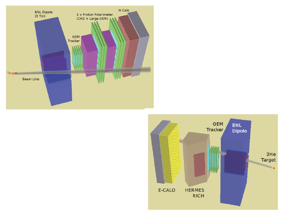
Hall A - Two High Resolution Spectrometers QDQ - Momentum Range: 0.3 –4 GeV/c p/p : 1 x 10⁻⁴ – p = =-5% - – mr 1 (+1) Cherenkov threshold aerogels + RICH in the hadron spectrometer + septum magnet
 Cherenkov threshold aerogels + RICH in the hadron spectrometer + septum magnet")
8
Spectroscopy analysis of 12 B : Aerogel vs. RICH K-selection Aerogel Kaon selection RICH Kaon selection 12 C(e,e’K) 12 B Freon/CsI RICH detector (like ALICE) Hermes areogel RICH
 12 B Freon/CsI RICH detector (like ALICE) Hermes areogel RICH.")
12
HEALTH-2009-1.2-3 Novel MR-compatible PET detectors for simultaneous PET/MRI imaging FP7-HEALTH-2009-single-stage The focus should be to develop novel magnetic-field-compatible nuclear detectors for PET imaging, aimed at maximizing the benefits of simultaneous PET/MRI acquisition, which can also be used efficiently and implemented in stand alone PET or SPECT applications. These detectors should operate in high magnetic fields, as used in MRI, without performance degradation, and have high spatial and time resolution. A dedicated integrated readout of high quality should also be developed. The full detector should be compact so as to allow good integration with an MRI system. Globally, it should allow fully exploiting the advantages of both PET and MR technologies in a simultaneous imaging modality and for implementation in both preclinical and clinical/human PET stand-alone systems beyond the state-of-theart. Active participation of industry, especially SMEs, could lead to an increate impact of the research proposed, and this will be considered in the evaluation of the proposal. Funding scheme: Collaborative Project (Large scale integrating project).
..")
13
Participants in Large-scale integrating projects are required to conclude a consortium agreement Large-scale integrating projects: the requested EC contribution shall be over € 6 million and not exceed € 12 million unless otherwise indicated in the topic description

14
Molecular Imaging Modalities CT Tissue Density, Z A 20-50 µm -galactocidase 0.1 µmole H / µmole 31 P MRIA H Concentration MF BOLD, DCE 0.1 mm UltrasoundStructure A F Doppler Optical (Bioluminescence, fluorescence) A Topography M ~10 3 cells quantitative µm to mm PET/SPECTRadiotracer M ~1-2 mm <10 -12 mole = quantitative F Unique !!
 A Topography M ~10 3 cells quantitative µm to mm PET/SPECTRadiotracer M ~1-2 mm < mole = quantitative F Unique !!")
15
what we shoudl do to be successfull ? - good (new???) detectors - “perfect” integration in multimodality (hardware and software) - loking at applications (“added value” by combining the modalities) - PET/SPECT high field and low field - “smart layout (s)” - What to propose ? - TOF – PET scintillator, electronics, sensor - Management and careful writing of the proposal (only 1/3 of the evaluation is on “technical/scientific)!!
 detectors - perfect integration in multimodality (hardware and software) - loking at applications ( added value by combining the modalities) - PET/SPECT high field and low field - smart layout (s) - What to propose . - TOF – PET scintillator, electronics, sensor - Management and careful writing of the proposal (only 1/3 of the evaluation is on technical/scientific)!!.")
16
What we have to study design What we have to build PET-SPECT MRI Low field High field - a prototype of SPECT/PET – MRI (low field) for - breast - small animal - a prototype of SPECT/PET – MRI (high field) for - brain - heart
 for - breast - small animal - a prototype of SPECT/PET – MRI (high field) for - brain - heart")
18
Dual Modality: PET / SPECT (Use SPECT Camera for PET) Less Expensive, But Not Optimized for PET SPECT cameras optimized to image 140 keV (not 511 keV) photons. Detectors are “thin” (0.8 attenuation lengths) NaI:Tl. lower efficiency higher scatter fraction Large gaps in angular coverage rotate for complete sampling lower solid angle coverage. Detector area large dead time effects Old slide from B. Moses
 NaI:Tl. lower efficiency higher scatter fraction Large gaps in angular coverage rotate for complete sampling lower solid angle coverage. Detector area large dead time effects Old slide from B. Moses.")
19
The Big Question: PET / SPECT Performance is Inferior to PET, but Is It Clinically Useful??? Dedicated PET PET / SPECT *Data courtesy of Tom Lewellen, University of Washington

20
Time-of-Flight Tomograph Can localize source along line of flight. Time of flight information reduces noise in images. Time of flight tomographs have been built with BaF 2 and CsF. Difficult to keep all detectors in accurate time coincidence. c = 1 foot/ns 500 ps timing resolution 8 cm localization Variance Reduction Given by 2D/c t 500 ps Timing Resolution 5x Reduction in Variance! Variance Reduction Given by 2D/c t 500 ps Timing Resolution 5x Reduction in Variance! D These scintillators force other tradeoffs that reduce performance. Not Compelling with Present Technology... Old slide from B. Moses

21
Benefit of Time-of-Flight in PET: Experimental and Clinical Results Joel S. Karp, Suleman Surti, Margaret E. Daube-Witherspoon, and Gerd Muehllehner Department of Radiology, School of Medicine, University of Pennsylvania, Philadelphia, Pennsylvania

22
Time Resolution (ns) x (cm) SNR improvement (20 cm object ) SNR improvement (40 cm object) 0.11.53.75.2 0.34.52.13.0 0.57.51.62.3 1.218.01.11.5 Time-of-Flight and SNR
 x (cm) SNR improvement (20 cm object ) SNR improvement (40 cm object) Time-of-Flight and SNR")
23
Detector Requirements Detect 511 keV Photons With (in order of importance): >85% efficiency <5 mm spatial resolution “low” cost (<$100 / cm 2 ) “low” dead time (<1 µs cm 2 ) <5 ns fwhm timing resolution <100 keV energy resolution Based on Current PET Detector Modules Patient port ~60 cm diameter 24 to 48 layers, covering 15 cm axially. 4–5 mm fwhm spatial resolution. ~2% solid angle coverage $1 – $2 million dollars.

26
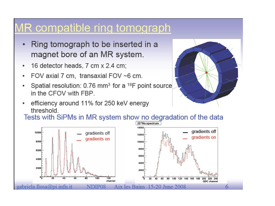
Llossa et al.. (Del Guerra)
")
28
DOI mandatory

29
SPECT/PET - mixed ring, “trivial” - full ring - at the same time? reducing too much the sensitivity - consecutively? How to build this? Scintillator LaBr3 - fast - light yield but - hygroscopic - density Different layout possible for A and B - continuous single slice (for Pet) or several slices (with equal or increasing thickness) - pixellated - modules of 50 x 50 x 20 (30) mm3 with pixels 2 x2 or 3 x3 or 4 x 4 mm2 (problems with DOI) - same scheme - modules of 50 x 50 mm2 with pixel “cubes” of 2 x2 x2 mm3 (or 3 x 3 x 3 mm3) (brute force) (diverging number of channels?) SPECT PET 5 mm 15-25 mm AB
 or several slices (with equal or increasing thickness) - pixellated - modules of 50 x 50 x 20 (30) mm3 with pixels 2 x2 or 3 x3 or 4 x 4 mm2 (problems with DOI) - same scheme - modules of 50 x 50 mm2 with pixel cubes of 2 x2 x2 mm3 (or 3 x 3 x 3 mm3) (brute force) (diverging number of channels ) SPECT PET 5 mm mm AB.")
31
brain - 6 sides (at least) to be “small” and compatible with the SPECT layout (FOV > 20 x 20 cm2 Small animal (and breast) 25 -30 cm 5 cm spect pet
 to be small and compatible with the SPECT layout (FOV > 20 x 20 cm2 Small animal (and breast) cm 5 cm spect pet")
Similar presentations


 Vertex Detector for SuperBelle Ariane Frey, Max-Planck-Institut für Physik München Contents: Software framework Simulation.>")


 511 Shibata Lab 11R50047 Jennifer Newsham YSEP.>")


>")

 reaction Graduate school of science, Tohoku University Toshiyuki Gogami for HES-HKS.>")


