Download presentation
Presentation is loading. Please wait.

1
Distinguishing Pigmented Skin Lesions and Melanoma
Toby Maurer, MD University of California, San Francisco

3
Epidemiology of Melanoma
Lifetime risk of an American developing melanoma 1935: 1 in 1500 1980: 1 in 250 2002: 1 in 68

4
Melanoma Statistics Not an old person’s disease
1 in 4 persons w/melanoma are under 40 Most common cancer in women ages 25-29 2nd most common cancer for women age 30-34

5
Risk Factors Red/blond hair
Family history of melanoma-specific gene mutations found-testing for research purposes only Sun exposure in childhood is risk factor Intermittent sun exposure more important than total lifetime exposure Sun exposure PLUS genetics Multiple nevi-typical and atypical in fair-skinned persons Melanoma- Miller AJ-NEJM July 2006-good review re: nevus to melanoma-what it takes

6
Survival In 1940’s 5 year survival was 40%, now 90%
Survival assoc. with tumor thickness-early detection is what has changed statistic not the treatment

7
Melanoma Survival Rates

8
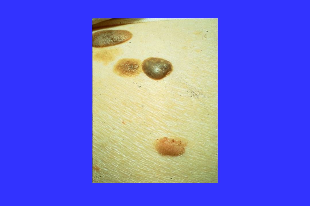
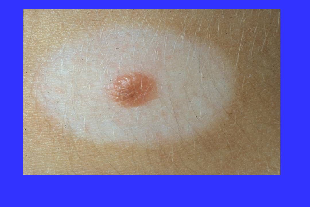
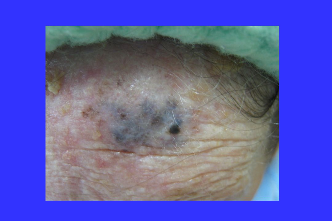
Melanoma Clinical Features (ABCDE’s)
Asymmetry: bisected halves of lesion are NOT identical Border: irregular, notched, vague Color: variegation of browns, red, blue, dark black Diameter: > 6mm in any dimension Evolution/enlargement: any change in nevus Melanoma may have 1 or more of the ABCDE’s

13
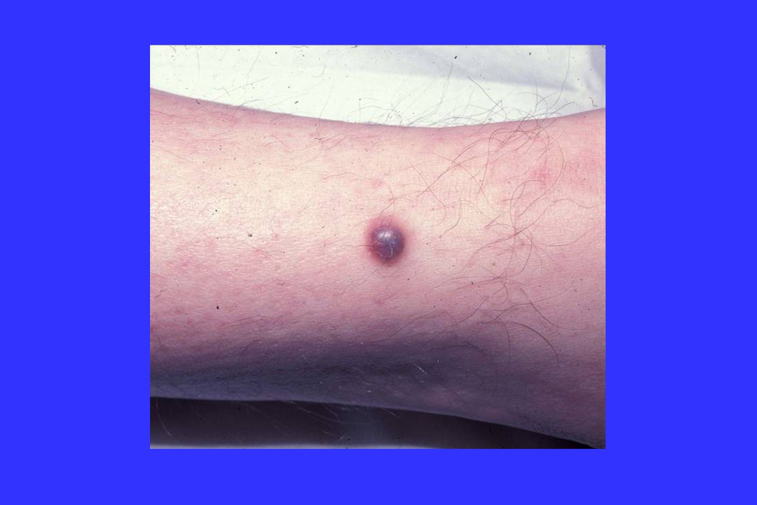
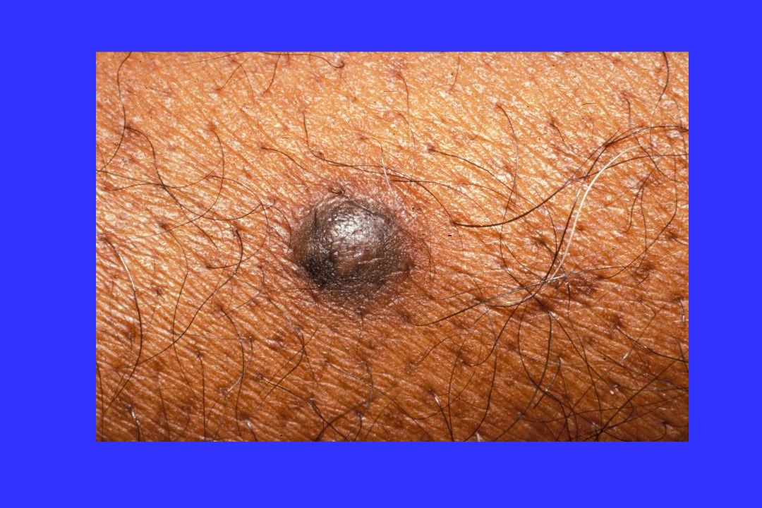
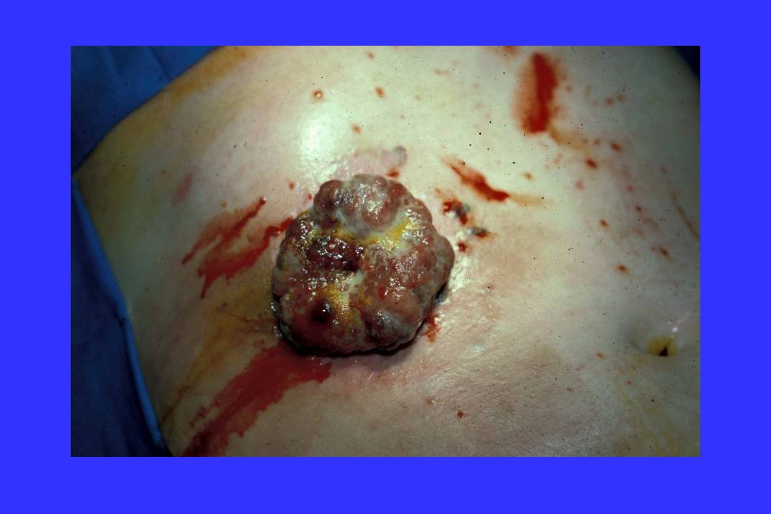
Specific Types of Melanoma
Lentigo maligna Nodular Melanoma Acral Melanoma Amelanotic Melanoma

19
Doc, I’m here for a skin check
1)Personal or family history of melanoma 2) History of atypical nevus that has been removed 3) Presence of new or changing mole- i.e. change in size or color
Personal or family history of melanoma. 2) History of atypical nevus that has been removed. 3) Presence of new or changing mole- i.e. change in size or color.")
20
Melanoma Melanoma may be INHERITED or occur SPORADICALLY
10% of melanomas are of the INHERITED type Familial Atypical Multiple Mole-Melanoma Syndrome (FAMMM)
")
21
Risk Factors for Sporadic (Nonhereditary) Melanoma
Numerous normal nevi, some atypical nevi Sun sensitivity, excessive sun exposure

24
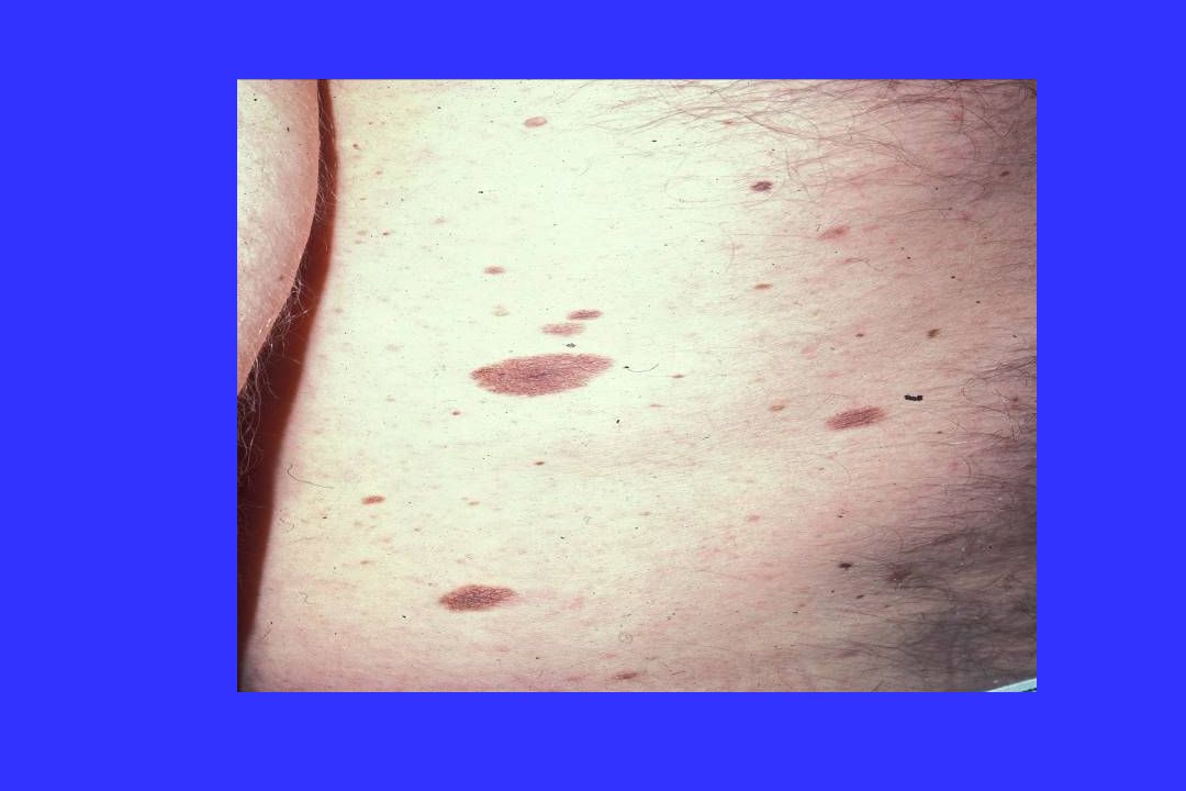
Clinical Features of FAMMM
Often numerous nevi ( ) Nevi > 6mm in diameter New nevi appear throughout life (after age 30) Nevi in sun-protected areas (buttocks, breasts of females) Family history of atypical nevi and melanoma
 Nevi > 6mm in diameter. New nevi appear throughout life (after age 30) Nevi in sun-protected areas (buttocks, breasts of females) Family history of atypical nevi and melanoma.")
25
CDKN2A best understood of hereditary genes-67% lifetime risk of developing melanoma
Testing not recommended outside research trials-penetrance not understood, role of outside factors, risk of other cancers not clearly defined, meaning of negative test

28
Risk Categories (Lifetime Risk)
Very low risk: pigmented races (Latino,African American ,Asian,etc.) Low risk: Caucasian = 1% Intermediate risk: Caucasian w/additional risk factors = 2% - 10% High risk: FAMMM Syndrome up to 100%
 Low risk: Caucasian = 1% Intermediate risk: Caucasian w/additional risk factors = 2% - 10% High risk: FAMMM Syndrome up to 100%")
30
Prevention Self examination for low-risk individuals
Self examination and regular physician examination (yearly to every several years) for intermediate risk individuals Self examination and examination by a dermatologist every 3-12 months for FAMMM kindred
 for intermediate risk individuals. Self examination and examination by a dermatologist every 3-12 months for FAMMM kindred.")
31
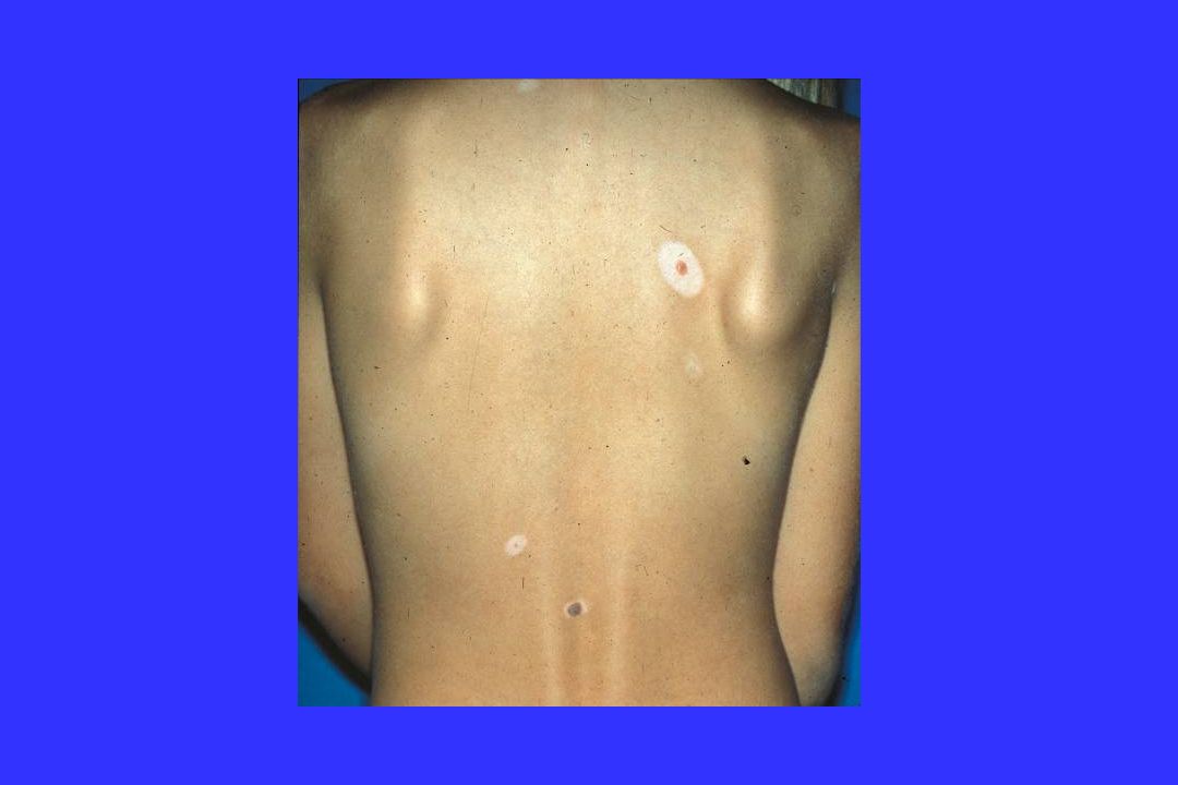
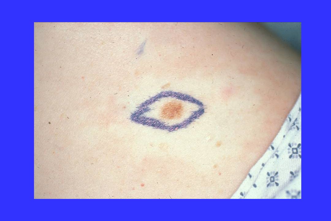
Tools to improve the Art
Photography- available at pigmented nevus centers Involves mapping of nevi, far and close up photos Set of photos for pt and provider About $200.00 Dermoscopy-magnified view of lesion-a science being developed and validated-needs lots of training; better developed in Europe Confocal microscopy-looking at lesions in the human at the bedside

34
Genomic Hybridization
Based on theory that with cancer there are alterations of the genome of the cancer cells DNA can be extracted from paraffin fixed blocks to assess the genome of the specimen Melanoma has specific alterations in genome and differs from nevi In cases where histology is difficult-this can be helpful Certain genomic alterations in melanoma may help stratify who is at higher risk for recurrence

35
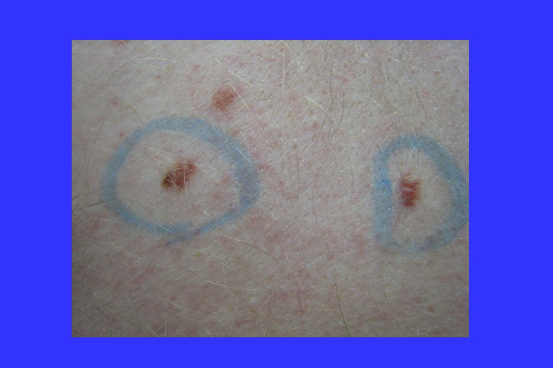
Differential Diagnosis
Seborrheic keratosis Nevus, blue nevus, halo nevus Solar (senile) lentigo Pigmented BCC Dermatofibroma
 lentigo. Pigmented BCC. Dermatofibroma.")
47
How to Diagnose If melanoma is suspected, an excisional biopsy is recommended If the lesion is too large to excise, an incisional biopsy may be done to include any nodules, dark-black areas and white areas

49
Why Excisional Biopsy? The diagnosis and prognosis of melanoma is dependent on the depth of the lesion Send your pathologist the whole thing Dermatopathologist or general pathologist?

51
What to do if Melanoma Staging workup for melanomas > 0.7 mm
Re-excise all melanomas with wider margins

52
What to Do if Melanoma Dx
Depth is key < 0.7 *mm *- Close clinical f/u and labs > 0.7 *mm* - CT scans of chest, pelvis, MRI/PET scan brain & sentinel nodes to stage Melanoma center at least once (or call for latest guidelines) Prognositc Importance of Sentinel Lymph Node in Thin Biopsies of Melanoma-Ranier JM et al. Ann Surg Oncol July 2006 Management of Cutaneous Melanomas-Tsao, et al. NEJM Sept 2004-good review
 Prognositc Importance of Sentinel Lymph Node in Thin Biopsies of Melanoma-Ranier JM et al. Ann Surg Oncol July Management of Cutaneous Melanomas-Tsao, et al. NEJM Sept 2004-good review.")
53
If Melanoma: Re-excise area with larger surgical margins: size of re-excision dependent on the original depth of melanoma Original melanoma in-situ-Excise 0.5 cm margin Original melanoma < 1 mm-Excise 1.0 cm margin Original melanoma >1 mm-Excise 2.0 cm margin Coordinate with surgeon in the know and someone who can do nuclear scan/sentinal node at time of the re-excision if indicated.

54
Primary care follow-up
For the first two years after diagnosis-see patient back q 6 months for total body exam Looking for local recurrence, in-transit metastases, lymph node involvement and second melanomas. Q yr CBC, LFT’s including LDH for lymph node involvement or ulcerative lesion CXray-controversial

55
Follow-up for Melanomas
Second melanomas 1% after 1 year, 2% at 5 yrs, 3% at 10 yrs and 5% at 20 yrs-regular f/u for LIFE (Cancer 97,2003) Developing new risk trees for patients with thinner melanomas Also look for non-melanoma skin cancer and non-Hodgkin’s lymphoma (higher risk is those who had primary melanoma) Melanoma risk is 5 x’s higher in renal transplant recipients
 Developing new risk trees for patients with thinner melanomas. Also look for non-melanoma skin cancer and non-Hodgkin’s lymphoma (higher risk is those who had primary melanoma) Melanoma risk is 5 x’s higher in renal transplant recipients.")
56
New Directions in Therapy
Surgical excision is our therapy Very little to offer re: metastatic disease-6-9 month survival . Current chemo extends life to 1.3 yrs Rational therapy that targets genes and interrupts signalling pathways for metastases Chudnovsky Y, Khavari P, Adams A. J. Clin Investigations April 2005

57
Special Cases Genital pigmented lesions Congenital nevi Pregnancy

58
Genital Pigmented Lesions
Follow the same rules as other pigmented lesions 15% had family history of melanoma

60
Congenital Nevi < 1 cm - 1% Lifetime risk of melanoma
1-5 cm - Unknown risk > 5 cm - 10% Lifetime risk Have congenital nevi evaluated once by a dermatologist

61
Pregnancy Nevi change during pregnancy New ones appear
Should people who have had melanoma get pregnant? Depends on depth of melanoma Call Central Melanoma Center for advice

Similar presentations