Download presentation
Presentation is loading. Please wait.

1
CHOLEDOCHAL CYSTS Aswad Habeeb Hameed Al-Obeidy FICMS GE & Hep

2
CHOLEDOCHAL CYSTS Congenital anomalies of the biliary tract that manifest as cystic dilatation of the extrahepatic and intrahepatic bile ducts The incidence rate of choledochal cysts is 1 in 13,000 to 15,000 in Western countries and as high as 1 in 1000 in Japan These cysts are not familial Females are more commonly affected than males Cases have been described in utero and in elderly patients Approximately two thirds of patients come to medical attention before age 10 years

3
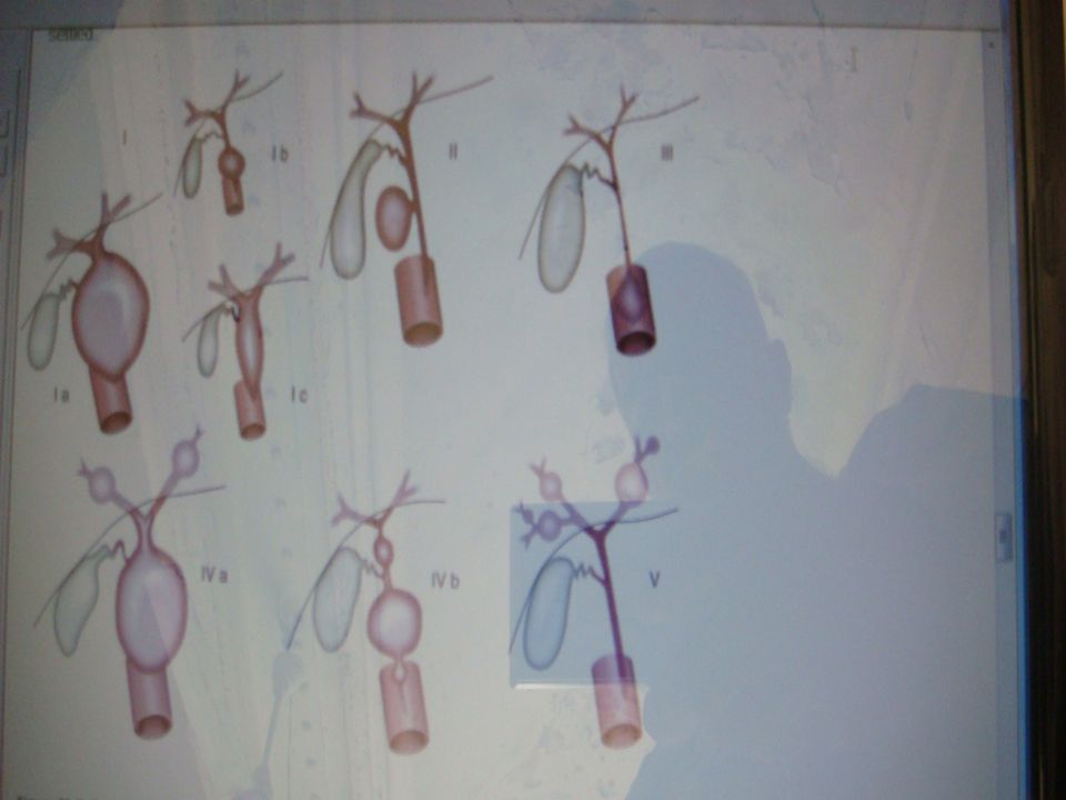
Classification The classification proposed by Todani and colleagues Several varieties of type I cysts, accounting for 80% to 90% of cases Exhibit segmental or diffuse fusiform dilatation of the common bile duct Ia, common type; Ib, segmental dilatation; Ic, diffuse dilatation Type II cysts consist of a true choledochal diverticulum Type III cysts consist of dilatation of the intraduodenal portion of the common bile duct, or choledochocele Type IV cysts may be subdivided into type IVa, multiple intrahepatic and extrahepatic cysts, and type IVb, multiple extrahepatic cysts V, or Caroli's disease, which consists of a single or multiple dilatations of the intrahepatic ductal system, should be viewed as a form of choledochal cyst is not settled

5
Etiology The cause of choledochal cysts has not been established Congenital weakness of the bile duct wall, a primary abnormality of epithelial proliferation during embryologic ductal development Congenital obstruction of bile ducts have been suggested A relationship to other obstructive cholangiopathies, such as biliary atresia, has been proposed but not proven Reovirus RNA has been detected by reverse transcriptase– polymerase chain reaction methodology in hepatic or biliary tissues of 78% A high frequency (40%) of an anomalous junction of the pancreatic and common bile ducts
 of an anomalous junction of the pancreatic and common bile ducts")
6
Pathology The cysts are composed of a fibrous wall There may be no epithelial lining or a low columnar epithelium Mild chronic inflammation may be present Complete, in-flammatory obstruction of the terminal portion of the common bile duct is common in infants who have a choledochal cyst Liver histology in the affected neonate shows typical features of large duct obstruction Portal tract edema, bile ductular proliferation, and fibrosis may be prominent A pattern of biliary cirrhosis may be observed in older patients with long-standing biliary obstruction Carcinoma of the cyst wall may occur by adolescence

7
Clinical Features Disease often appears during the first months of life As many as 80% of patients have cholestatic jaundice and acholic stools Vomiting, irritability, and failure to thrive may occur Physical examination shows hepatomegaly and, in approximately one half of patients, a palpable abdominal mass Spontaneous perforation of a choledochal cyst may occur In older patients, epigastric pain, which may result from pancreatitis, is the most common symptom Intermittent jaundice and fever may result from recurrent episodes of cholangitis The classic triad, consisting of abdominal pain, jaundice, and a palpable abdominal mass, is observed in less than 20% of patients

8
Diagnosis The diagnosis of a choledochal cyst is best established with ultrasonography Several reports have demonstrated that antenatal ultrasonography can be used to detect a choledochal cyst in the fetus Sequential ultrasonographic examinations have allowed study of the evolution of choledochal cysts during pregnancy In the older child, percutaneous transhepatic cholangiography or ERCP may help define the anatomic features of the cyst MRCP is being used increasingly to evaluate the extent of the cyst and defects within the biliary tree and to detect an anomalous junction of the pancreaticobiliary ducts In practice, most pediatric surgeons rely on an operative cholangiogram to define the extent of intrahepatic and extrahepatic disease

9
Treatment Preferred treatment for choledochal cyst is surgical excision of the cyst with reconstruction of the extrahepatic biliary tree Biliary drainage is usually accomplished by a choledochojejunostomy with a Roux-en-Y anastomosis Excision of the cyst reduces bile stasis and the risk of cholangitis and malignancy Simple decompression and internal drainage should be done only when the complicated anatomic characteristics do not allow complete excision Long-term follow-up is essential, because recurrent cholangitis, lithiasis, anastomotic stricture, and pancreatitis may develop years after the initial operation

10
Caroli's Disease Caroli's disease is a subtype of choledochal cyst characterized by diffuse intrahepatic dilatation Classically segmental and saccular and is associated with stone formation and recurrent bacterial cholangitis May manifest as cholangitis, abscesses, jaundice, or cirrhosis A more common type, Caroli's syndrome, is associated with a portal tract lesion typical of CHF Renal disease occurs in both forms, renal tubular ectasia occurs with the simple form, and both conditions can be associated with autosomal recessive polycystic renal disease The gene encodes a large protein (4074 amino acids), which has been called fibrocystin to reflect the main structural abnormalities in liver and kidney The intrahepatic cysts are in continuity with the biliary tract and are lined by epithelium that may be ulcerated and hyperplastic The cysts may contain inspissated bile, calculi, and purulent material
, which has been called fibrocystin to reflect the main structural abnormalities in liver and kidney The intrahepatic cysts are in continuity with the biliary tract and are lined by epithelium that may be ulcerated and hyperplastic The cysts may contain inspissated bile, calculi, and purulent material")
11
Caroli's Disease During childhood and adolescence because of hepatomegaly and abdominal pain The disorder appears in the neonate as renal disease or cholestasis Stagnation of bile, leading to formation of biliary sludge and intraductal lithiasis Fever and intermittent jaundice may occur during episodes of bacterial cholangitis Hepatospleno-megaly is found in cases associated with CHF; affected patients may exhibit bleeding esophageal varices Ultrasonography, MRC, and computed tomography are of great value Percutaneous or endoscopic cholangiography usually demonstrates a normal common duct with segmental, saccular dilatations of the intrahepatic bile ducts Hepatic resection is indicated for disease limited to a single lobe Therapy with ursodeoxycholic acid, 10 to 15 mg/kg/day in individual doses Liver transplantation is an option in pati-ents who have extensive disease and frequent complications

Similar presentations