Download presentation
Presentation is loading. Please wait.

1
Chapter 10: Cell Growth and Division
Mitosis/Cancer

2
Why is it necessary for cells to divide?
DNA Overload-not enough information for the cell as it grows larger in size To improve material exchange Surface area to volume-not enough cell membrane surface for exchange that’s required of larger volume cell as it grows

3
Ratio of Surface Area to Volume in Cells
Section 10-1 Cell Size Surface Area (length x width x 6) Volume (length x width x height) Ratio of Surface Area to Volume
 Volume. (length x width x height) Ratio of Surface Area to Volume.")
4
What is Cell Division (M phase)?
process where a cell divides into two new daughter cells Before cell division takes place, the cell must copy or replicate its DNA. Each daughter cells gets a complete copy of the original DNA Cell division has 2 parts Mitosis: division of nucleus and DNA Cytokinesis: division of cytoplasm and organelles

5
Mitosis (1st stage of cell division)
4 parts of mitosis Prophase Metaphase Anaphase Telophase Mitosis is followed by 2nd part of cell division; cytokinesis

6
Interphase Prophase Anaphase Telophase/Cytokinesis

7
Preparation for division-organelle replication
Cell Growth-protein and organelle production DNA Replication Preparation for division-organelle replication

8
Figure 10–4 The Cell Cycle G1 phase M phase S phase G2 phase

9
Interphase G1 phase: Cell Growth S phase: DNA replication
G2 phase: Preparation for Mitosis Longest phase of cell cycle

10
Figure 10–5 Mitosis and Cytokinesis
Spindle forming Centrioles Nuclear envelope Chromatin Centromere Centriole Chromosomes (paired chromatids) Interphase Prophase Spindle Cytokinesis Centriole Metaphase Telophase Individual chromosomes Anaphase Nuclear envelope reforming
 Interphase. Prophase. Spindle. Cytokinesis. Centriole. Metaphase. Telophase. Individual chromosomes. Anaphase. Nuclear envelope reforming.")
11
Prophase-Phase #1 of Mitosis
Longest Phase-50% to 60% of total time to complete mitosis Chromatin condenses into Chromosomes Centromeres connect sister chromatids Centrioles separate to opposite poles Spindle is organized Nucleolus disappears and nuclear envelope breaks down.

12
Figure 10–5 Mitosis and Cytokinesis
Spindle forming Centrioles Nuclear envelope Chromatin Centromere Centriole Chromosomes (paired chromatids) Interphase Prophase Spindle Cytokinesis Centriole Metaphase Telophase Individual chromosomes Anaphase Nuclear envelope reforming
 Interphase. Prophase. Spindle. Cytokinesis. Centriole. Metaphase. Telophase. Individual chromosomes. Anaphase. Nuclear envelope reforming.")
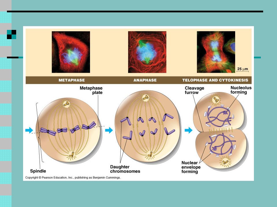
13
Metaphase-Phase #2 of Mitosis
Centromeres attach to spindle fibers Chromosomes line up across the equator of the cell-metaphase plate

14
Figure 10–5 Mitosis and Cytokinesis
Spindle forming Centrioles Nuclear envelope Chromatin Centromere Centriole Chromosomes (paired chromatids) Interphase Prophase Spindle Cytokinesis Centriole Metaphase Telophase Individual chromosomes Anaphase Nuclear envelope reforming
 Interphase. Prophase. Spindle. Cytokinesis. Centriole. Metaphase. Telophase. Individual chromosomes. Anaphase. Nuclear envelope reforming.")
15
Anaphase-Phase #3 of Mitosis
Sister chromatids separate becoming individual chromosomes and moving to opposite poles of cell

16
Figure 10–5 Mitosis and Cytokinesis
Spindle forming Centrioles Nuclear envelope Chromatin Centromere Centriole Chromosomes (paired chromatids) Interphase Prophase Spindle Cytokinesis Centriole Metaphase Telophase Individual chromosomes Anaphase Nuclear envelope reforming
 Interphase. Prophase. Spindle. Cytokinesis. Centriole. Metaphase. Telophase. Individual chromosomes. Anaphase. Nuclear envelope reforming.")
17
Telophase-Phase #4 of Mitosis
Chromosomes disperse into chromatin Nuclear envelope re-forms around each cluster of chromatin Spindle breaks apart Nucleolus visible in each new daughter cell (2 new nuclei form)
")
18
Figure 10–5 Mitosis and Cytokinesis
Spindle forming Centrioles Nuclear envelope Chromatin Centromere Centriole Chromosomes (paired chromatids) Interphase Prophase Spindle Cytokinesis Centriole Metaphase Telophase Individual chromosomes Anaphase Nuclear envelope reforming
 Interphase. Prophase. Spindle. Cytokinesis. Centriole. Metaphase. Telophase. Individual chromosomes. Anaphase. Nuclear envelope reforming.")
19
Cytokinesis Division of cytoplasm and organelles
Animal Cells: Cleavage Furrow-cell membrane pinches inward Plant Cells: Cell Plate-develops into separating membrane (cell wall appears shortly after) 2 new daughter cells each with nucleus and identical chromosomes
 2 new daughter cells each with nucleus and identical chromosomes.")
20
Figure 10–5 Mitosis and Cytokinesis
Spindle forming Centrioles Nuclear envelope Chromatin Centromere Centriole Chromosomes (paired chromatids) Interphase Prophase Spindle Cytokinesis Centriole Metaphase Telophase Individual chromosomes Anaphase Nuclear envelope reforming
 Interphase. Prophase. Spindle. Cytokinesis. Centriole. Metaphase. Telophase. Individual chromosomes. Anaphase. Nuclear envelope reforming.")
21
Cytokinesis

22
Longest Phase-50% to 60% of total time to complete mitosis-Prophase

24
Prophase Interphase

25
Metaphase Prophase

26
Anaphase

27
Telophase

28
Cyclins: proteins that regulate timing of cell cycle
Two types of regulatory proteins Internal regulators: proteins that respond to events inside the cell (ex. No mitosis until all chromosomes are replicated) External regulators: Proteins that respond to events outside cell Speed up, or slow down cell cycle Ex: wound healing and embryonic development
 External regulators: Proteins that respond to events outside cell. Speed up, or slow down cell cycle. Ex: wound healing and embryonic development.")
29
Figure 10–8 Effect of Cyclins-
The sample is injected into a second cell in G2 of interphase. A sample of cytoplasm is removed from a cell in mitosis. As a result, the second cell enters mitosis.

30
Cancer: Disorder in which some cells lose ability to control growth
Density-Dependent Inhibition- ability to respond to signals that regulate the growth of cells Cancer cells do not exhibit this characteristicform tumors Carcinogen- cancer causing agent (ex. Cigarettes, UV radiation from sun)
")
31
Cancer Divide uncontrollably and form tumors
Damage surrounding tissues P53 Gene: stops cell cycle until replication is complete defect in this gene present in a large number of cancers

32
Tumors Masses of cells that can damage surrounding tissue
Benign-non-cancerous growth Localized and not spread Malignant-cancerous growth Invade and damage nearby tissues and organs Metastasis-spread of cancer

33
Regulating the Cell Cycle

34
Why didn’t the cells keep dividing until they spilled over the edge of the petri dish?
What would happen if the cells continued to divide?

36
Asymmetrical, Borders, Color, Diameter, Elevation
Skin Cancer : Melanoma Asymmetrical, Borders, Color, Diameter, Elevation

Similar presentations