Download presentation
Presentation is loading. Please wait.

1
Chemistry of Proteins

2
Chemistry of Proteins Definition:
Proteins are organic compounds with a high molecular weight formed of carbon, oxygen, hydrogen and nitrogen and may also contain sulfur, phosphorus coloring non-protein organic groups and metal ions. They are polymers formed of subunits called amino acids linked together by peptide linkage.

3
Biological importance of proteins
Nutritional role: Provide the body with essential amino acids, nitrogen and sulfur. Catalytic role: All enzymes are proteins in nature. Hormonal role: Most of hormones and all receptors are protein in nature. Defensive role: The antibodies (immunoglobulins) that play an important role in the body’s defensive mechanisms are proteins in nature.
 that play an important role in the body’s defensive mechanisms are proteins in nature.")
4
5-Plasma proteins are responsible for most effective osmotic pressure of the blood. This osmotic pressure plays a central role in many processes, e.g., urine formation. Blood clotting factors are proteins. 6-Transport role: Proteins carry lipids in the blood forming lipoprotein complexes. Proteins also carry, hormones, e.g., thyroid hormones and minerals, e.g., calcium, iron and copper. Hemoglobin (a chromo-protein) carries O2 from the lung to tissues is a protein. 7-Structural role: Proteins are the main structural component in bone, muscles, cyto-skeleton and cell membrane. 8-Control of gene expression: Most factors required for DNA replication, transcription and mRNA translation are protein in nature.
 carries O2 from the lung to tissues is a protein. 7-Structural role: Proteins are the main structural component in bone, muscles, cyto-skeleton and cell membrane. 8-Control of gene expression: Most factors required for DNA replication, transcription and mRNA translation are protein in nature.")
5
Amino Acids Amino acids are organic acids that contain NH2 group. They are the structural units of proteins and are obtained from them by hydrolysis. The general formula of any amino acid is as follows: All these amino acids are alpha-amino acids and the metabolizible form of them are L-amino acids. Alpha-amino acid means that the amino group is attached to the -carbon atom,

6
Each amino acid (except proline and hydroxyproline) has a carboxyl group (COOH), an amino group (NH2) and a characteristic side chain (R). All amino acids (except glycine) are optically active, i.e., can rotate plane polarized light. This is because the 4 groups attached to -carbon are different. In glycine, the -carbon is attached to 2 hydrogen atoms, therefore, is optically inactive.
 are optically active, i.e., can rotate plane polarized light. This is because the 4 groups attached to -carbon are different. In glycine, the -carbon is attached to 2 hydrogen atoms, therefore, is optically inactive.")
7
Classification of Amino Acids
Amino acids can be classified by one of three methods: I-Chemical classification: Based upon the number of amino groups or carboxyl groups in the amino acid: Neutral amino acids (mono-amino, mono-carboxylic). - Acidic amino acids (mono-amino, dicarboxylic). - Basic amino acids (diamino, mono-carboxylic).
. - Acidic amino acids (mono-amino, dicarboxylic). - Basic amino acids (diamino, mono-carboxylic).")
8
II-Biological classification: Based upon whether the amino acids can be synthesized in human body or not: Essential amino acids: Not synthesized in the body and must be supplied in the diet. Non-essential amino acids: Can be synthesized in the body and is not essential to be present in diet.

9
III-Metabolic Classification: Based upon the fate of amino acid inside the body:
Glucogenic amino acids, that can be converted to glucose. Ketogenic amino acids, that can be converted to ketone bodies. Mixed function amino acids, i.e., can be converted to both glucose and ketone bodies

10
I-Chemical classification
(According to number of carboxyl and amino groups) Amino acids can be classified into: a) Neutral amino acids. b) Acidic amino acids. c) Basic amino acids.
 Amino acids can be classified into: a) Neutral amino acids. b) Acidic amino acids. c) Basic amino acids.")
11
A) Neutral amino acids They contain one amino group and one carboxyl group. They have 5 types: 1-Aliphatic amino acids: e.g.,

12
2. Hydroxy amino acids: e.g.,
serine, threonine,.

13
3. Aromatic amino acids: e.g.,
phenylalanine and tyrosine . Tyrosine is synthesized from phenyl alanine and both give triiodothyronine and thyroxin, adrenaline and noradrenaline. Melanin pigment and cresol ,phenol in the body, e.g.,

14
4-Sulfur-containing amino acids: e.g.,
Cysteine gives cystine and its SH group is very essential in activity of many proteins particularly the active sites of enzymes.

15
5-Heterocyclic amino acids: e.g.,
Histidine gives histamine a very important inflammatory mediator. Proline gives hydroxyproline that is essential for collagen cross-linking. Tryptophan gives nicotinic acid, melatonin, serotonin and indican in the body.

16
B) Acidic amino acids They contain 2 carboxyl groups and one amino group, e.g., glutamic acid and asparatic acid. These acidic amino acids can occur in the tissue in the form of amides, e.g., glutamic acid glutamine and asparatic acid asparagine.

17
C) Basic amino acids They contain 2 amino groups and one carboxyl group, e.g., Ornithine and Arginine. Ornithine does not enter in the synthesis of proteins and is usually present in the free form. It is synthesized from arginine. Citrulline is formed from ornithine during urea synthesis

18
Lysine and Hydroxy lysine: They participate in protein cross-linking.

19
II-Metabolic classification:
Amino acids may be classified into A-glucogenic amino acids, i.e., those which can be converted into glucose, B-ketogenic amino acids, i.e., those which can be converted into ketone bodies C-mixed amino acids, i.e., those which can be converted into both glucose and ketone bodies.

20
Ketogenic & glucogenic
Leucine Lysine Rest of amino acids Isoleucine Tyrosine Tryptophan Phenyl alanine

21
III-Biological or Nutritional Classification:
Some amino acids can not be synthesized inside the body. If these amino acids are not taken in diet they will affect the growth and the health. Thus, amino acids may be classified into: A- Essential amino acids: These are amino acids that can not be synthesized in the human body and should be taken in the diet, otherwise their deficiency will lead to a nutrition deficiency disease that affect both growth and health.

22
Valine Isoleucine Threonine Tryptophan Arginine Leucine Lysine Methionine Phenylalanine Histidine B- Non essential amino acids: - The rest of amino acids can be synthesized inside the human body and their deficiency in diet does not affect the growth or the health.

23
I. Physical properties of amino acids: 1. Solubility:
All amino acids are soluble in water, diluted acids and alkalis. 2. Optical activity: All amino acids, except glycine, are optically active, i.e., they contain asymmetric carbon atom (-carbon), thus they can deviate the plane polarized light either to the right or to the left. 3. Absorption of ultraviolet light: Aromatic amino acids (tryptophan, tyrosine and phenylalanine) can absorb ultraviolet light.
, thus they can deviate the plane polarized light either to the right or to the left. 3. Absorption of ultraviolet light: Aromatic amino acids (tryptophan, tyrosine and phenylalanine) can absorb ultraviolet light.")
24
Structure of Proteins There are 4 levels or orders of organization of the structure protein molecule: primary, secondary, tertiary and quaternary structures. This complication gives the molecule its functional domain to explain its structure-function requirements that if changes due to mutation will give non-functional protein and, therefore, a disease.

25
1. Primary structure: Primary structure is the linear form of the polypeptide illustrating the total number, chemical nature, and linear order of all of the amino acid residues in the polypeptide chain or chains of a protein and position of disulfide bonds if present. The peptide bonds (primary bond) are responsible for the primary structure.
 are responsible for the primary structure.")
26
2. Secondary structure: It is the fine folding of polypeptide chain into specific regular coiled structure or irregular random coiling held together by hydrogen, ionic and disulfide bonds. It is due to the interaction of amino acids located very close to each other.

27
3. Tertiary structure: It is the final three-dimensional form due to the more complicated course folding and super-folding of the polypeptide chain in its secondary level into globular or fibrous form of different size. It is due to interaction of amino acids located far apart (away from each other). It is the biologically active conformation of the polypeptide and therefore, is the most liable to denaturation. The bond stabilizing the tertiary structure are disulfide bonds, hydrogen bonds and ionic bonds
. It is the biologically active conformation of the polypeptide and therefore, is the most liable to denaturation. The bond stabilizing the tertiary structure are disulfide bonds, hydrogen bonds and ionic bonds.")
28
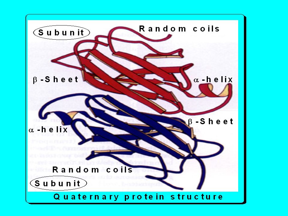
4. Quaternary structures:
Proteins consist of two or more polypeptide chains in their tertiary structure united by forces other than peptide bonds are said to possess a quaternary structure. Quaternary structure therefore, is the positional relationship between individual polypeptides associating to form one protein molecule. The bonds responsible for the quaternary structure are not disulphid or peptide bonds

30
Classification of Proteins
I. According to the biological importance of the protein: 1) Proteins of high biological value: These are all proteins of animal origin (with a few exceptions) and some proteins of plant origin that contain all the 10 essential amino acids in well balanced amounts and are easily digestible. Examples of animal proteins include; milks and its products, egg, liver, fishes, red and while meats. Examples of the few plant proteins of high biological value are lentils and broad beans.
 Proteins of high biological value: These are all proteins of animal origin (with a few exceptions) and some proteins of plant origin that contain all the 10 essential amino acids in well balanced amounts and are easily digestible. Examples of animal proteins include; milks and its products, egg, liver, fishes, red and while meats. Examples of the few plant proteins of high biological value are lentils and broad beans.")
31
2) Proteins of low biological value:
These are proteins that are deficient in one or more of the essential amino acids or containing very little amount of one of them or are indigestible. Most of plant protein are of low biological value and a very few animal proteins are also of low biological value such are collagen because is deficient in tryptophan and cysteine and keratins because they are indigestible. This does not imply that a person should eat only a protein of high biological value to avoid deficiency of essential amino acids, but this can be also avoided by eating two or more proteins of low biological value that complete each other’s deficiency.

32
II. According to the axial ratio of the protein molecule:
Studies on the shape of the protein molecule using ultramicroscope indicates that there are two types of proteins in nature: 1. Fibrous proteins. 2. Globular proteins.

33
Fibrous proteins: They have an axial ratio of more than 10. Axial ratio = Length/Width of the protein molecule. They are fairly stable proteins. Examples, a. Keratin proteins in hairs, wool, skin, and most cells. In its native state, it is present in the form of coiled polypeptide chains called -keratin. It can be stretched by denaturation forming -keratin. b. Myosin is the major protein of muscles. During muscle relaxation it is called -myosin but during muscle contraction, it undergoes a change in its structure and it becomes -myosin

34
2. Globular proteins: Their axial ratio is less than 10. Their peptide chains are folded or coiled on themselves in a very compact manner. They are less stable than fibrous proteins. Examples are albumin, globulins, and insulin

35
III. According to the composition of the Protein:
There are 3 main groups: A) Simple proteins. B) Conjugated or conjugated proteins. C) Derived proteins.
 Simple proteins. B) Conjugated or conjugated proteins. C) Derived proteins.")
36
A) Simple Proteins These are proteins which on hydrolysis produce amino acids only. Simple proteins are subdivided according to their physical properties, solubility, molecular weight and amino acid composition into: 1-Albumin and globulins . 2-Basic proteins (Histones & Protamines). 3-Acidic proteins (Glutelins & Gliadins ). 4-Scleroproteins(Keratin&Collagen&Elastin)
. 3-Acidic proteins (Glutelins & Gliadins ). 4-Scleroproteins(Keratin&Collagen&Elastin)")
37
- Soluble in water and salt solution - Soluble in salt solution.
1-Albumin and globulins . Albumin Globulin - Soluble in water and salt solution - Soluble in salt solution. - Coagulated by heat. - The same as albumin -They are of high biological value: -The same as albumin. - Contain all essential amino acids. - Easily digested. - M.W.: 68 KDa. - M.W.: 150 KDa. -Precipitated by full saturation with ammonium sulfate. -Precipitated by half saturation with ammonium sulfate. -They are present in: serum albumin,egg albumin, milk: lactalbumin. They are present in: serum globulin, milk Lactglobulin, egg globulin. - It functions as transporting protein for elements, vitamins, and hormones other than keeping blood osmosis. - It functions in transport also but its major function is being antibodies.

38
2-Basic proteins(rich in basic amino acids)
A-Protamines: Protamines are the simplest natural proteins. Their molecule is small and contains not more than 20 amino acid residues Protamines are present in sperms and ova Protamines are water-soluble and ammonia soluble proteins, non-coagulable . They also exist in combination with nucleic acids in the nucleus. In plants, protamines are present in pollen grains. Protamines are strongly basic, due to the presence of large amount of basic amino acids specially arginine.

39
B-Histones: (compare to protamines)
Histones are basic proteins, which are characterized, by being soluble in water, dilute acids, and insoluble in dilute ammonia. Their insolubility in ammonia differentiates them from protamines. They are usually present associated with nucleic acid and porphyrins. They are not present in plants. The most important amino acids entering in the structure of histones are arginine, histidine and less lysine. The amount of their basic charges is less than protamines. Histones are present in glandular tissues as liver and spleen. The protein globin, which enters in the formation of the hemoglobin of blood, is similar to histones.

40
The examples of prolamines are gliadin of wheat and zein of maize.
3-Acidic proteins A-Gliadins or Prolamines: Gliadins are never present in animals but only present in plant kingdom. They are present in high concentrations in cereals and have been obtained from all grains except rice. Gliadins are also called prolamines due to the presence of a high percentage of the amino acids proline (10-14%) and glutamine. The amino acid lysine is deficient in gliadins. Gliadins are proteins, which are insoluble in water and saline, but soluble in 70-80% alcohol due to the presence of excess proline. The examples of prolamines are gliadin of wheat and zein of maize.
 and glutamine. The amino acid lysine is deficient in gliadins. Gliadins are proteins, which are insoluble in water and saline, but soluble in 70-80% alcohol due to the presence of excess proline. The examples of prolamines are gliadin of wheat and zein of maize.")
41
B-Glutelins: They are plant proteins. They are soluble in diluted acids but not in water or diluted salt solutions. They are very rich in glutamic acid. They have very large molecular weight and are heat coagulable. Examples are oryzenin of rice and glutelin of wheat.

42
4-Scleroproteins (Albuminoids)
Scleroproteins are characterized by their extreme insolubility in water, dilute acids and the most common reagents. They are strong fibrous structural proteins that are rich in sulfur containing amino acids and hence disulfide bonds. Their main function is the protection of the body. Hairs, nail and connective tissues, contain scleroproteins and are never present in plants. The main important groups of scleroproteins are: 1. Elastins Collagens Keratin

43
A-Elastins: They are present in the yellow fibers of the connective tissues in lungs, uterine wall during pregnancy, tendons and ligaments. Elastins are also present in the elastic tissues of tendons and big arteries. It is rich in alanine, leucine, valine and proline but deficient in cysteine, methionine, lysine and histidine. Boiling scleroproteins with strong acids or strong alkalis or their digestion by elastase leads to their hydrolysis to free amino acids

44
B-Collagens: They are present in white fibrous connective tissues, tendons and bones. Collagen is insoluble in water, dilute acids and alkalis. From this point of view it is similar to elastins. Collagen is resistant to peptic and trypsin digestion, but when boiled for a long time with water, dilute acids or alkalis, it changes to gelatin. Thus, gelatin is a derived protein obtained from the partial hydrolysis of collagen. Both gelatin and collagen are of little nutritive value because they contain about 40% of the non-essential amino acid glycine. Collagen is rich in glycine, proline and hydroxy proline but low in sulfur containing amino acids, tryptophan and cysteine

45
Gelatins: It is the product of prolonged boiling of collagen in water. It is easily digested and has the property of forming a gel on cooling (gel formation). Gelatin is a very good diet for patients because it is an appetizer and easily digested. Gelatin is deficient in certain essential amino acids. So it is not an adequate protein diet, as it is deficient in tryptophan and cysteine and contains very small amounts of methionine (protein of low biological value).
. Gelatin is a very good diet for patients because it is an appetizer and easily digested. Gelatin is deficient in certain essential amino acids. So it is not an adequate protein diet, as it is deficient in tryptophan and cysteine and contains very small amounts of methionine (protein of low biological value).")
46
C-Keratins Keratins are highly insoluble compounds. They are insoluble in all protein solvents, and are not digestible by proteolytic enzymes (e.g pepsin and trypsin). Keratins are hydrolyzed by prolonged boiling with alkalis. The sulfur content of keratin is high. It is present in the form of cystine, which is responsible for the stability and insolubility of keratins. Most keratins yield histidine, lysine and arginine amino acids on hydrolysis
. Keratins are hydrolyzed by prolonged boiling with alkalis. The sulfur content of keratin is high. It is present in the form of cystine, which is responsible for the stability and insolubility of keratins. Most keratins yield histidine, lysine and arginine amino acids on hydrolysis.")
47
Keratins are present in hairs, nails and superficial layer of the skin.
Barium sulfide is one of the important substances that dissolve keratins. For this reason this material enters in the formation of cosmetics dealing with removal of hair. Keratin is a typical fibrous protein. It consists of long peptide chains. The peptide chains may be coiled or reset in spiral or helix types.

48
B) Conjugated proteins
On hydrolysis, they give amino acids and prosthetic group (i.e., a non-protein group). They include: 1. Phosphoproteins: These are proteins conjugated with phosphate. Phosphate is attached to OH group of serine, tyrosine or threonine present in protein. They are found in: a. Casein: milk protein. b. Vitellin: Egg yolk protein.
. They include: 1. Phosphoproteins: These are proteins conjugated with phosphate. Phosphate is attached to OH group of serine, tyrosine or threonine present in protein. They are found in: a. Casein: milk protein. b. Vitellin: Egg yolk protein.")
49
2. Lipoproteins: These are proteins conjugated with lipids converting them into water soluble substances. Present in blood, brain and egg. Cell membrane is of lipoprotein. Examples: Plasma lipoproteins: see lipid chemistry. 3. Glycoproteins: These are proteins conjugated with carbohydrates in varying amounts attached as short or long chains. Examples: Mucous secretion of gastrointestinal tract and Glycoproteins of cell wall.

50
4. Metalloproteins: These are proteins conjugated with metals such as; Iron: e.g., ferritin is intracellular iron-binding protein and Transferrin is an iron-binding transport protein in the blood. Zinc: e.g., Insulin hormone present in crystals containing zinc. Copper: e.g., Ceruloplasmin: is a protein present in blood. It is responsible for the oxidation of Fe2+ ions to Fe3+ ions.

51
5. Chromoproteins: These are proteins conjugated with colored pigment. Example; Hemoglobin and cytochrome enzyme present in mitochondria contains haem pigment, which is red in color. 6. Nucleoproteins: These are proteins (protamines or histones) conjugated with nucleic acids (DNA or RNA). Examples; Chromosomes: These are proteins conjugated with DNA. Ribosomes: They are proteins conjugated with RNA.
 conjugated with nucleic acids (DNA or RNA). Examples; Chromosomes: These are proteins conjugated with DNA. Ribosomes: They are proteins conjugated with RNA.")
52
C) Derived Proteins They include: A- Denatured protein: e.g., coagulated albumin or globulin. B. Hydrolytic product of protein: e.g., Protein Proteoses Peptone Polypeptide. 1-Proteoses, they are soluble in water, not coagulable and precipitated by saturated salt solution. 2-Peptones: are soluble in water and not coagulable by heat, not precepitated by saturated salt solution.

53
3-Peptides: are soluble in water and salt solution, not coagulable by heat. They are formed from 2 or more amino acids. The amino acids in any polypeptide chain are arranged so that the first amino acid has a free amino group (N-terminal end) and the last one has a free carboxyl group (C-terminal end). So the tripeptide alanine-cysteine-tryptophan is different from the tripeptide tryptophan-cysteine-alanine.
 and the last one has a free carboxyl group (C-terminal end). So the tripeptide alanine-cysteine-tryptophan is different from the tripeptide tryptophan-cysteine-alanine.")
54
Bonds responsible for protein structure
I-Strong bonds: 1. Peptide bonds (primary bond): A peptide bond is a covalent bond formed by a reaction between amino group of one amino acid and a carboxylic group of the next amino acid with the loss of H2O that required ATP.
: A peptide bond is a covalent bond formed by a reaction between amino group of one amino acid and a carboxylic group of the next amino acid with the loss of H2O that required ATP.")
55
2. Disulfide bonds (secondary bond):
The disulfide bond is formed between the SH groups of two cysteine residues within same (intra-chain) or two different polypeptide chains (inter-chain). It maintain secondary structure of a peptide chain or connects two polypeptide chains together in the tertiary structure. It follows the peptide bond in strength but liable to denaturation.
 or two different polypeptide chains (inter-chain). It maintain secondary structure of a peptide chain or connects two polypeptide chains together in the tertiary structure. It follows the peptide bond in strength but liable to denaturation.")
56
II- Weak bonds: 1. Hydrogen bonds: Hydrogen bond is a weak bond formed between the hydrogen atom of –NH of a peptide bond on one peptide chain and the oxygen of C=O of another peptide bond on an adjacent peptide chain or a loop belongs to same peptide chain.

57
2. Hydrophobic bonds: The non polar side chains of neutral amino acids tend to associate in hidden core of protein molecule away from solvent.

58
3. Electrostatic bonds: These are salt bonds formed between oppositely charged groups in the side chains of amino acids e.g. -amino group of lysine and the carboxyl group of asparatic acid.

59
Denaturation of Proteins
Definition: It is the loss of the native form of the protein leading to disruption of its secondary, tertiary and quaternary structure with the changes in their physical and chemical characteristics and loss of their biological activity.

60
Causes of Denaturation
Physical causes: As shaking (mechanical effect), high temperature, X-rays and atomic radiations. Chemical causes: As organic solvents (e.g. acetone), strong alkalis and acids and agents that irreversibly precipitate proteins.
, high temperature, X-rays and atomic radiations. Chemical causes: As organic solvents (e.g. acetone), strong alkalis and acids and agents that irreversibly precipitate proteins.")
61
Effects of Denaturation
Physical changes: Increase in viscosity, decreased solubility and decreased diffusiblility. Chemical changes: leads to loss of hydrogen, hydrophobic and electrostatic bonds but not peptide and disulfide bonds.The result are loss of secondary, tertiary and quaternary structures but not of the primary structure. Biological changes: which include loss of enzymatic, hormonal and other biological properties of proteins. Denatured proteins (e.g cooking), are easily digested than native proteins.
, are easily digested than native proteins.")
62
Significance and Application of denaturation:
Denatured proteins, e.g., cooked meat are easily digested. Avoidance of denaturation is important for biological samples used for determination of enzymatic, hormonal or protein contents. This is done by proper sample collection and storage because if denaturation occurs false results will be obtained. Blood samples to be analyzed for small molecules, e.g., uric acid and glucose are first treated with acid such as trichloroacetic acid or phosphotungestic acid to precipitate the plasma proteins (by denaturation). Detection of albumin in urine by heat coagulation test is based on denaturation by heat. Several approaches for stoppage of bleeding and treatment of burns is based on precipitation and denaturation of a superficial protein layer.
. Detection of albumin in urine by heat coagulation test is based on denaturation by heat. Several approaches for stoppage of bleeding and treatment of burns is based on precipitation and denaturation of a superficial protein layer.")
63
Protein Separation Protein separation is based on Protein solubility
Size of protein molecule Charge of the molecule Methods of protein separation Chromatography. Salting out Electrophoresis. Dialysis. Ultracentrifugation.

64
1- Salting out It depends on elemination of water from the solution. e.g. albumin by full saturation with Amm. sulphate globulin by half saturation with Amm. Sulphate 2-Chromatography Chromatography is a group of separation techniques, where a mixture of molecules is separated. The separated molecules are divided between a stationary sold phase and liquid mobile phase. The separation process depends on the tendency of one type of molecules in the mixture to associate more strongly with one phase than the other.

65
3- Electrophoresis It is movement of charged particles in an electric field towards the oppositely charged electrode. By electrophoresis a mixture of amino acids, polypeptides or proteins can be separated into distinct bands by using electric current.

66
4-Dialysis Dialysis means separation of colloids from crystalloids. Proteins have a high molecular weight that forms a colloidal solution. If there is a mixture of proteins (colloids) and salts (crystalloids) they can be separated by dialysis, i.e., by using a semi-permeable membrane. Crystalloids with very small molecular weight can pass through this membrane, while colloids can not due to the large size of their partic
 and salts (crystalloids) they can be separated by dialysis, i.e., by using a semi-permeable membrane. Crystalloids with very small molecular weight can pass through this membrane, while colloids can not due to the large size of their partic.")
67
5-Ultracentrifugation:
Using high speed centrifuge, a mixture of proteins is separated into different fractions according to their densities. Lipoproteins can be separated by ultracenrifugation to chylomicrons, VLDL, LDL, and HDL.

Similar presentations


![7.5: PROTEINS Proteins Function Structure. Function 7.5.4: State four functions of proteins, giving a named example of each. [Obj. 1] Proteins are the.](/23/6900992/big_thumb.jpg "7.5: PROTEINS Proteins Function Structure. Function 7.5.4: State four functions of proteins, giving a named example of each. [Obj. 1] Proteins are the.>")


