Download presentation
Presentation is loading. Please wait.

1
Bio 321 Neuroanatomy Dr. M. Yu

2
Nervous System Introduction Bio 321 Neuroanatomy

3
Medical Significance The brain and nervous system control all other functions of the body. The extreme importance of the nervous system in medicine is based on the serious nature of the many disorders affecting its structures (more than 1000 disorders). Causes more hospitalization than any other diseases, including heart diseases and cancers. Neurological diseases affect 50 million Americans and costs us about $400 billions annually
. Causes more hospitalization than any other diseases, including heart diseases and cancers. Neurological diseases affect 50 million Americans and costs us about $400 billions annually.")
4
Introduction In this country alone, the numbers are overwhelming: –1. Cerebrovascular Disease - is the 3rd ranking cause of death - vascular conditions of brain & spinal cord annually kill ~500,000 –2. Epileptics seizures ~ 1,500,000 –3. Movement disorders affect another one million people –4. There are ~ 2 million totally blind individuals; & over 13 million with visual impairments –5. There are ~ 17 million totally or partially deaf persons

5
Introduction –6. Over 3 million people are afflicted with Alzheimer’s disease –7. At least 700,000 have cerebral palsy –8. More than 250,000 have multiple sclerosis –9. In addition, there are over 500,000 accidental head and spine injuries annually; fortunately only a minority of which actually injure the brain or spinal cord –10. Acute head injury is the leading cause of death or disability between ages 2 & 40 (as of 1995)
.")
6
Cellular Components of the Nervous System Neurons - the primary functional cells in the nervous system (- approx. 100 Billion in CNS) –1. responsible for initiating & conducting electrical signals by which nervous system communicates –2. size & shape varies greatly between regions of the nervous system & with respective functions –3. mature neurons do NOT divide or replicate, do NOT regenerate following injury
 –1. responsible for initiating & conducting electrical signals by which nervous system communicates –2. size & shape varies greatly between regions of the nervous system & with respective functions –3. mature neurons do NOT divide or replicate, do NOT regenerate following injury.")
11
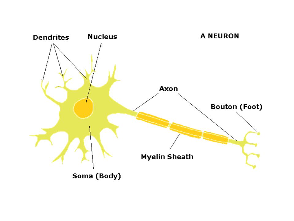
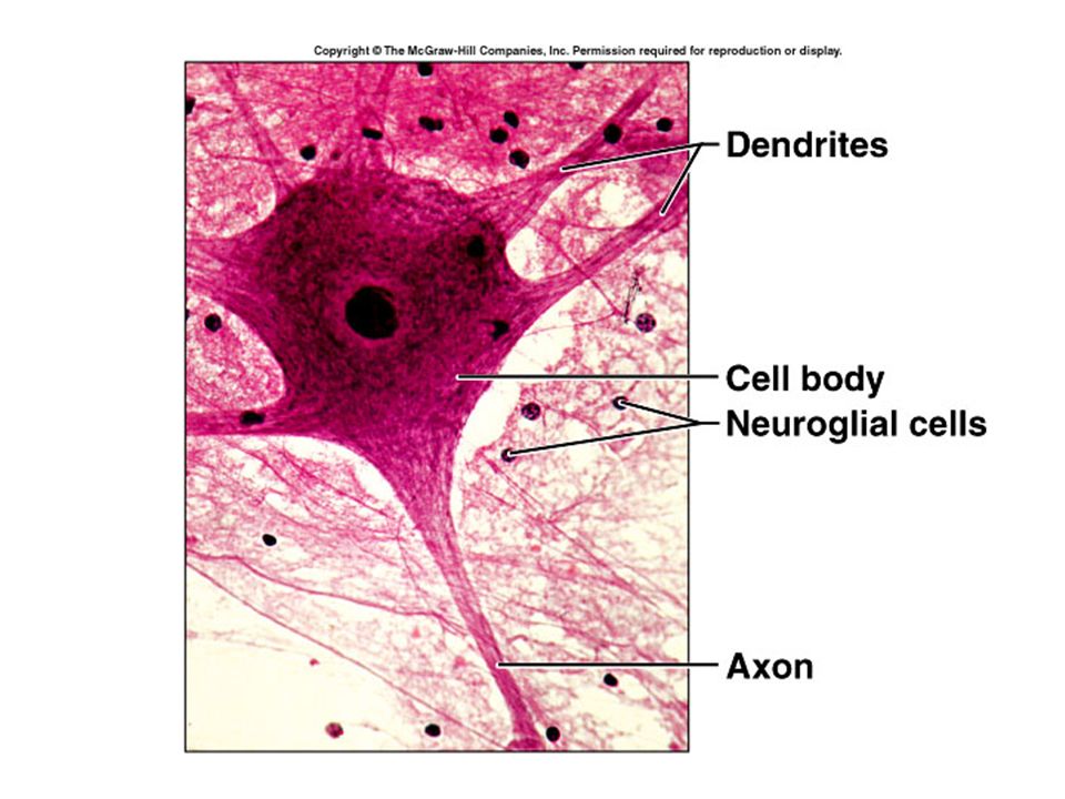
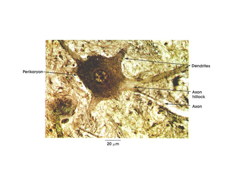
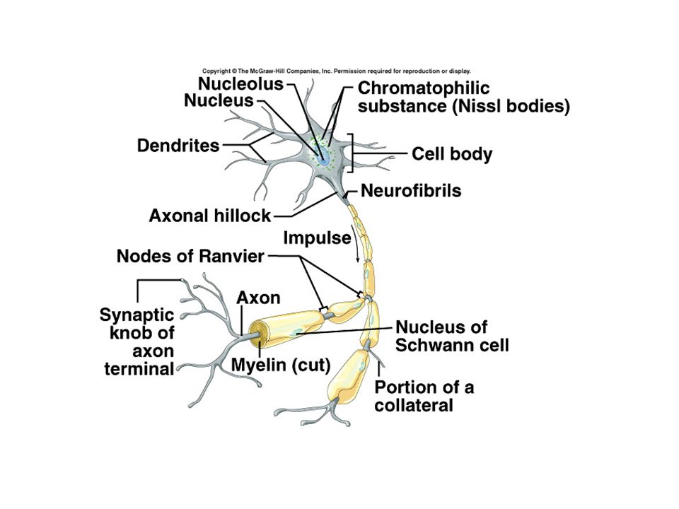
Anatomic features (common to all neurons) 1. Soma - cell body –metabolic center of cell –typical cellular organelles: large clear nucleus, nucleolus, mitochondria, ER, Golgi Apparatus, lysosomes 2. endoplasmic reticulum - a characteristic feature of neurons: - high concentration of RER (granules, form Nissl substance, stain basic)
.")
12
Neurons 3. Cytoplasmic fibrils - another distinguishing feature of neurons, make up cytoskeleton –a. Neurofilaments: intermediate cytoskeletal filaments - abundant, found throughout soma & along processes (10 nm in diameter) – a principle support system component –b. Neurotubules (Microtubules): especially abundant in dendrites - found in cytoplasmic, axonal transport, movement of organelles throughout cell - (20-30 nm in diameter) = microtubules, oriented lengthwise –c. Microfilaments: thinnest, associated with external membrane & dendritic spines - anchor membrane constituents, hold Receptors in place - (5 nm in diameter) = microfilaments in other cells Processes of neurons
 – a principle support system component –b. Neurotubules (Microtubules): especially abundant in dendrites - found in cytoplasmic, axonal transport, movement of organelles throughout cell - (20-30 nm in diameter) = microtubules, oriented lengthwise –c. Microfilaments: thinnest, associated with external membrane & dendritic spines - anchor membrane constituents, hold Receptors in place - (5 nm in diameter) = microfilaments in other cells Processes of neurons.")
14
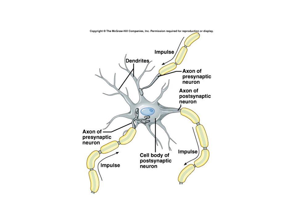
Dendrites Dendrites - number, length, degree of ramification varies between neuronal types –a. usually many per neuron, typically branch extensively –b. carry incoming signals toward soma –c. neuron signal receptors (the dendritic field of a cell = its receptive field)
.")
15
Axon Axon - one per neuron –a. conducting process - carries info away from soma, toward other neurons or effectors –b. length varies greatly, can be very long - sciatic axons > 1 meter (spine -> foot) –c. no ribosomes, no protein synthesis, depend on axonal cytoplasmic transport from soma –d. collaterals = branches of axon, degree of branching varies greatly
 –c. no ribosomes, no protein synthesis, depend on axonal cytoplasmic transport from soma –d. collaterals = branches of axon, degree of branching varies greatly.")
16
Axon Axon Hillock = Initial segment (of axon) –a. base of axon as it leaves soma - looks pale (no Nissl staining) –b. specialized segment of membrane for action potential initiation due to lower threshold than rest of cell –c. also has an extremely high concentration of voltage- gated ion channels –d. allows action potential conduction to travel in ONE direction
 –b. specialized segment of membrane for action potential initiation due to lower threshold than rest of cell –c. also has an extremely high concentration of voltage- gated ion channels –d. allows action potential conduction to travel in ONE direction.")
18
Terms 1. Nucleus - a group of neuronal cell bodies within the central nervous system, eg lateral geniculate nucleus 2. Ganglion (ganglia, pl) - group of cell bodies in peripheral nervous system 3. Nerve - collection of axons in the PNS (tract, fasciculus = a bundle of nerve fibers) 4. White matter - axons, myelinated 5. Gray matter - concentrations of cell bodies and unmyelinated dendrites
 - group of cell bodies in peripheral nervous system 3. Nerve - collection of axons in the PNS (tract, fasciculus = a bundle of nerve fibers) 4. White matter - axons, myelinated 5. Gray matter - concentrations of cell bodies and unmyelinated dendrites.")
19
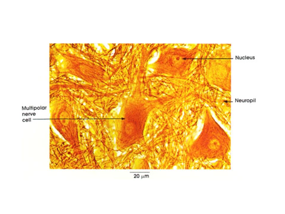
Classification of neurons A. based on processes: –1. Unipolar neurons a. a single primary process extends from soma, which can branch into dendrites & axon b. the principle neuronal cell type of invertebrates c. Pseudounipolar neurons, e.g. dorsal root ganglion cells, appear unipolar, in embryo are actually bipolar; axon & dendrite extensions fuse –2. Bipolar neurons - e.g. bipolar cells in retina - axon & dendrite both extend from soma, from opposite ends –3. Multipolar neurons a. multiple dendritic branches from all parts of soma, & one axon b. many variations due to length, number of dendrites, length of axon c. e.g. pyramidal neuron, spinal motor neuron, purkinje neuron

21
Classification of neurons B. Based on functions –Sensory neurons: carry sensory information from –Motor neurons: carry motor information to muscle sand glands –Interneurons: in between sensory and motor –Projection neuron / relay neuron: has long fibers and project information from on region to another

22
Classification of neurons C. Based on orientation –1. Afferent - refers to the neuron or process extending toward the cell in question –2. Efferent - refers to the neuron or process extending away from the cell in question

23
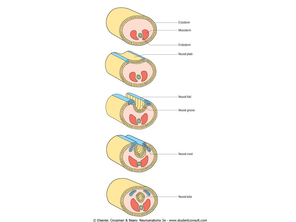
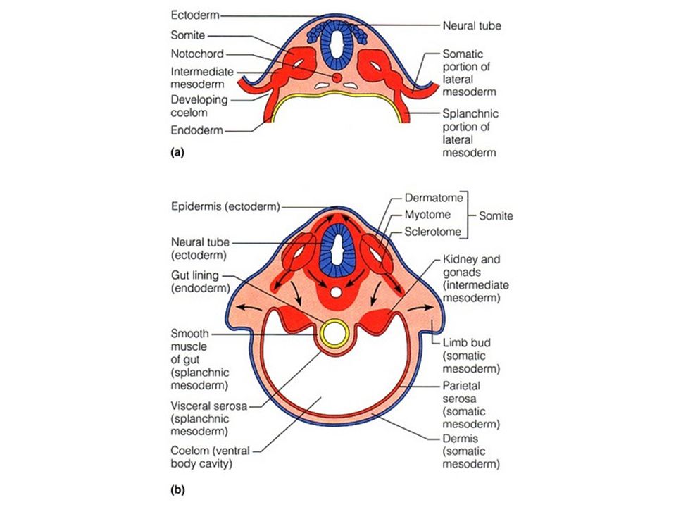
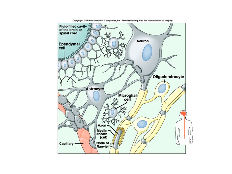
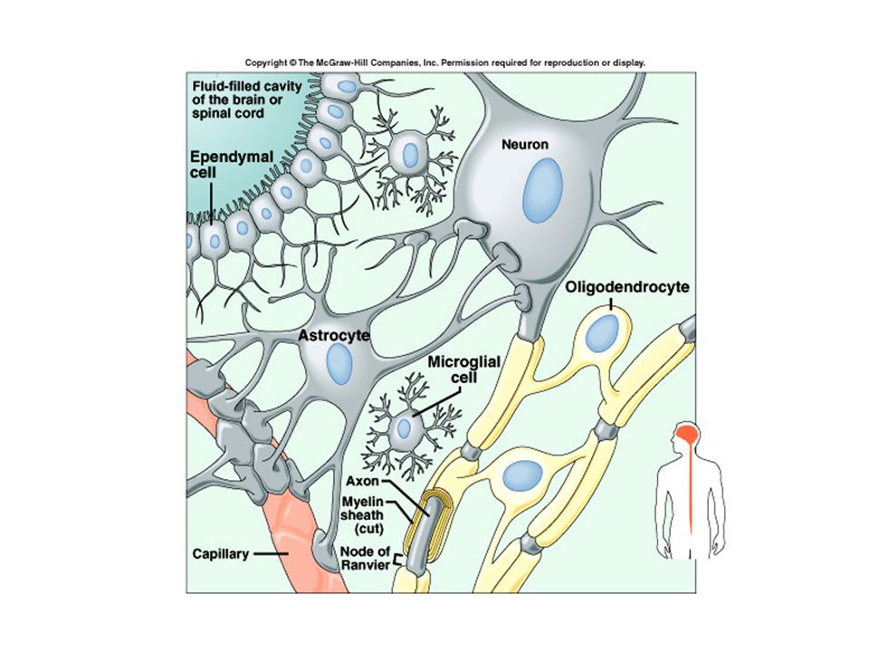
Neuroglial (Glia) supporting cells - Do not conduct action potential, body has 10-15 times more glia cells than neurons (about one trillion) Derived from Neuroectodermal and mesodermal origin
 supporting cells - Do not conduct action potential, body has times more glia cells than neurons (about one trillion) Derived from Neuroectodermal and mesodermal origin")
26
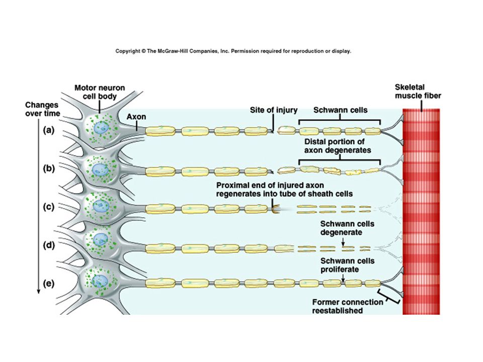
Neuroectodermal origin: #1-4 –1. Schwann cells - form myelin sheath which insulates an axon in peripheral nerves - cell winds around axon, inside its own layers, piling up layers of lipid/protein cell membranes - one Schwann cell associates with and myelinates a segment of only one axon - Schwann cell, myelin, axon are all surrounded by a basement membrane (covers whole unit) - help to buffer excess extracellular K+ (prevent rampant depolarization) - myelin sheath insulation greatly speeds conduction
 - help to buffer excess extracellular K+ (prevent rampant depolarization) - myelin sheath insulation greatly speeds conduction.")
28
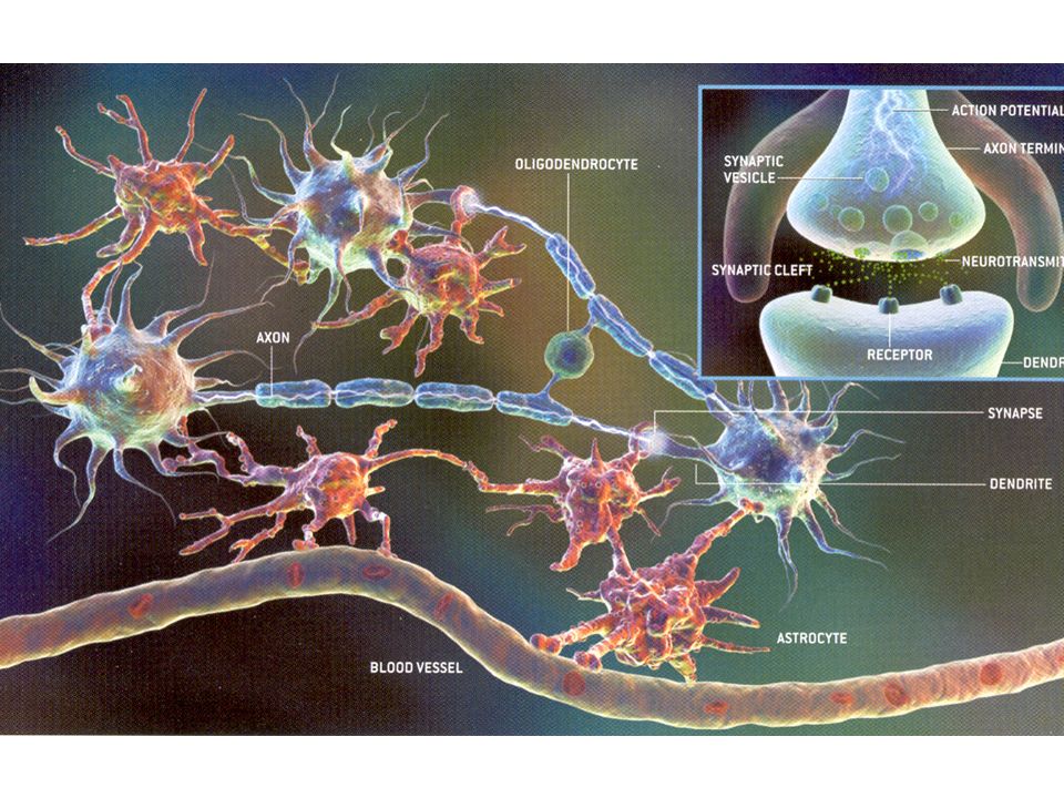
2. Oligodendroglia –same function as Schwann cells, but for axons of neurons in central nervous system –have small, round, dense nuclei –unlike Schwann cells, can myelinate a segment of several axons –no basement membrane surrounds the axon like Schwann cells do in PNS –these features affect ability of CNS cells to regenerate following injury (vs. PNS)
.")
31
3. Astrocytes –named from ‘star’ appearance of processes radiating out from soma –oval nuclei (larger & much less dense than oligodendroglia)
.")
32
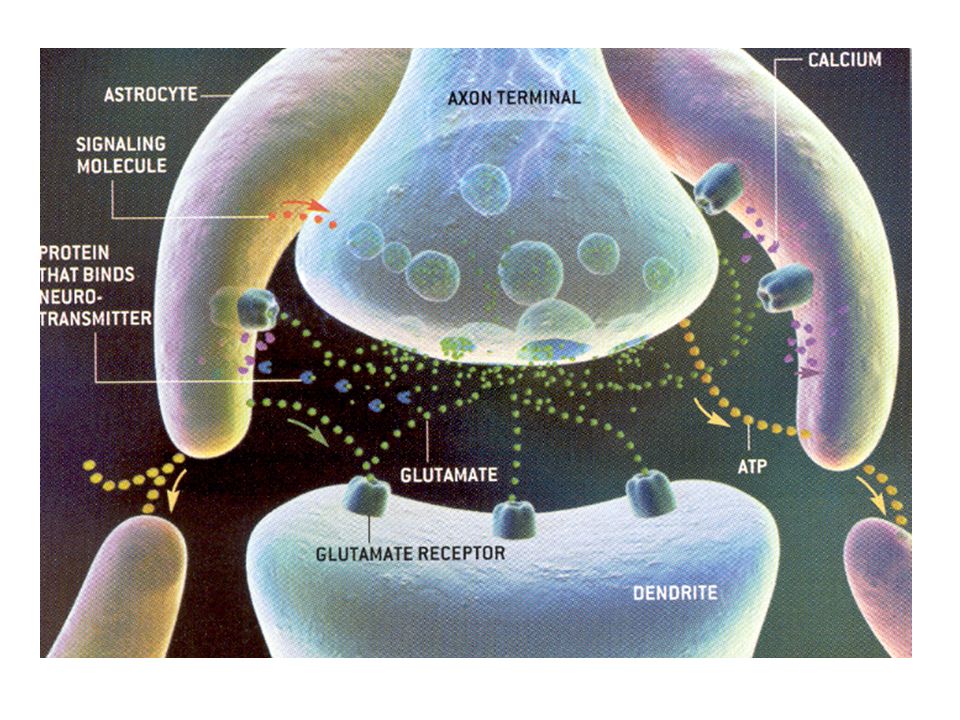
Function of Astrocytes –i. nutritive: form significant portion of brain blood barrier (BBB), surrounding CNS vasculature thought to be important communicators between neurons and capillaries transport nutrients, ions, molecules from capillary to neurons –ii. healing: act as scavengers, remove neuronal debris, seal off area for healing –iii. buffers excess extracellular K+, preventing excess depolarization –iv. remove chemical neurotransmitters from synaptic clefts - high affinity for specific neurotransmitters –v. support: provide structure, stability, act similarly to connective tissues –vi. in development: act as guides for outgrowth & migration in the developing NS
, surrounding CNS vasculature thought to be important communicators between neurons and capillaries transport nutrients, ions, molecules from capillary to neurons –ii. healing: act as scavengers, remove neuronal debris, seal off area for healing –iii. buffers excess extracellular K+, preventing excess depolarization –iv. remove chemical neurotransmitters from synaptic clefts - high affinity for specific neurotransmitters –v. support: provide structure, stability, act similarly to connective tissues –vi. in development: act as guides for outgrowth & migration in the developing NS.")
33
Types of astrocytes Distinguished by localization, not on any functional difference –i. fibrous astrocytes - name for those in, around white matter (areas rich in axons) –ii. protoplasmic astrocytes - those in gray matter, near synapses, dendrites
 –ii. protoplasmic astrocytes - those in gray matter, near synapses, dendrites.")
34
4. Ependymal cells –layer of ciliated columnar epithelial cells with tight junctions which line cavities of the neural tube (cerebral ventricles, spinal cord central canal) a. this layer forms a selective barrier between nervous tissue & ventricular fluid b. also forms choroid plexus - produces cerebral spinal fluid by filtering its components from blood
 a. this layer forms a selective barrier between nervous tissue & ventricular fluid b. also forms choroid plexus - produces cerebral spinal fluid by filtering its components from blood.")
35
Glial cells of mesodermal origin Microglia - cells from mesoderm which migrate into CNS - normally in small numbers, proliferate as needed - become macrophages in response to injury or damage, - act as scavengers, have a phagocytic role in removing debris, damaged cells

37
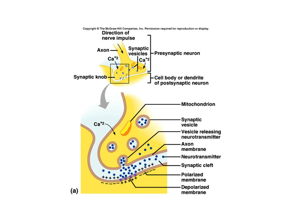
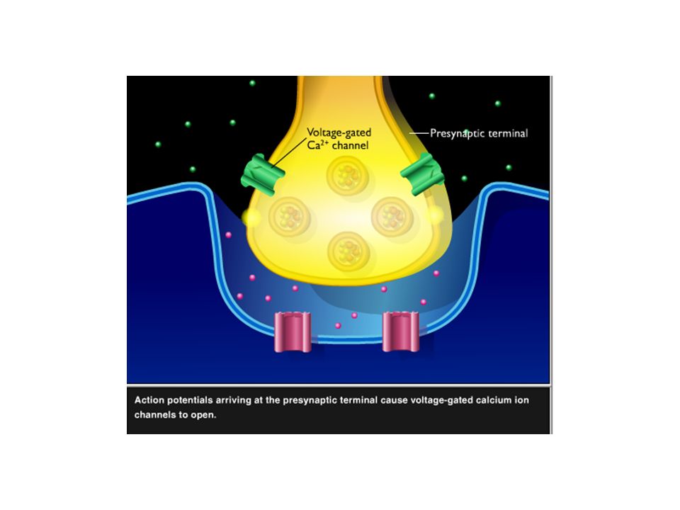
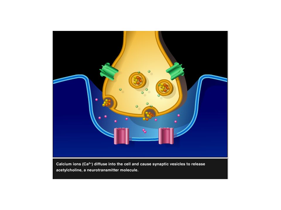
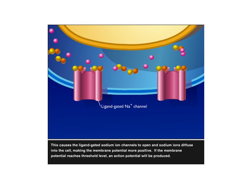
Synapse A. Chemical synapse –Presynaptic membrane, postsynaptic membrane, and synaptic cleft –can also be classified as: axodendritic, axosomatic, axoaxonal, and dendrodendritic –neurotransmitter (adrenaline, acetylcholine, dopamine, serotonin, glutamic acid…) –neuro-modulators (excitable or inhibitive neurotransmitters) B. Electrical synapse –neuron close together, rare in mammalian nervous system
 –neuro-modulators (excitable or inhibitive neurotransmitters) B. Electrical synapse –neuron close together, rare in mammalian nervous system.")
45
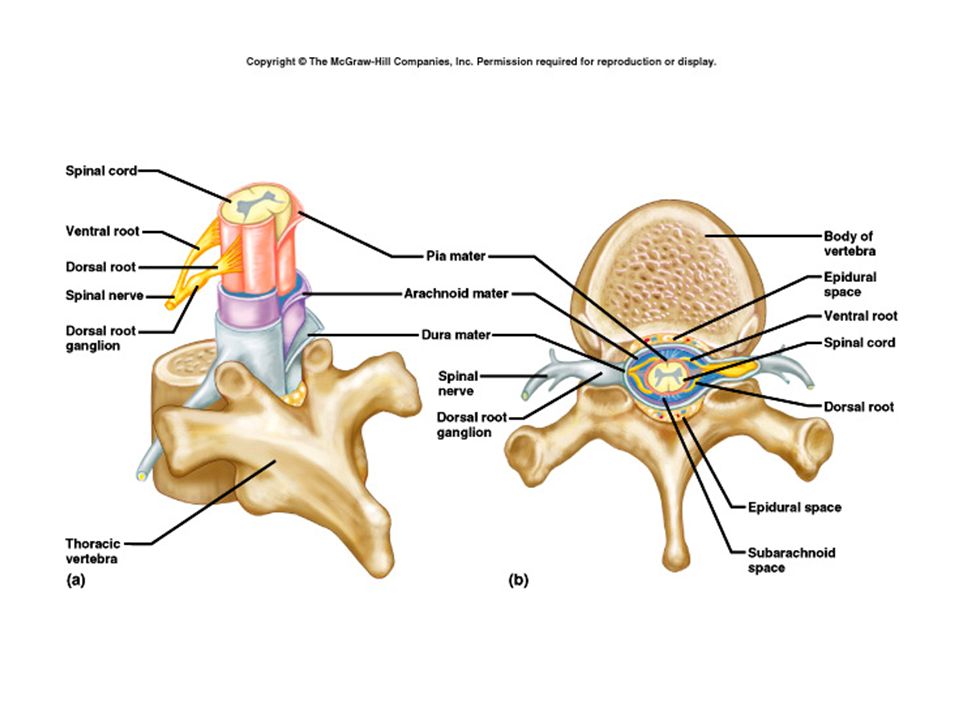
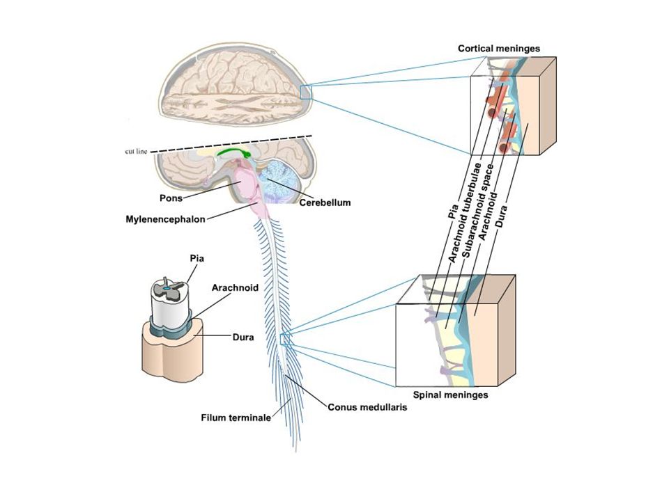
Meninges There is very little connective tissue in brain tissue, except in linings of vasculature –A. CNS = meninges: Connective tissue membranes surrounding the NS 1. pia mater - immediately next to the nervous tissue, thin & delicate 2. arachnoid - middle layer 3. dura mater - outermost meninges, thicker & very tough

48
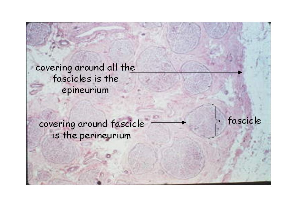
PNS: 1. endoneurium: thin collagenous layer, immediately surrounds a myelinated nerve fiber 2. perineurium: Connective tissue covering surrounding a fascicle of nerve fibers 3. epineurium: thick connective tissue layer surrounding many fascicles which make up a nerve trunk * the 3 CNS meningial layers are continuous with the connective tissue layers around PNS nerves

51
Axonal Transport Fast Axonal Transport - involves microtubules as a track -some organelles move along in stepwise fashion - especially synaptic vesicles, precursors, etc - requires oxidative metabolism, energy utilization

52
1. Anterograde: = orthograde, from cell body to terminal –a. mechanism by which synaptic vesicles move from soma to terminal along microtubules –b. speed = 410 mm/day 2. Retrograde: from terminal to soma –a. mechanism for returning worn out materials to cell body for degradation or recycling –b. returns to cell body information from, about the axon - communicates with neighbors –c. nerve growth factor (NGF) travels from source to soma, stimulates neuronal growth –d. toxin transport, e.g. neurotropic viruses get to CNS: herpes simplex, rabies, polio –e. speed = approx 1/2 - 2/3 that of anterograde transport
 travels from source to soma, stimulates neuronal growth –d. toxin transport, e.g. neurotropic viruses get to CNS: herpes simplex, rabies, polio –e. speed = approx 1/2 - 2/3 that of anterograde transport.")
53
Axonal Transport Slow Axoplasmic Flow –1. predominant means of organelle movement within the neuron –2. movement is through cytoplasm of axon, not along microtubules –3. speed = approx. 10 mm/day

54
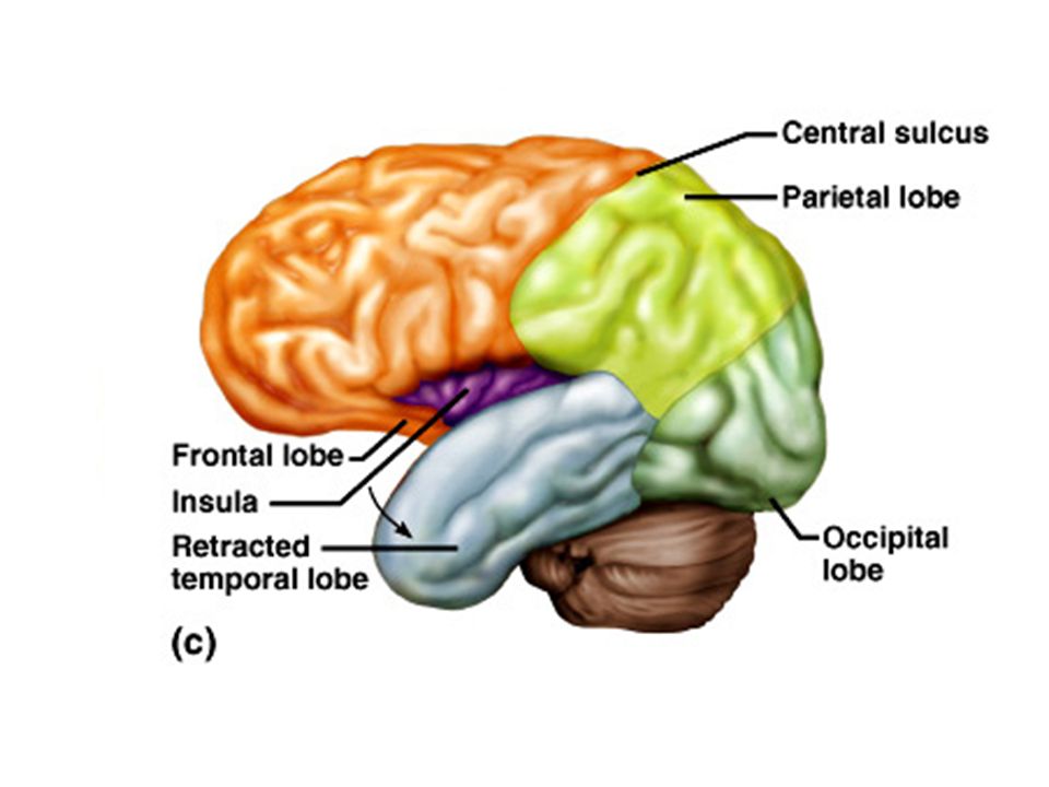
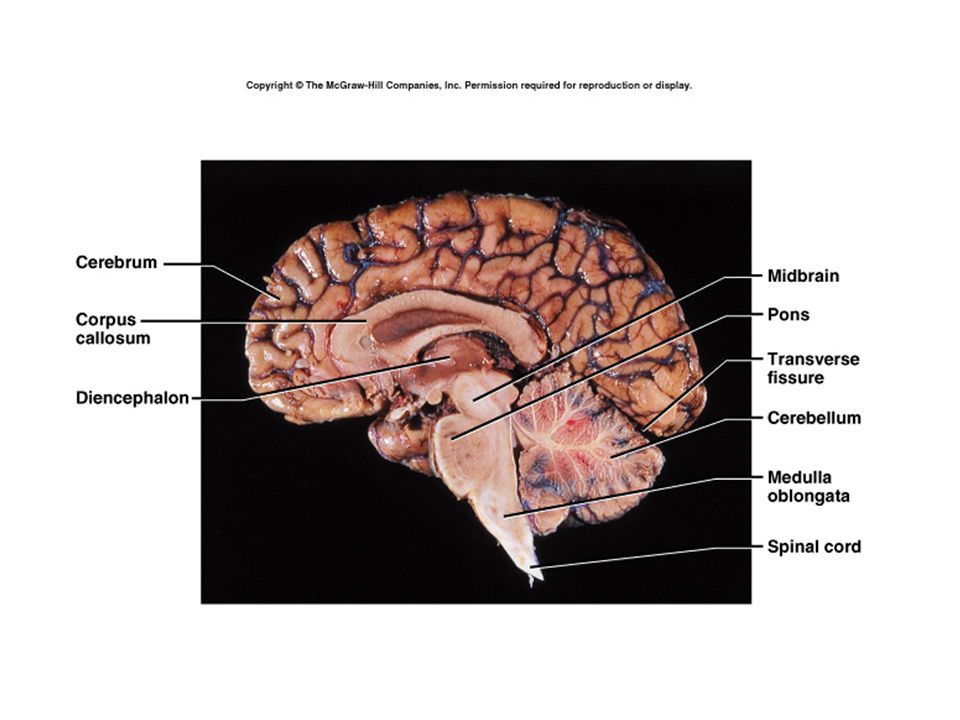
Main regions of the central nervous system Cerebrum –Frontal, parietal, occipital, temporal and insula –Interpretation –initiating voluntary movements –storing memory –retrieving memory –Reasoning –center for intelligence and personality Cerebellum –Large in human, receiving information from sensory systems and the cerebral cortex, main function to maintain balance, posture, mostly not initiated from the cerebral cortex.

56
Brain stem Medullla, pons and midbrain are generally referred as brain stem Medulla Oblongata –Continuation from spinal cord into the brain, part of the brain stem. Pons –Dorsal part consists of sensory and motor tracts –Ventral part contains connection between two hemispheres, contribute to motor efficiency Midbrain –Involved with visual and auditory system, red nucleus and substantia nigra are also located here (more later)
.")
58
Diencephalon: –Forms the central core of the cerebrum, including thalamus, hypothalamus, epithalamus, and subthalamus, controls automatic nervous system, endocrine function via hormones and nervous impulses. Telencephalon –Includes cerebral cortex, corpus striatum, and medullary center. Areas of cerebral cortex (paleocortex) receive primitive function from olfactory system, which is common to lower vertebrates. Other areas are called archicortex, includes limbic system (emotions, and some memories, early vertebrates). Most areas of the cerebral cortex (90%) in human are referred as neocortex, which controls all sensations (except smell), involves emotions, memories and intellectual activities.
 receive primitive function from olfactory system, which is common to lower vertebrates. Other areas are called archicortex, includes limbic system (emotions, and some memories, early vertebrates). Most areas of the cerebral cortex (90%) in human are referred as neocortex, which controls all sensations (except smell), involves emotions, memories and intellectual activities..")
Similar presentations


 and gathers information.>")


>")
 1.Microglial cells –Scattered throughout CNS –Support neurons.>")


 from one part of the body to another. ◦ Major regions.>")


 2008, 2005 by Mosby, Inc., an affiliate of Elsevier Inc. All rights reserved. Copyright © 2005, Elsevier, Inc. All rights reserved. Slide.>")