Download presentation
Presentation is loading. Please wait.

1
Chair : Prof. Dr. B. Jayakumar
STROKE IN THE YOUNG Chair : Prof. Dr. B. Jayakumar

2
INTRODUCTION Young stroke is stroke occurring between 15 and 45 years of age Differential diagnosis for potential etiologies is broader Even after extensive investigations, the cause may remain elusive in 20-50% cases Prognosis depends on the underlying factor

3
Responsible for about 5% of all cases of stroke
Incidence is much higher in developing countries like India. Above the age of 30 years stroke is more common in males whereas below that female predominance is seen.

4
Etiology of young stroke
ISCHEMIC Large artery disease Premature atherosclerosis Dissection (spontaneous or traumatic) Inherited metabolic diseases (homocysteinuria, Fabry’s disease, pseudoxanthoma elasticum, MELAS syndrome) Fibromuscular dysplasia Vasculitis Moyamoya disease Radiation Toxic (drug induced)
 Inherited metabolic diseases (homocysteinuria, Fabry’s disease, pseudoxanthoma elasticum, MELAS syndrome) Fibromuscular dysplasia. Vasculitis. Moyamoya disease. Radiation. Toxic (drug induced)")
5
Cardioembolic disease
Small vessel disease Vasculopathy (infectious, non infectious, microangiopathy) Cardioembolic disease Rheumatic heart disease Congenital heart disease Arrhythmias Bacterial and non-bacterial endocarditis Mitral valve prolapse Patent foramen ovale Atrial myxoma Cardiac surgeries and procedures
 Cardioembolic disease. Rheumatic heart disease. Congenital heart disease. Arrhythmias. Bacterial and non-bacterial endocarditis. Mitral valve prolapse. Patent foramen ovale. Atrial myxoma. Cardiac surgeries and procedures.")
6
Hematologic disease Migraine Sickle cell disease Leukemia
Hypercoagulable states (antiphospholipid antibody syndromes, deficiency of antithrombin III or protein S or C, resistance to activated protein C, increased factor VIII) Disseminated intravascular coagulation Thrombocytosis Polycythemia vera Thrombotic thrombocytopenic purpura Venous occlusion Migraine
 Disseminated intravascular coagulation. Thrombocytosis. Polycythemia vera. Thrombotic thrombocytopenic purpura. Venous occlusion. Migraine.")
7
Subarachnoid hemorrhage Intraparenchymal hemorrhage
HEMORRHAGIC Subarachnoid hemorrhage Cerebral aneurysm Intraparenchymal hemorrhage Arteriovenous malformation Neoplasm (primary central nervous system, metastatic, leukemia) Hematological disorders (sickle cell disease, neoplasm, thrombocytopenia) Moyamoya disease Drug use (warfarin, amphetamines, cocaine, phenylpropanolamine) Iatrogenic (peri-procedural)
 Hematological disorders (sickle cell disease, neoplasm, thrombocytopenia) Moyamoya disease. Drug use (warfarin, amphetamines, cocaine, phenylpropanolamine) Iatrogenic (peri-procedural)")
8
Premature atherosclerosis
Premature atherosclerosis is the single most important cause of stroke as age advances Incidence is 7-30% below the age of 50 years. It is presumed in all undiagnosed cases with more than two risk factors.

9
Risk factors for atherosclerosis
Male sex Systemic hypertension Diabetes mellitus Dyslipidemia (low HDL cholesterol, hypertriglyceridemia) Cigarette smoking Alcohol abuse Ischemic heart disease Recent infection Oestrogen related stroke including oral contraceptives
 Cigarette smoking. Alcohol abuse. Ischemic heart disease. Recent infection. Oestrogen related stroke including oral contraceptives.")
10
NON ATHEROSCLEROTIC VASCULOPATHIES
Cervicocephalic arterial dissections Traumatic cerebrovascular disease Radiation-induced vasculopathy Moyamoya disease Fibromuscular dysplasia Vasculitis Migrainous infarction

11
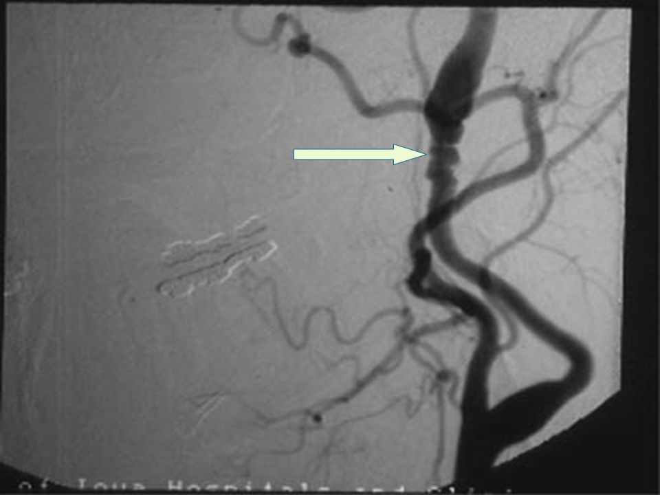
Cerviocephalic arterial dissections
Subintimal penetration of blood with subsequent longitudinal extension of the hematoma between its layers Common sites Extracranial segment of internal carotid artery Extracranial vertebral arteries Recurrence rate is 1%, more in young and in those with positive family history

12
Causes Spontaneous Secondary Blunt or penetrating trauma
Fibromuscualar dysplasia Ehlers’ Danlos syndrome type IV Marfan’s syndrome Pseudoxanthoma elasticum Coarctation of aorta Menke’s disease α1-antitrypsin deficiency Cystic medial degeneration Osteogenesis imperfecta Adult polycystic kidney disease Homocystinuria Luteic arteritis

13
Dissection results in ischemic symptoms due to arterial occlusion or secondary embolism
Diagnosis Arteriography – Elongated, irregular, narrow column of dye, ‘string sign’ High resolution MRI MR angiography CT angiography Doppler ultrasound of the neck especially for carotid diseection

14
Internal carotid artery dissection

16
Treatment Anticoagulation with heparin should be started followed by warfarin therapy for 3–6 months Antiplatelet therapy Surgical therapy indicated in the presence of pseudoaneurysms and if there is no response to medical treatment Anticoagulation should be withheld in intracranial dissection since there is a risk of subarachnoid haemorrhage

17
Trauma Blunt or penetrating trauma can produce arterial dissection, rupture, thrombosis, pseudoaneurysm formation and AV fistula. Can occur during sports, violent coughing, vigorous nose blowing, neck manipulation, anesthesia administration etc. Cervical rotation or extension compresses cervical carotid artery against transverse processes of upper cervical vertebra Angiography and surgical repair is the treatment.

18
Radiation vasculopathy
Accelerated atherosclerosis occurs especially in those with dyslipidemia Radiation results in damage to the endothelial cells producing complications months to years later Lesions occur at unusual sites The amount of damage depends on Radiation dose Age at the time of therapy

19
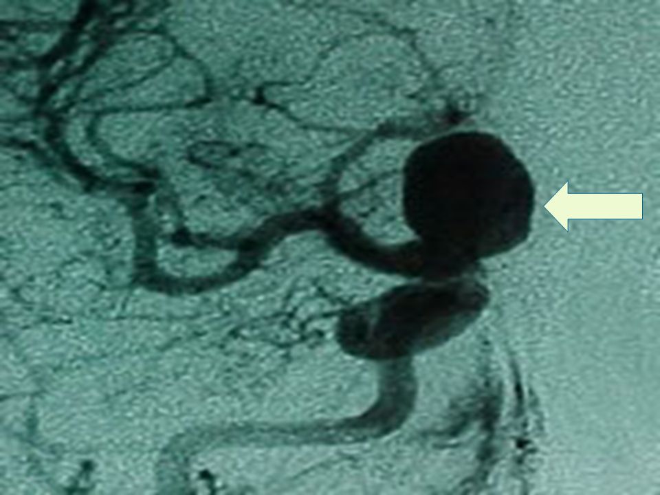
Moyamoya disease Moyamoya is a Japanese word meaning ‘puff of smoke’
It is a chronic progressive non-amyloid non-atherosclerotic, non-inflammatory occlusive arteriopathy of unknown cause. Characterized by progressive bilateral stenosis of distal ICA extending to proximal ACA and MCA with involvement of circle of Willis and development of extensive collateral network (parenchymal, leptomeningeal or transdural) at the base of the brain like a puff of smoke. Intracranial aneurysms are seen, especially in posterior circulation.
 at the base of the brain like a puff of smoke. Intracranial aneurysms are seen, especially in posterior circulation.")
20
Pathology Clinical features
Fibrocellular intimal thickening, smooth muscle proliferation and increased elastin accumulation leading to stenosis of suprasellar intracranial ICA Thinning of media with tortuous and multilayered internal elastic lamina Clinical features TIA, seizures, headache, movement disorders, mental deterioration, cerebral infarction , intracranial hemorrhage TIAs are precipitated by crying, blowing and hyperventilation

21
Bimodal age distribution is seen
First decade – ischemic events more common Fourth decade – Hemorrhagic commoner Diagnosis is established by arteriography Suzuki’s six arteriographic changes Stenosis of carotid fork Initial appearance of moyamoya vessels at the base of brain Intensification of moyamoya vessels Minimization of moyamoya vessels Reduction of moyamoya vessels Disappearance of the vessels

22
MCA block with multiple collateral moyamoya vessels

23
Treatment Ischemic Moyamoya Hemorrhagic Moyamoya
Platelet antiaggregants, vasodilators, calcium channel blockers and corticosteroids have been tried. Anticoagulants are not useful Surgical revascularization techniques like superficial temporal to MCA anastamosis have produced good results. Hemorrhagic Moyamoya No established therapy to prevent rebleed

24
Fibromuscualar dysplasia
Segmental, non-atheromatous, dysplastic, non-inflammatory angiopathy Commonly affects young and middle aged women White race more affected Etiology Unknown May be related to immunological mechanisms, estrogenic effects, α1-antitrypsin deficiency Familial association seen

25
Most often extracranial carotids involved Bilateral in two thirds
Renal arteries most commonly involved. Cervicocephalic involvement in less than 1% Most often extracranial carotids involved Bilateral in two thirds Four histologic types Intimal hyperplasia Medial hyperplasia Medial fibroplasia: Commonest Perimedial dysplasia Most of the patients are asymptomatic

26
Diagnosis Treatment Cervical angiography
‘String of beads’ appearance in medial fibroplasia Treatment Benign natural history Platelet antiaggregants used Surgical intervention seldom needed

28
Types of fibromuscular dysplasia

29
Cerebral autosomal dominant arteriopathy with subcortcal infarcts and leucoencephalopathy (CADASIL)
Familial nonarteriosclerotic, nonamyloid microangiopathy Characteristic features Migraine with aura (CADASILM) Recurrent subcortical ischemic strokes from mid-adulthood Pseudobulbar palsy, cognitive decline, subcortical dementia Early white matter hyperintensities in MRI
 Recurrent subcortical ischemic strokes from mid-adulthood. Pseudobulbar palsy, cognitive decline, subcortical dementia. Early white matter hyperintensities in MRI.")
30
Genetics Missense mutations or small deletions in Notch 3 gene on chromosome 19q12 Codes for transmembrane receptor Notch 3 Granular eosinophilic material deposited in arterial walls, including dermal arteries

31
Binswanger disease Widespread degeneration of cerebral white matter of vascular causation Associated with hypertension, atherosclerosis of small blood vessels and multiple strokes. Radiological picture of leucoareosis – less intense appearance of periventricualar tissues in chronically hypertensive patients

32
Mitochondrial myopathy, lactic acidosis and strokes (MELAS)
Mitochondrial disorder which may manifest at any age,usually in childhood Clinical features Proximal myopathy, exercise intolerance Recurrent migraine-type headaches Hemiparesis, hemianopsia or cortical blindness Precipitated by exercise or infection Serum and CSF lactate concentrations are elevated

33
Migrainous infarction
Migraine commonly affects women and starts during childhood or adolescence Rare association of migraine and ischemic stroke seen in young women particularly below 35 years of age. Pathogenesis is not completely known Migrainous infarctions are mostly cortical and involve PCA territory Usually there is gradual build up of unilateral throbbing headaches with visual phenomena occurring in both visual fields simultaneously, in one of which the visual loss becomes permanent.

34
Definite migrainous infarction – all criteria satisfied
Diagnostic critreria Definite diagnosis of migraine with aura in the past One or more of the migrainous aura symptoms must be present and not fully reversed within 7 days from the onset, with neuroimaging confirmation of ischemic infarction Clinical manifestations should be those typical of previous attacks Other causes of infarction should be excluded Definite migrainous infarction – all criteria satisfied Possible – only some criteria satisfied Increases risk for recurrent stroke

35
CEREBRAL VASCULITIDES
Infectious vasculitis Bacterial, fungal, parasitic, spirochetal, viral, rickettsial, mycobacterial Necrotising vasculitis Classic polyateritis nodosa, Wegener’s granulomatosis, allergic angitis and granulomatosis, lymphomatoid granulomatosis Vasculitis associated with collagen vascular disease SLE, Rheumatoid arthritis, Scleroderma, Sjogren’s syndrome Giant cell arteritides Takayasu’s arteritis, temporal arteritis

36
Vasculitis associated with other systemic diseases
Behcet’s disease, Ulcerative colitis, Sarcoidosis, Relapsing polychondritis, Kohlmeier-Degos disease Hypersensitivity vasculitis Henoch-Schonlein’s purpura, Drug-induced vasculitis, Essential mixed cryoglobulinemia Miscellaneous Vasculitis associated with neoplasia and radiation, Cogan’s syndrome, Dermatomysoitis polymyositis, X-linked lymphoproliferative syndrome, TAO, Kawasaki’s syndrome, Primary central nervous system vasculitis

37
Inflammatory vasculitis should be considered in
Young patients with stroke Recurrent strokes Stroke with encephalopathic features Stroke with fever Multifocal neurological events Mononeuritis multiplex, palpable purpura or abnormal urinary sediment Diagnosis usually requires confirmation with arteriography or biopsy

38
Meningovascular syphilis
Patients usually have prodromal symptoms Vasculitis can cause cerebral infarctions, commonly in MCA territory or spinal cord infarction. May be associated with headache, meningismus, mental status abnormalities or cranial nerve abnormalities.

39
Diagnosis CSF study reveals lymphocytic pleocytosis, elevated protein control and positive VDRL test Treatment Aqueous Penicillin G x days Luteic aneurysms of the ascending aorta can extend to the root of great vessels causing stroke

40
Neurotuberculosis Usually affects basilar meninges resulting in basilar meningitis which traps 3, 4 and 6 cranial nerves causing their palsy. Basilar arteriolitis commonly involves the penetrating branches of ACA, MCA or PCA Risk factors include alcoholism, substance abuse, corticosteroid use or HIV CSF study shows increased protein and decreased glucose levels and lymphocytic and mononuclear pleocytosis % of the CSF smears show AFB.

41
HIV/AIDS Cerebral infarction on AIDS can result from
Vasculitis, meningovascular syphilis, varicella-zoster virus vasulitis, opportunistic infections, infective endocarditis, aneurysmal dilatation of major cerebral arteries, nonbacterial thrombotic endocarditis, aPL antibodies, or other hypercoagulable states, hyperlipidemia induced by protease inhibitors, HIV-1 related malignancy, cancer chemotherapy and TTP.

42
Other infectious agents are varicella zoster, coxsackie 9 virus, California encephalitis, mumps paramyxovirus, hepatitis C virus, Mycoplasma pneumoniae, Borrelia burgdorferi, Rickettsia typhi, cat-scratch disease, Trichinella infection, cysticercus of Taenia solium and free living amoeba

43
Takayasu’s arteritis Chronic inflammatory arteriopathy of aorta, its major branches and pulmonary artery More common in women Probable immune mechanism Slow progression of stenosis, occlusion, aneurysmal dilatation and coarctation of the involved vessels

44
Two phases Acute or prepulseless phase – non-specific systemic symptoms Occlusive phase – multiple arterial occlusions Neurological symptoms usually result from CNS or retinal ischemia due to stenosis or occlusion of the aortic arch and arch vessels, or arterial hypertension resulting from coarctation of aorta or renal artery stenosis

45
Diagnosis Treatment MR angiography Aortogram
In active disease, treatment is with oral glucocorticoids; cyclophosphamide, azathiprine or methotrexate used rarely Surgical treatment of severely stenotic vessels

46
Drug induced vasculitis
Drugs implicated Amphetamines, cocaine, phencyclidine, phenylpropanolamine, pentazocine with pyribenzamine, heroin, anabolic steroids and glue sniffing. Mechanisms Foreign body embolization, vasculitis, vasospasm, acute onset of arterial hypertension or hypotension, endothelial damage, accelerated atherosclerosis, hyper or hypocoagulability, cardiac arrhythmias, emboism from MI or AIDS.

47
CARDIOGENIC EMBOLISM High embolic potential Other causes
Rheumatic mitral valve Acute myocardial infarction Infective endocarditis Mechanical prosthetic valves Dilated cardiomyopathy Cardiac tumours Cardiac arrhythmias – Atrial fibrillation Other causes Mitral valve prolapse Mitral annulus calcification Aortic valve calcification

48
Non bacterial thrombotic endocarditis
Filamentous strands of mitral valve Giant Lambl’s excresences Aneurysms of Sinus of Valsalva Intracardiac defects with paradoxical embolism Patent foramen ovale, atrial septal aneurysm, atrial septal defect Cyanotic congenital heart disease Iatrogenic embolism Cavopulmonary anastamosis, coronary artery bypass grafting, pacing, heart transplantation, artificial hearts, cardioversion for atrial fibrillation, balloon angioplasty, ventricular support devices, extracorporeal membrane oxygenator

49
Cardiac sources account for 15 to 20% of all ischemic strokes.
The emboli usually consist of platelet, fibrin, platelet-fibrin, calcium, microorganisms, or neoplastic fragments. Cardioembolic cerebral infarcts – large, multiple, bilateral, wedge shaped Features of cardioembolic stroke Worse at onset, fast recovery Presence of Wernicke’s aphasia, homonymous hemianopia, ideomotor apraxia Involvement of posterior division of MCA, ACA or cerebellar involvement Involvement of multiple vascular territories Hemorrhagic component of the infarction

50
Acute myocardial infarction
1% of patients with acute MI have embolic stroke LV thrombi are commonly associated with recent anterior wall transmural MI Usually embolism occurs within first 3 months, 85% within 4 weeks Decreased ejection fraction – independent predictor of risk of embolism There is more chance of embolism following thrombolysis with tpA

51
Dilated cardiomyopathy
Akinesia of the chambers produces stasis of blood and thrombus formation Associated LV failure and atrial fibrillation increase the risk 18% of the non- anticoagulated patients develop embolism Mitral stenosis Rheumatic mitral stenosis associated with atrial fibrillation has high potential for embolisation. 9-14% have systemic emboli of which 60–75% have cerebral ischemia.

52
Endocarditis Infective endocarditis – vegetations may cause systemic (left sided) or pulmonary (right sided) embolism. If vegetations are detectable by transthoracic echocardiogram, there is increased risk of embolism. Non bacterial thrombotic endocarditis – multiple small sterile thrombotic vegetations occur commonly involving the mitral and aortic valves Prosthetic valves – mechanical valves in mitral position are more prone. Filamentous strands attached to the mitral valve concede increased risk.
 or pulmonary (right sided) embolism. If vegetations are detectable by transthoracic echocardiogram, there is increased risk of embolism. Non bacterial thrombotic endocarditis – multiple small sterile thrombotic vegetations occur commonly involving the mitral and aortic valves. Prosthetic valves – mechanical valves in mitral position are more prone. Filamentous strands attached to the mitral valve concede increased risk.")
53
Atrial fibrillation Sick sinus syndrome More common in older adults
Risk for AF and embolism increases with age High risk for embolism exists with rheumatic AF and AF in hyperthyroidism Sick sinus syndrome Maximum risk is associated with bradytachyarrhythmias, LA spontaneous echocardiographic contrast, and decreased atrial ejection force

54
Congenital heart disease
Intracardiac tumours Atrial myxomas are the commonest tumours in adults. Embolic complications are the presenting symptom in one-third of patients. Peripheral ad multiple cerebral arterial aneurysms are remote associations. Cardiac rhabdomyomas and mitral valve fibroelastomas are rarely associated with embolism. Congenital heart disease Common cause of stroke in children. Risk is increased with arterial hypertension, atrial fibrillation, history of phlebotomy and microcytosis. Low Hb is associated with arterial stroke and high Hb with cerebral venous thrombosis.

55
Spontaneous echo cardiographic contrast
Paradoxical embolism Higher rate of cerebral ischemia is reported in persistent foramen ovale and atrial septal aneurysms. A demonstrable source of embolism should be present. Antiplatelet therapy, anticoagulant thearapy, transcatheter or surgical closure of PFO is the treatment. Spontaneous echo cardiographic contrast Associated with elevated fibrinogen levels and plasma viscosity and is a potential risk factor for stroke

56
Post operative Causes Hypoperfusion Ventricular thrombi Emboli Posterior circulation stroke is more common after cardiac catheterisation

57
INHERITED AND MISCELLANEOUS DISORDERS
Homocystinuria Fabry’s disease Marfan’s syndrome Ehler-Danlos’ syndrome Pseudoxanthoma elasticum Sneddon’s syndrome Rendu-Osler-Weber’s syndrome Neoplastic angioendotheliomatoisis Susac’s syndrome

58
Eales’ disease Reversible cerebral segmental vasoconstriction Hypereosinophilic syndrome Cerebral amyloid angiopathy Coils and kinks Arterial dolichoectasia Complications of coarctation of aorta Air, fat, amniotic fluid, bone marrow, and foreign particle embolism

59
Homocystinuria Inborn error of amino acid metabolism.
Any of the three enzymes deficient Cystathionine β-synthetase Homocysteine methyl transferase Methylene tetrahydrofolate reductase This leads to accumulation of homocysteine in the blood – resulting in endothelial injury and premature atherosclerosis. Elevated levels of homocysteine is an independent risk factor for development of cerebrovascular disease, coronary and peripheral arterial occlusive disease.

60
Homocysteine metabolism

61
Clinical features Marfanoid habitus, malar flush, liveo reticularis, ectopia lentis, myopia, glaucoma, optic atrophy, optic atrophy, psychiatric manifestations, mental retardation, spasticity, seizures, osteoporosis and a propensity for intracranial arterial or venous thrombosis Homocysteine levels may be reduced by administration of folic acid, with pyridoxine and vitamin B12, choline, betaine, estrogen and N-acetyl cysteine.

62
Fabry’s disease X-linked disorder of glycosphingolipid metabolism
Deficient lysosomal α-galactosidase activity Ceramide trihexosidase accumulate in the endothelial and smooth muscle cells Clinical features Painful peripheral neuropathy, hypertension, cardiomegaly, renal dysfuction, autonomic dysfunction, corneal opacifications Angiokeratoma corporis diffusum

63
Marfan’s syndrome Autosomal dominant
Quantitative and qualitative defects of fibrillin Variety of skeletal, ocular and cardiovascular manifestations Cystic medial necrosis of the aortic segments occur Dilatation of the aortic root dissection of the ascending aorta ischemia of brain, spinal cord and peripheral nerves Saccular intracranial aneurysms or dissection of the carotid artery can occur Annual echocardiograms should be done

64
Ehler-Danlos’ syndrome
Inherited connective tissue disorder Characterized by hyperextensibility of skin, hypermobile joints and vascular fragility Arterial complications occur predominantly with type IV disease Complications include arterial dissections, arteriovenous fistulae and aneurysms. Arteriography should be avoided if possible.

65
Pseudoxanthoma elasticum
Inherited disorder of elastic tissue Clinical features Loose skin, orange-yellowish papules of intertriginous areas, arterial hypertension, angiod streaks, retinal hemorrhages, arterial occlusive disease and arterial dissection. High risk for arterial occlusion – coronary artery disease and stroke. Women should avoid estrogens

66
Sneddon’s syndrome Unknown etiology
Frequent correlation with hypertension, smoking and OCP Occurs in young women Charaterized by widespread livedo reticularis and ischemic cerebrovascular infarcts in the carotid artery territory Histology reveals perivascular lymphocytic infiltration on the skin arteries with proliferation of smooth muscle fibres on the internal elastic lamina without inflammation.

67
Atheromatous emboli Cholesterol embolisation usually occurs following manipulation of an atherosclerotic aorta during catheterisation or surgery. Patient may present with TIAs, strokes, retinal embolism, pancreatitis, renal failure, livedo reticualris and purple toes. Patients have eosinophilia, anemia, elevated ESR and elevated serum amylase. Anticoagulation should be avoided.

68
Air embolism Accidental introduction of air into systemic circulation can occur during surgical procedures and scuba diving. Usually causes cerebral and retinal ischemia Symptoms include seizures and multifocal neurological findings such as cerebral edema, confusion, memory loss and coma. CT scan can visualize gaseous bubbles. Treatment includes resuscitative measures, placement in left lateral position, inotropic agents, anticonvulsants, anti-edema agents and hyperbaric agents.

69
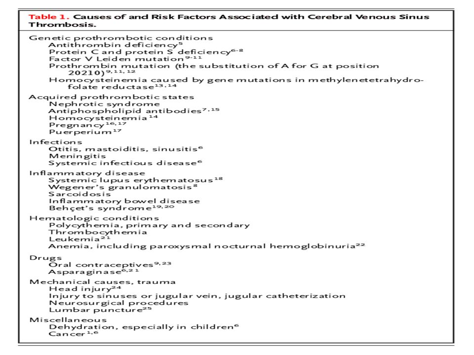
HYPERCOAGULABLE DISORDERS
Primary hypercoagulable states Antithrombin III deficiency Protein C deficiency Protein S deficiency Activated protein C resistance Prothrombin G20210 mutation Afibrinogenemia Hypofibrinogenemia Hypoplasminogenemia Plasminogen activators deficiency Lupus anticoagulant and anticardiolipin antibodies

70
Secondary hypercoagulable states
Malignancy Pregnancy/Puerperium Oral contraceptive use/ Other hormonal treatments Ovarian hyperstimulation syndrome Nephrotic syndrome Polycythemia vera Essential thrombocythemia Paroxysmal nocturnal hemoglobinuria Diabetes mellitus Heparin induced thrombocytopenia Homocysteinuria Sickle cell disease Thrombotic thrombocytopenic purpura Chemotherapeutic agents

71
Inherited thrombophilias suspected if
Recurrent episodes of deep venous thrombosis Recurrent pulmonary emboli Family history of thrombotic events Unusual sites of venous (mesenteric, portal or cerebral) or arterial thrombosis Thrombotic events in childhood, adolescence or early adulthood More than half of the events occur spontaneously Risk is increased with additional risk factors like pregnancy, surgery, trauma or OCP
 or arterial thrombosis. Thrombotic events in childhood, adolescence or early adulthood. More than half of the events occur spontaneously. Risk is increased with additional risk factors like pregnancy, surgery, trauma or OCP.")
72
Coagulation cascade

73
Antithrombin III deficiency
Autosomal dominant inheritance Three classes of inherited deficiency Classic or type I – decreased immunological and biological activity of antithrombin III Type II – low biological activity, normal immunological activity Type III – normal activity in the absence of heparin, but reduced in heparin dependent assays Acquired deficiency can occur in acute thrombosis and DIC

74
A normal level of AT-III during an acute event excludes primary deficiency.
Treatment In acute thrombosis – Heparin with or without AT III concentrate Recurrent thrombosis – Long term warfarin therapy to keep INR at 2 - 3

75
Protein C deficiency Autosomal dominant inheritance Acquired form
Homozygous – Purpura fulminans neonatalis Heterozygous – Recurrent thrombosis especially venous Acquired form Administration of L-aparaginase, warfarin, liver disease, DIC, postoperatively, bone marrow transplantation and ARDS Assays should be done after discontinuing anticoagulation for at least a week. Initial treatment with heparin followed by incremental doses of warfarin ( to avoid skin necrosis)
")
76
Protein S deficiency Exists in free form (40%) and bound to binding proteins Autosomal dominant inheritance, patients prone for recurrent thromboembolism Acquired deficiency Pregnancy, acute thromboembolic episodes, DIC, nephrotic syndrome, SLE, OCP, anticoagulants and L-asparaginase Total and free protein S levels and functional assay of protein S is done after discontinuation of anticoagulants. Treatment with heparin; warfarin in case of recurrent thrombosis

77
Activated protein C resistance
Commonest inherited thrombotic disorder Autosomal dominant, usually associated with a single point mutation in factor V gene (Arg to Gln at position 506) Prone for venous thrombosis Assay is done after discontinuation of the anticoagulants
 Prone for venous thrombosis. Assay is done after discontinuation of the anticoagulants.")
78
Antiphospholipid antibody syndrome
Antiphospholipid antibodies may be IgG, IgA or IgM Different antibodies are anticardiolipin antiphosphatidyl ethanolamine antiphospahtidyl serine antiphosphatidyl choline. Characterized by Recurrent arterial or venous thrombosis Recurrent fetal loss Livedo reticualris

79
Livedo reticularis

80
β2 glycoprotein in plasma is needed to bind to cardiolipin. Occurs as
Secondary to SLE, other autoimmune diseases, Sneddon’s syndrome, acute and chronic infections, neoplasias, IBD, drugs, severe pre eclampsia, liver transplantaion Primary APLA syndrome Treatment High dose warfarin to keep INR above 3 with or without aspirin In pregnancy, low dose aspirin and prednisone is given

81
Infarcts of undetermined cause (Cryptogenic stroke)
In many cases the cause of the ischemic event is not identified even after after an extensive work up Occurs in 20 to 50% of people with young stroke Recurrence risk in such cases is less

82
INTRACEREBRAL HEMORRHAGE
Vascular malformations Intracranial tumours Bleeding disorders, anticoagulant and fibrinolytic treatment Cerebral amyloid angiopathy Granulomatous angitis of the central nervous system Hemorrhagic infarction Trauma

83
Vascular malformations
The vascular malformation may be Saccular or mycotic aneurysms Arteriovenous malformations Cavernous angiomas Intracerebral hemorrhages caused by small lesions are characterized by Located in the subcortical white matter Hematoma is smaller Symptoms develop slowly Usually subarachnoid hemorrhage seen Younger patients with female preponderance MRI or histological examination needed for diagnosis

84
Aneurysms On the basis of morphology, aneurysms are classified as saccular, fusiform or dissecting. Saccular aneurysms are more often acquired than congenital They tend to occur at the branching points in the circle of Willis and proximal cerebral arteries (40% in anterior communicating artery). Usually presents as SAH; less commonly as ICH, space occupying lesion producing compression, seizures, embolism from thrombus, hydorocephalus
. Usually presents as SAH; less commonly as ICH, space occupying lesion producing compression, seizures, embolism from thrombus, hydorocephalus.")
85
Associations of intracranial saccular aneurysms
Polycystic kidney disease Fibromuscular dysplasia Cervical artery dissection Coarctation of the aorta Intracranial vascular malformations Marfan’s syndrome Ehler-Danlos syndrome Pseudoxanthoma elasticum Hereditary hemorrhagic telangiectasia Moyamoya syndrome Klinefelter’s syndrome Progeria

86
Types of aneurysms

88
Arteriovenous malformations
Abnormal fistulous connections between one or more hypertrophied feeding arteries and dilated draining veins Diagnosis suspected in Ct scan. Non-enhanced scan shows calcification and non-specific hypo- or hyperdensity. Contrast CT scan shows dilated veins of large malformations. MRI or angiogram may be needed to confirm diagnosis.

90
Cavernous hemangioma Detected using MRI
Shows a central nidus of irregular bright signal intensity mixed with mottled hypointensity, surrounded by a peripheral hypointense ring Hemosiderin deposits in periphery due to prior bleeding Usually single lesions Predominantly supratentorial, presents as seizures

91
Intracranial tumours Bleeding into tumour occurs with
Glioblastoma multiforme Metastasis from melanoma, bronchogenic carcinoma, renal cell carcinoma, choriocarcinoma Relatively rare complication If suspected, search for primary or secondary brain tumour and systemic focus Cerebral angiography and craniotomy for biopsy of hematoma wall may be needed Extremely poor prognosis

92
Suspicion of an underlying tumour should arise when
Presence of papilloedema at presentation Rare location, e.g., corpus callosum Presence of ICH in multiple sites Ring of high density hemorrhage surrounding a low density centre in non-enhanced CT scan Disproportionate surrounding edema and mass effect Enhancing nodules adjacent to h’ages in contrast CT scan MRI showing heterogeneous signal lesions within a mass lesion , surrounded by hemosiderin hypointense ring and bright signal edema at periphery in T2

93
Bleeding disorders Hemophilia A Immune mediated thrombocytopenia
Platelet count < 10,000/μl Acute leukemia Acute lymphocytic leukemia Acute promyelocytic leukemia – due to DIC

94
Anticoagulants Risk factors for IC bleeding
Advanced age Hypertension Preceding cerebral infarction Head trauma Excessive prolongation of prothrombin time Severe leukoareosis in CT scan Slowly progressive course, larger collections causing high mortality

95
Fibrinolytic agents Thrombolysis with streptokinase or t-PA for MI and use of intravenous t-PA or intraarterial prourokinase for ischemic stroke associated with ICH in 0.6% and 6.4% respectively. Complication more in those with pre-existing vasculopathies

96
Risk factors for ICH in thrombolysis of cerebral infarct
Severe neurological deficit at presentation Documentation of hypodensity or mass effect on CT before treatment Hyperglycemia pretreatment Microhemorrhages detected in gradient- echo MRI sequences after thrombolysis H’ages occur at the site of preceding cerebral infarct Dismal prognosis

97
Cerebral amyloid angiopathy
Occurs in the elderly Selective deposition of amyloid in cerebral vessels, primarily small and medium-sized arteries of cortex and leptomeninges Recurrent and multiple lobar hemorrhages Associated with Alzheimer’s disease Histology – congo red positive, birefringent amyloid material in the media an dadventitia of arteries

98
Sympathomimetic agents
Cocaine, amphetamine and phenylpropanolamine implicated Risk increased with heavy alcohol intake Mechanism – Hypertension and drug induced vasculitis

99
Conditions producing both ischemic and h’agic strokes
Hypertension Moyamoya disease Vasculitis Cocaine and other sypathomimetic drugs

101
Causes of arterial and venous thrombosis
Homocysteinemia APLA syndrome Protein S deficiency

102
APPROACH TO A YOUNG PATEINT WITH STROKE

103
History Presentation similar (R/o multiple sclerosis and malignancy)
Presence of risk factors H/o drug intake, hematologic disordrs, cardiac disease, vasculitis, infections, radiation

104
Physical examination Ocular findings
Corneal arcus (hypercholesterolemia) Corneal opacity (Fabry’s disease) Lisch nodules, optic atrophy (Neurofibromatosis) Lens subluxation (Marfan’s, homocystinuria) Retinal perivasculitis (sickle cell disease, syphilis, connective tissue disease, IBD) Retinal occlusions (emboli) Retinal angioma (cavernous malformation) Hamartoma (tuberous sclerosis) Roth spots (infective endocarditis)
 Corneal opacity (Fabry’s disease) Lisch nodules, optic atrophy (Neurofibromatosis) Lens subluxation (Marfan’s, homocystinuria) Retinal perivasculitis (sickle cell disease, syphilis, connective tissue disease, IBD) Retinal occlusions (emboli) Retinal angioma (cavernous malformation) Hamartoma (tuberous sclerosis) Roth spots (infective endocarditis)")
105
Dermatologic examination
Splinter hemorrhages, Osler’s nodes, Janeway lesions (endocarditis) Xanthoma (hyperlipidemia) Café-au-lait spots, neurofibromas (neurfibromatosis) Purpura (coagulopathy) Capillary angiomata (cavernous malformation) Cardiovascular examination
 Xanthoma (hyperlipidemia) Café-au-lait spots, neurofibromas (neurfibromatosis) Purpura (coagulopathy) Capillary angiomata (cavernous malformation) Cardiovascular examination.")
106
Approach to investigations
CT scan brain /MRI brain Urine routine Hb, TC, DC, ESR, Platelet count PCV, Peripheral smear Blood sugar, renal function, electrolytes

107
Premature atherosclerosis
Blood sugar Lipid profile Urine homocysteine Lipoprotein (a) Serum fibrinogen level Cystathionine synthetase level in cultures of fibroblasts or liver biopsy
 Serum fibrinogen level. Cystathionine synthetase level in cultures of fibroblasts or liver biopsy.")
108
Coagulation profile PCV, platelet count Red cell mass PT, INR, aPTT
Antiphospholipid antibody Protein C, protein S assay Activated protein C resistance Sickle cell preparation, Hemoglobin electrophoresis Serum viscosity, fibrinogen levels Ham test, sucrose lysis test Bone marrow study Prothrombin mutation G20210A testing

109
Cardiac study ECG Chest X ray
Transthoracic or transesophageal echocardiography 24 hour Holter monitoring Coronary angiography Gum or rectal biopsy for amyloid (cardiomyopathy)
")
110
Vasculitis Miscellaneous ESR Autoantibody profile VDRL, HIV, HBsAg
Mantoux test, sputum AFB CSF study Leptomeningeal biopsy Miscellaneous Toxiclogical studies Serum lactate

111
Further imaging studies
MRI brain MRI with diffusion weighted imaging and perfusion imaging Extracranial (carotid-vertebral) doppler ultrasound MR angiogram Cerebral arteriography
 doppler ultrasound. MR angiogram. Cerebral arteriography.")
112
Treatment The management in the acute stage of stroke is similar to that of usual atherosclerotic CVD Further management depends upon the underlying cause Prognosis is usually much better than strokes in older individuals Chance of recurrence high if the primary cause is not corrected

113
Summary Stroke in young individuals is a common phenomenon
The differential diagnosis of the etiology is wider than for strokes in older individuals Patients should be judiciously investigated depending on other clinical features Prognosis is usually better

114
Bibliography Neurology in clinical practice – Bradley, 4th edition
Principles of neurology – Adams, 6th edition Merrit’s textbook of neurology – 6th edition Brain’s diseases of nervous system – 10th edition Harrison’s principles of internal medicine – 16th edition Stroke – Journal of American Heart Association

115
THANK YOU

Similar presentations


 Dr. Raid Jastania. Vasculitis Inflammation of the walls of the vessels Causes of inflammation: –Infectious, physical, chemical,>")


 Intracerebral hemorrhage (hemorrhagic stroke)>")

