Download presentation
Presentation is loading. Please wait.

1
بنام خدا

2
Blood transfusion by Dr.Jarahzadeh
Intensivist

3
Blood Therapy Indications for Transfusion Compatibility Testing
Crossmatch Emergency Transfusion Storage of Blood Complications Transfusion Reactions Infectivity of Blood Other Adverse Effects of Blood Transfusion Leukoreduction of Red Blood Cell Transfusions

4
Blood Component Therapy
Packed Red Blood Cells Platelet Concentrates Fresh Frozen Plasma Cryoprecipitate Prothrombin Complex Single-Donor Plasma Other Options to Reduce Infectivity Albumin and Plasma Protein Preparations

5
Synthetic Colloid Solution Therapy
Synthetic Hydroxyethyl Starch Dextrans Hypertonic Saline, PossiblywithDextran Synthetic Oxygen-Carrying Substances

7
Expert on BLOOD THERAPY
According to a survey conducted by the Committee on Blood and Blood Products of the American Society of Anesthesiologists, I much of all blood given to patients is during the perioperative period. Expert on -Implications -Complications associated with blood transfusions -A leader of acute transfusion medicine in the hospital setting

8
Indications for Transfusion
*Allogeneic (Homologous) Blood Blood transfusions are given to increase oxygen-carrying capacity and intravascular volume. Increasing oxygen-carrying capacity is the only real indication for blood transfusions. I think the basis for determining the transfusion the hemoglobin concentration should provide requirements for each patient.
 Blood. Blood transfusions are given to increase oxygen-carrying. capacity and intravascular volume. Increasing oxygen-carrying capacity is the only real. indication for blood transfusions. I think. the basis for determining the transfusion the hemoglobin concentration should provide requirements for each patient.")
9
cardiovascular status, age Anticipated additional blood loss
Hemoglobin value at which blood should be given will have to be a clinical judgment based on many factors, cardiovascular status, age Anticipated additional blood loss Arterial oxygenation Mixed venous oxygen tension Cardiac output, blood volume Oxygen extraction ratio

10
Two complementary recommendations are blood given.
The American Society of Anesthesiologists Practice Guideline 1. Transfusion is rarely indicated when the hemoglobin concentration is greater than 10 g/dL and is almost always indicated when it is less than 6 g/dL, especially when the anemia is acute. 2. The determination of whether intermediate hemoglobin concentrations (6 to 10 g/dL) justify or require RBC transfusion should be based on the patient's risk for complications of inadequate oxygenation
 justify or require RBC transfusion should be based on the patient s risk for complications of inadequate oxygenation.")
11
3. The use of a single hemoglobin "trigger" for all patients
and other approaches that fail to consider all important physiologic and surgical factors affecting oxygenation is not recommended. 4. When appropriate, preoperative autologous blood donation, intraoperative and postoperative blood recovery, acute normovolemic hemodilution, and measures to decrease blood loss (i.e., deliberate hypotension and pharmacologic agents) may be beneficial. 5. The indications for transfusion of autologous RBCsmay be more liberal than those for allogeneic RBCsbecause of less frequent (but still significant) risks associated with the former.
 may be beneficial. 5. The indications for transfusion of autologous RBCsmay. be more liberal than those for allogeneic RBCsbecause. of less frequent (but still significant) risks associated. with the former.")
13
Habibi and colleagues in 1998, the following indications were recommended,
1. Blood loss greater than 20% of blood volume when more than 100 mL 2. Hemoglobin level less than 8 g/dL 3. Hemoglobin level less than 10 g/dL with major disease (e.g., emphysema, ischemic heart disease) 4. Hemoglobin level of less than 10 g/dL with autologous blood 5. Hemoglobin level less than 12 g/dL and ventilator Dependent.
 4. Hemoglobin level of less than 10 g/dL with autologous. blood. 5. Hemoglobin level less than 12 g/dL and ventilator. Dependent.")
15
Autologous blood Autologous blood is assumed to be much safer than allogeneic blood, Complications associated with autologous blood transfusions include the following: 1. Anemia 2. Preoperative myocardial ischemia from anemia 3. Administration of the wrong unit (1:100,000) 4. Need for more frequent blood transfusions 5. Febrile and allergic reactions.
 4. Need for more frequent blood transfusions. 5. Febrile and allergic reactions.")
16
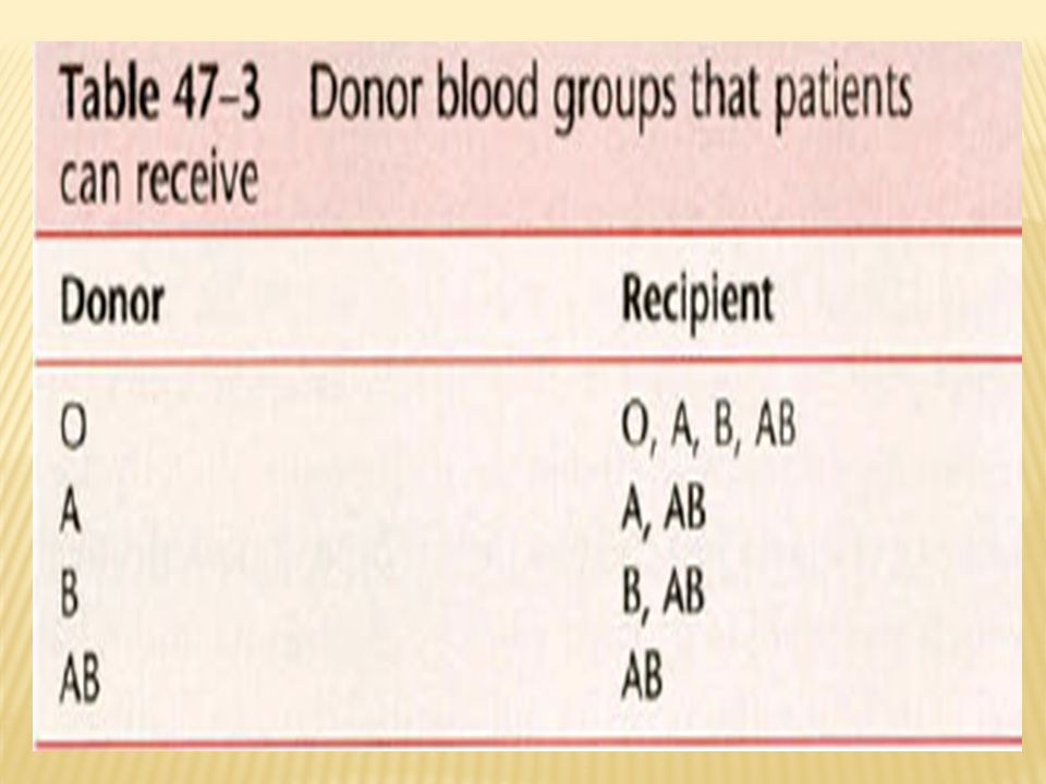
Compatibility Testing
The ABO-Rh type, cross match, and antibody screen are frequently referred to as compatibility tests 1-ABO-Rh Typing Rh(D) antigen. Rh+ 85%. Rh- 15%
 antigen. Rh+ 85%. Rh- 15%")
17
Crossmatch The crossmatch can be completed in approximately 45 to 60
Donor RBCsare mixed with recipient serum The crossmatch can be completed in approximately 45 to 60 minutes and is carried out in three phrases: Immediate phase At room temperature Antibodies in the MN, P, and Lewis systems. 1 to 5 minutes to complete Incubation phase Albumin or low-ionic strength salt solution.The incubation of 30 to 45 minutes in albumin and of 10 to 20 minutes in low-ionic-strength salt solution in this phase is of sufficient duration to allow antibody uptake. Antiglobulin phase Most incomplete antibodies in the blood group systems, including the Rh, Kell, Kidd and Duffy blood group systems

19
Antibody Screening Type and Screen
The antibody screen is also carried out in three phases and is similar in length to the crossmatch. The screen,however, is a trial transfusion between the recipient's serum and commercially supplied RBCs that are specifically selected to contain optimal numbers of RBC antigens, Type and Screen Elimination of the crossmatch The type and screen without crossmatch determines the ABO-Rh of the patient and the presence of the most commonly found unexpected Antibodies ,patient's serum is screened for the presence of unexpected antibiotics by incubating it with selected reagent RBCs (screen cells)..
..")
21
Is the Crossmatch Really Needed?
In previously transfused or pregnant patients, only about 1 patient in 100 may have an irregular antibody other than the anti-A and anti-B antibodies.However, some of these irregular antibodies are reactive order of probable significance,anti-Rh(D), Kell, C, E, and Kidd are the most common of clinically significant antibodies. After anti-A and anti-B,anti-Rh(D) is the most common significant antibody. ABO-Rh typing alone results in a 99.8%chance of a compatible transfusion, the addition of an antibody screen increases the safety to 99.94%and a crossmatch increases this to 99.95%
, Kell, C, E, and Kidd are. the most common of clinically significant antibodies. After anti-A and anti-B,anti-Rh(D) is the most common significant antibody. ABO-Rh typing alone. results in a 99.8%chance of a compatible transfusion, the addition of an antibody screen increases the safety to 99.94%and a crossmatch increases this to 99.95%")
22
Emergency Transfusion
Type-Specific, Partially Crossmatched Blood . Type-Specific, Uncrossmatched Blood previously received transfusions or have had pregnancies. Type 0 Rh-Negative (Universal Donor),Uncrossmatched Blood Type 0 blood lacks the A and B antigens and consequently cannot be hemolyzed by anti-A or anti-B antibodies in the recipient's blood some type 0 donors produce high titers of hemolytic IgG, IgM, anti-A, and anti-B antibodie . Whole blood/ PRBC Emergency transfusion of more than 2 units of type 0 Rh-negative,
,Uncrossmatched Blood. Type 0 blood lacks the A and B antigens and consequently. cannot be hemolyzed by anti-A or anti-B antibodies in. the recipient s blood. some type 0 donors produce high titers of. hemolytic IgG, IgM, anti-A, and anti-B antibodie. . Whole blood/ PRBC. Emergency transfusion of more than 2 units of type 0 Rh-negative,")
23
Specific Recommended Protocol
The following steps are recommended for patients who are hypovolemic and require blood transfusion: 1. Infuse crystalloids or colloids. 2. Draw a blood sample for typing and cross matching. 3. If cross matched blood is not ready to give, use type specific or type 0 Rh-negative cells or type 0 Rhpositive cells for males or postmenopausal females without a history of transfusions 4- Type-specific, partially cross matched blood; or type-specific, cross matched blood.

24
The shelf life can be extended to 42 days when
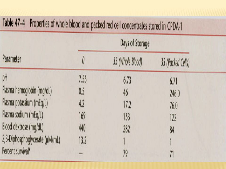
Storage of Blood Citrate phosphate dextrose adenine (CPDA-l) is an anticoagulant preservative in which blood is stored at l°C to 6°C. Citrate is an anticoagulant, phosphate serves as a buffer, Dextrose is a red cell energy source. The addition of adenine to CPD solution allows RBCs to resynthesize adenosine triphosphate (ATP), which extends the storage time from 21 to 35 days. The shelf life can be extended to 42 days when AS-1(Adsol), AS-3 (Nutricel), or AS-5 (Optisol) is used Adsol contains adenine, glucose, mannitol, and sodium chloride Nutricel contains glucose, adenine, citrate,phosphate, and sodium chloride. Optisol only contains dextrose, adenine, sodium chloride, and mannitol.
 is an anticoagulant. preservative in which blood is stored at l°C to 6°C. Citrate is an anticoagulant, phosphate serves as a buffer, Dextrose is a red cell energy source. The addition of adenine to CPD solution allows RBCs to resynthesize adenosine triphosphate (ATP), which extends the storage time from 21 to 35 days. The shelf life can be extended to 42 days when. AS-1(Adsol), AS-3 (Nutricel), or AS-5 (Optisol) is used. Adsol contains adenine, glucose, mannitol, and sodium chloride. Nutricel contains glucose, adenine, citrate,phosphate, and sodium chloride. Optisol only contains dextrose, adenine, sodium chloride, and mannitol.")
25
During storage of whole blood and PRBC,
During storage, RBCs metabolize glucose to lactate, hydrogen ions accumulate, and plasma pH decreases. The storage temperatures of 1°C to 6°C stimulate the sodium-potassium pump, and RBCs lose potassium and gain sodium The osmotic fragility of RBCs increases during storage, and some cells undergo lysis, resulting in elevated plasma .hemoglobin levels Storage is associated with progressive decreases in RBC concentrations of ATP and 2,3-diphosphoglycerate(2,3-DPG). Packed RBCs have a slightly lower survival than whole blood .although values for hemoglobin and potassium concentrations may appear somewhat high in 35-day stored RBC concentrates. However, it should be remembered that the total plasma volume in the concentrates is only 70 mL.
. Packed RBCs have a slightly lower survival than whole. blood .although values for hemoglobin and potassium concentrations may appear somewhat high in 35-day stored RBC concentrates. However, it should be remembered that the total plasma volume in the concentrates is only 70 mL.")
26
Frozen Storage Satisfactory storage of RBCs in the frozen state became possible when these cells, mixed with glycerol, could be frozen and thawed without damage. RBCs previously frozen to 79°C in glycerol survive well in humans.RBCs must be free from glycerol before being transfused, There are several advantages for frozen and thawed RBCs. 1- Blood of rare types can be stored for long periods, increasing viability and eliminating outdating. 2-Frozen,reconstituted blood is believed to be safer in patients who are especially susceptible to allergic reactions, because the freezing and washing process reduces sites with histocompatible antigens. 3-Frozen, washed blood may reduce risk of transfusion hepatitis. 4-Frozen blood, low in fibrin and leukocytic aggregates, would be safer in patients requiring massive blood transfusion, 5- frozen RBCs may be desirable in clinical conditions requiring prompt tissue oxygenation because normal levels of 2,3-DI'G are retained in frozen RBC .

29
Complications 1-Changes in Oxygen Transport
Changes in Oxygen Transport RBCs are transfused primarily to increase transport of oxygen to tissues. An increase in the circulating red cell mass produces an increase in oxygen uptake in the lung and a corresponding probable increase in oxygen delivery to tissues. The respiratory function of red cells may be impaired during preservation, making it difficult fo them to release oxygen to the tissues immediately after transfusion. 2-Review of the Oxygen Dissociation Curve P5o, which is the partial pressure of oxygen at which hemoglobin is half saturated with oxygen at 37°C and pH 7.4. A low P50 indicates a left shift in the oxygen dissociation curve and an increased affinity of hemoglobin for oxygen .in other words

31
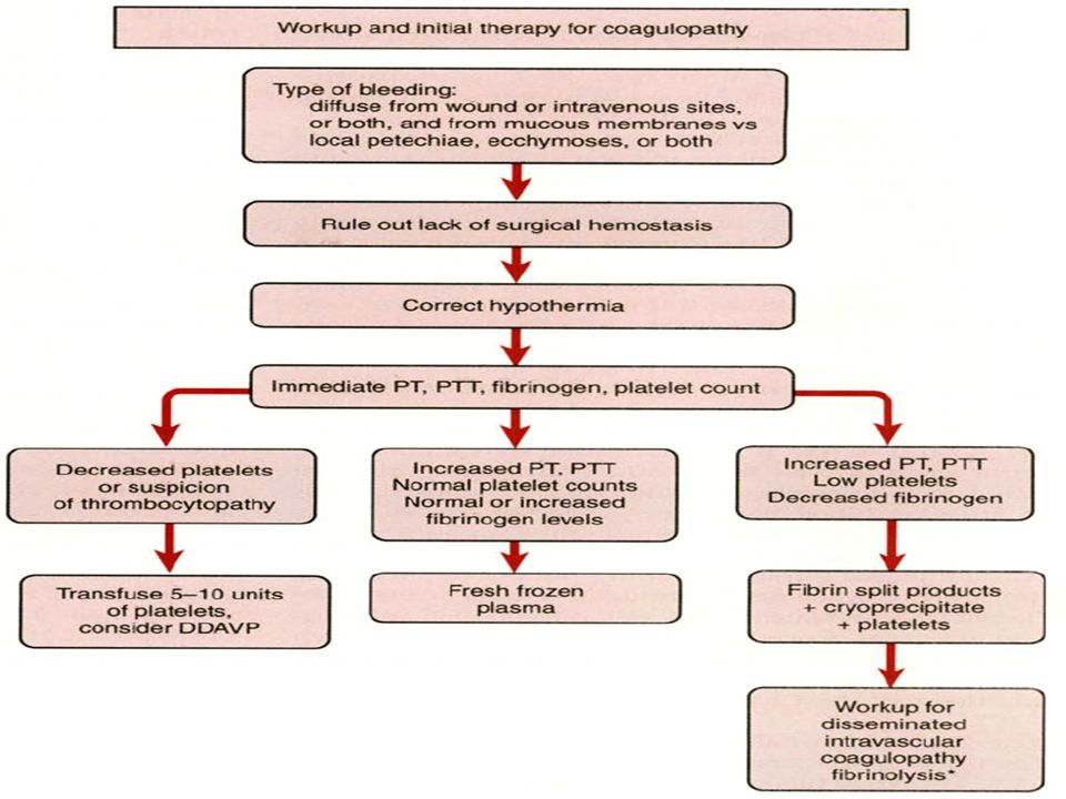
3-Coagulation Unless a patient has a preoperative coagulopathy (aspirin, antiplatelet drugs hemophilia), A transfusion induce coagulopathy usually occurs only after a large amount of blood has been given (6 to 10 units of PRBCs). This coagulopathy is caused by a combination of factors, of which the most important are the volume of blood given and the duration of hypotension or hypo perfusion. The patient who is hypotensive and has received many units of blood probably has a coagulopathy from a condition that resembles disseminated intravascular coagulation (DIC) and dilution of coagulation factors from stored bank blood. When such bleeding occurs, the differential diagnosis for a patient who did not have a pretransfusion coagulopathy (hemophilia) is dilutional thrombocytopenia, low factors V and VIII, DIC-like syndrome, hemolytic transfusion reaction. Clinical manifestations include oozing into the surgical field, hematuria, gingival bleeding, petechial bleeding from venipuncture sites, and ecchymoses.
. This coagulopathy is caused by a combination of factors, of which the most important are the volume of blood given and the duration of hypotension or hypo perfusion. The patient who is hypotensive and has received many. units of blood probably has a coagulopathy from a condition that resembles disseminated intravascular coagulation (DIC) and dilution of coagulation factors from stored bank blood. When such bleeding occurs, the differential diagnosis for a patient who did not have a pretransfusion coagulopathy (hemophilia) is dilutional thrombocytopenia, low factors V and VIII, DIC-like syndrome, hemolytic transfusion reaction. Clinical manifestations include oozing into the surgical field, hematuria, gingival bleeding, petechial bleeding from venipuncture sites, and ecchymoses.")
32
4-Dilutional Thrombocytopenia
Considering survival time and viability, total platelet activity is only 50% to 70% of the original in vivo activity after 6 hours of storage in bank blood at 4°c.After 24 or 48 hours of storage, platelet activity is only about 10%or 5% of normal, Platelets should not be given to treat laboratory evidence of thrombocytopenia unless clinical coagulopathy is also present. Treating laboratory numbers without correlation with the clinical status is fundamentally contrary to good medical practice; transfusion medicine is no exception. When the platelet count is less than 50,000 to 75,000/mm3, a bleeding problem is likely and is probably a combination of dilutional thrombocytopenia and Die.Platelet therapy would be appropriate in this situation patients with chronic thrombocytopenia or leukemia are commonly known to survive and not have a hemorrhagic diathesis with a platelet count lower than 15,000 cells/mm3.

36
6-Low levels of Factors V and VIII
These factors gradually decrease to 15%and 50% normal, respectively, after 21 days of storage. 5% to 20% of factor V and 30% of factor VIII are needed for adequate hemostasis during surgery. In other words, in spite of a patient's receiving massive The following criteria should be used: 1. Generalized bleeding that cannot be controlled with surgical sutures or cautery 2. Partial thromboplastin time at least 1.5 times normal 3. Platelet count greater than 70,000/mm3 (to ensure that thrombocytopenia is not the cause of bleeding 7-Disseminated Intravascular Coagulation-like Syndrome

37
7-Hemolytic Transfusion Reaction
Diagnosis and Treatment of a Hemorrhagic Diathesis after Whole Blood Transfusions Provided that the partial thromboplastin time is 1.5 times normal or more increased and other tests are normal, the bleeding is probably a result of very low levels of factors V and VIII. This can be treated with FFP,which contains all the coagulation factors except platelets, or with cryoprecipitatea. Dilutional thrombocytopenia in association with DlC is the most likely cause of bleeding from blood transfusion When the platelet count is less than 100,OOO/mm], a bleeding problem is likely to develop .Platelets should be ordered after infusion of 9 or 10 units of blood when several more will probably be required. Ideally, the platelets are available when 20 to 25 units of blood have been administered Fresh blood «6 hours old) supplies the largest number of platelets per donation. More than 80% of the platelet can be given by platelet-rich plasma, which has one half of the volume of a unit of blood.
 supplies the largest number of platelets per donation. More than 80% of the platelet can be given by platelet-rich plasma, which has one half of the volume of a unit of blood.")
38
Drugs Used to Improve Hemostasis
E-Aminocaproic acid (EACA) inhibits the formation of plasmin and attenuates fibrinolysis. 1-deamino-8-D-arginine vasopressin (DDAVP), a synthetic analog of the antidiuretic hormone vasopressin.It increases the levels of factor VIII and von Willebrand DDAVI' can cause hypotension, hyponatremia, and increased platelet adhesion Aprotinin, a serine protease factor inhibitor that inhibits fibrinolysis and improves platelet function(CABG) Tranexamic acid, which is also an anti fibrinolytic drug.
 inhibits the formation of plasmin and attenuates fibrinolysis. 1-deamino-8-D-arginine vasopressin (DDAVP), a synthetic analog of the antidiuretic hormone vasopressin.It increases the levels of factor VIII and von Willebrand. DDAVI can cause hypotension, hyponatremia, and increased platelet adhesion. Aprotinin, a serine protease factor inhibitor that inhibits fibrinolysis and improves. platelet function(CABG) Tranexamic acid, which is also an anti fibrinolytic drug.")
39
Diagnosis and Treatment of Hemorrhagic Diathesis after Packed Red Blood Cell Transfusion Most studies have .examined the influenc With much less plasma, dilution of certain coagulation values may be more profound with the use of PRBCs rather than whole blood. With use of PRBCs, fibrinogen levels decreased significantly in contrast to use of whole blood, in which fibrinogen level remained unchanged unless DIC was present When PRBCs are used to replace major blood loss, the clinician may be tempted to give FFP prophylactically. PT and PTT>1.5 normal and fibrinogen<75mg/dl or 12 or more units of PRBCs or cell-saver blood had been given, coagulation factors (FFP) were Necessary.
 were Necessary.")
40
Citrate Intoxication and Hyperkalemia
Hypothermia, liver disease, liver transplantation, and hyperventilation increase the possibility of citrate intoxication. The appearance of severe hypocalcemia during liver transplantation Calcium infusions are common during liver transplantation The rate of citrate metabolism is decreased by 50%when body temperature is decreased from 37" to 31°C. Excluding these conditions, infusion of more than 1 unit of blood every 10 minutes or >50ml/min in neonate is necessary for ionized calcium levels to Even at these rates of infusion, ionized calcium levels do not decrease enough to cause bleeding.. Serum potassium levels may be as high as 19 to 30 mEq/L in blood stored for 21 days Although hyperkalemia is occasionally reported, large amounts of blood must be given.

41
bank blood must be given at a rate of 120 mL/min or more
bank blood must be given at a rate of 120 mL/min or more. The fact that such rapid infusion rates of blood are required for the production of hyperkalemia suggests that the potassium ion must leave by diffusion into extravascular spaces, by reuptake into RBCs the intravascular spaces Temperature believe that there are more subtle reasons for warming all blood, even in patients receiving only 1 to 2 units intra operatively. Because of the cool temperature of the operating room, body temperature often decreases, particularly in patients undergoing extensive abdominal surgery. A decrease in body temperature as small as 0.5 to I.0°C may induce shivering postoperatively; this may increase oxygen consumption by as much as 400%. To meet the demands of elevated oxygen consumption, cardiac .output must be Increase Perhaps the safest and most common method of warming blood is to pass it through plastic coils immersed in warm water (37 to 38°C) bath. With increased use of PRBCs (in contrast to whole blood), other method of warming blood have been suggested. For example,added normal saline warmed to 45°C to PRBCs.
 bath. With increased use of PRBCs (in contrast to whole blood), other method. of warming blood have been suggested. For example,added normal saline warmed to 45°C to PRBCs.")
44
Acid-Base Abnormalities.
The pH of most storage media is very acidotic (5.5 pH for CPD). When this solution is added to a unit of freshly drawn blood, the pH of the blood immediately decreases to approximately 7.0 to 7.1. the pH of bank blood continues to decrease to about 6.9 after 21 days of storage. A large portion of the acidosis can be accounted for by the Pco2 of 15O to 220 mm Hg. The Pco2 is high mainly because the plastic container of blood does not provide an escape mechanism for carbon dioxide. With adequate ventilation in the recipient, the high pco2 should be of little consequence.
. When this solution is added to a unit of freshly drawn blood, the pH of the blood immediately decreases to approximately 7.0 to 7.1. the pH of bank blood continues to decrease to about 6.9 after 21 days of storage. A large portion of the acidosis can be accounted for by the Pco2 of 15O to 220 mm Hg. The Pco2 is high mainly because the plastic container of blood does not provide an escape mechanism for carbon dioxide. With adequate ventilation in the recipient, the high pco2 should be of little consequence.")
45
Transfusion Reactions
Hemolytic Transfusion Reaction Such a reaction can occur from infusion of as little as 10 mL of blood. Between 20% and 60% of patients with severe symptomatic hemolytic reactions may die, and these deaths usually result from AB0 blood group incompatibility between the donor and the patient.

46
Haptoglobin, which is a protein that can bind about 100 mg of hemoglobin per 100 mL of plasma.
A sample of plasma that contains 2 mg/dL of hemoglobin is faintly pink or light brown. When the level of hemoglobin reaches 100 mg/dL, the plasma is red. When the level of plasma hemoglobin reaches 150 mg/dL, hemoglobinuria occur Treatment If a hemolytic reaction is suspected, blood and urine samples should be sent to the laboratory for examination. The blood bank should check all paperwork to ensure that the correct blood component was transfused to the patient. Laboratory tests should be performed to determin the presence of hemoglobinemia A direct antiglobulin test, repeat compatibility testing, repeat other serologic tests (ABO and Rh) and analysis of urine for hemoglobinuria
 and analysis of urine for hemoglobinuria.")
50
Delayed Hemolytic Transfusion Reaction
In many cases of hemolytic transfusion reaction, the transfused donor cells may survive well initially,but after a variable delay (2 to 21 days), they are hemolyzed. This type of reaction occurs mainly in recipients sensitized to RBC antigens by previous blood transfusions or pregnancy. These delayed reactions are often manifested only by a decrease in the post-transfusion hematocrit value. However, jaundice and hemoglobinuria can occur in these patients and can cause some impairment in renal function, but only rarely do they lead to death. Unlike immediate reactions, antibodies most commonly involved in delayed hemolytic reactions are those in the Rh and Kidd systems rather than the ABOsystem.
, they are hemolyzed. This type of reaction occurs mainly in recipients sensitized to RBC antigens by previous blood transfusions or pregnancy. These delayed reactions are often manifested only by a decrease in the post-transfusion hematocrit value. However, jaundice and hemoglobinuria. can occur in these patients and can cause some impairment in renal function, but only rarely do they lead to death. Unlike immediate reactions, antibodies most commonly involved in delayed hemolytic reactions are those in the Rh and Kidd systems rather than the ABOsystem.")
51
Nonhemolytic Transfusion Reactions
Occasionally, fever may be the first sign of a hemolytic reaction or of bacterial contamination. For less serious febrile reactions, the most common adverse reactions to blood transfusions are the febrile reactions. The symptoms consist of chills, fever, headache,myalgia, nausea, and nonproductive cough occurring shortly after blood transfusion. Less frequently, the patient may have hypotension, chest pain, vomiting, and dyspnea. Allergic transfusion reactions Are mild and are thought to be caused by the presence of foreign protein in the transfused blood. The most common symptom is urticaria associated with itching. Occasionally, the patient has facial swelling. Allergic reactions occur in about 3%all transfusions. When these reactions are accompanied by fever or any other symptoms suggestive of a serious hemolytic reaction,it is not necessary to discontinue the transfusion. Antihistamine are used

52
Anaphylaxis in transfusion
occurs in which the patient has dyspnea, hypotension, laryngeal edema, chest pain, and shock. These are anaphylactic reactions caused by the transfusion of IgA to patients who are IgA deficient and have formed anti-IgA. This type of reaction does not involve red cell destruction and occurs very rapidly, usually after the transfusion of only a few milliliters of blood or plasma.

53
INFECTIOUS COMPLICATIONS
Viral (Hepatitis 88% of per unit viral risk) Hepatitis B Risk 1/ 200,000 due to HBsAg, antiHBc screening (7-17 % of PTH) Per unit risk 1/63-66,000 0.002% residual HBV remains in ‘negative’ donors (window 2-16 weeks) Anti-HBc testing retained as surrogate marker for HIV
 Hepatitis B. Risk 1/ 200,000 due to HBsAg, antiHBc screening (7-17 % of PTH) Per unit risk 1/63-66, % residual HBV remains in ‘negative’ donors (window 2-16 weeks) Anti-HBc testing retained as surrogate marker for HIV.")
54
NANB and Hepatitis C Risk now 1/ 103,000 (NEJM 96) with 2nd/ 1/ 125,000 with 3rd generation HCV Ab/ HVC RNA tests Window 4 weeks 70 % patients become chronic carriers, % develop cirrhosis

56
HIV Current risk 1/ ,000 (95) With current screening (Abs to HIV I, II and p24 Ag), window 6-8 weeks (third generation ELISA tests in Europe) sero -ve window to < 16 days
, window 6-8 weeks (third generation ELISA tests in Europe) sero -ve window to < 16 days.")
57
HTLV I, II Only in cellular components (not FFP, cryo)
Risk 1/ 641,000 (window period unknown) Screening for antibody I may not pick up II
 Screening for antibody I may not pick up II.")
58
CMV Cellular components only
Problem in immunocompromised, although 80 % adults have serum Ab WBC filtration decreases risk of transmission CMV -ve blood: CMV -ve pregnant patients, LBW neonates, CMV -ve transplant recipient, CMV-ve/ HIV +ve

59
II. Bacterial III. Protozoal
Contamination unlikely in products stored for > 72 hours at C gram –ve, gram +ve bacteria most frequent – Yersinia enterocolitica Produced endotoxin Platelets stored at room temperature for 5 days, with infection rate of 0.25% III. Protozoal Trypanosoma cruzi (Chaga’s disease) Malaria Toxoplasmosis Leishmaniasis
 Malaria. Toxoplasmosis. Leishmaniasis.")
60
TRALI (Transfusion related acute lung injury )
Donor Ab reacts with recipient Ag (1/ 10,000) noncardiogenic pulmonary edema Supportive therap
 noncardiogenic pulmonary edema. Supportive therap.")
61
Transfusion-related Acute Lung Injury (TRALI)
Pathophysiology Leukocyte Ab in donor react with pt. leukocytes Activate complements Adherence of granulocytes to pulmonary endothelium with release of proteolytic enz.& toxic O2 metabolites Endothelial damage Interstitial edema and fluid in alveoli

63
Transfusion-related Acute Lung Injury (TRALI)
Acute and severe type of transfusion reaction Symptoms and signs Fever Hypotension Tachypnea Dyspnea Diffuse pulmonary infiltration on X-rays Clinical of noncardiogenic pumonary edema

64
Transfusion-related Acute Lung Injury (TRALI)
Therapy and Prevention Adequate respiratory and hemodynamic supportive treatment If TRALI is caused by pt. Ab use LPB If TRALI is caused by donor Ab no special blood components

65
BLOOD COMPONENT THERAPY
Packed Red Blood Cells PRBCs contain the same amount of hemoglobin as whole blood, but much of the plasma has been removed. The hematocrit value is 40%in whole blood and 70%in packed erythrocytes . Transfusion of whole blood is required primarily for blood loss acute enough to cause hypovolemic shock and actively bleeding and have sustained a loss of greater than 25% of their total blood volume. In other words, whole blood provides oxygen carrying capacity and intravascular blood volume expansion. Less severe degrees of hemorrhage may be effectively treated with PRBCs,

66
Packed Red Blood Cells The administration of PRBCs is facilitated by reconstituting them with a crystalloid or colloid; however, not all crystalloids are suitable. If the solution contains calcium,clotting occurs. Lactated Ringer's solution is usually not recommended for use as a diluent for PRBCs Conversely, using flow rates and clot formation.A more important factor may be whether the diluent is hypotonic with respect to plasma. If so, the RBCs will swell and eventually lyse.

67
PRBCs may cause low serum concentrations may be
tempted to use a plasma derivative, such as Plasmanate. However, these solutions also can cause hemolysis. The osmolality of Plasmanate is only 180 mOsm/kg. Solutions recommended for reconstituted packed erythrocytes are 5% dextrose in 0.4% saline, 5%>dextrose in 0.9% saline, 0.9%>saline, and Normosol-R with a pH of 7.4.

69
Platelet Concentrates
Platelet concentrates are prepared by differential centrifugation .from freshly drawn units of blood or from donor If platelets are stored at room temperature, they are satisfactory to use 5 days after collection Because use of multidonor platelet products stored for 5 days results in an incidence of sepsis five times higher than use of those stored for 4 days, shorter storage times are being emphasized The incidence of platelet-related sepsis is about 1 case in 12,000 people.. The increased risk of bacterial overgrowth is related to the storage temperature of 20°C to 24°C. Because there is no test to identify bacterially contaminated blood products,

71
1. Prophylactic platelet transfusion is ineffective and
The American Society of Anesthesiologists (ASA)Task Force 2 provided the following recommendations 1. Prophylactic platelet transfusion is ineffective and rarely indicated when thrombocytopenia is due to increased platelet destruction (ITP). 2. Prophylactic platelet transfusion is rarely indicated in surgical patients with thrombocytopenia due to decreased platelet production when the platelet count is greater than 100 x 109/L and is usually indicated when the platelet count is below 50 x 109/L. The determination of whether patients with intermediate platelet counts (50 to 100 x 109/L) require therapy should be based on the patient's risk of bleeding.
Task Force 2 provided the following recommendations. 1. Prophylactic platelet transfusion is ineffective and. rarely indicated when thrombocytopenia is due to. increased platelet destruction (ITP). 2. Prophylactic platelet transfusion is rarely indicated in surgical patients with thrombocytopenia due to decreased platelet production when the platelet count. is greater than 100 x 109/L and is usually indicated when the platelet count is below 50 x 109/L. The determination of whether patients with intermediate platelet counts (50 to 100 x 109/L) require therapy should be based on the patient s risk of bleeding.")
72
3. Surgical and obstetric patients with microvascular bleeding usually require platelet transfusion if the platelet count is less than 50x 109/L and rarely require therapy if it is greater than 100 x 109/L. With intermediate platelet counts (50to 100 x 109/L), the determination should be based on the patient's risk for more significant bleeding. 4. Vaginal deliveries or operative procedures ordinarily associated with insignificant blood loss may be undertaken in patients with platelet counts less than 50x 109/L 5. Platelet transfusion may be indicated despite an apparently adequate platelet count if there is known platelet dysfunction and microvascular bleeding. Patients with severe thrombocytopenia «20,000 cells/mm) and clinical signs of bleeding usually When possible, ABO-compatible platelets should beused
 and clinical signs of bleeding usually. When possible, ABO-compatible platelets should beused.")
73
one platelet concentrate usually produces an increase of about 7000 to 10,000 platelets/mm3 .1 hour after transfusion to the 70-kg adult. Ten units of platelet concentrates are required to increase the platelet count by 100,000 cells/mm3 ,However, many factors, including splenomegaly, previous sensitization, fever, sepsis, and active bleeding, may lead to decreased survivals and decreased recovery of transfused platelets Various different types of platelet concentrates have been proposed, including apheresis (collecting more platelets from one donor to avoid pooling of platelet from multiple donors), leukocyte-depleted platelets, an ultraviolet B-irradiated platelets.
, leukocyte-depleted platelets, an ultraviolet B-irradiated platelets.")
74
Fresh Frozen Plasma FFP is prepared at the time that blood is obtained from a donor. It contains all the plasma proteins, particularly factors V and VIII, which gradually decline during the storage of blood. The use of FFP carries with it certain inherent risks that are observed with the use of essentially any blood product. The major risk is transmission of infectious diseases, such as hepatitis H, hepatitis C, and AIDS Other risks include sensitization to foreign proteins the ASA Task Force 2 recommended the administration of FFP with the following guidelines: 1.urgent reversal of warfarin therapy 2. For correction of known coagulation factor deficiencies for which specific correlates are unavailable 3. For correction of microvascular bleeding in the presence of increased (>1.5 times normal) PT or PTT
 PT or PTT.")
75
4.For correction of microvascular bleeding secondary to coagulation factor deficiency in patients transfused with more than one blood volume and when prothrombin time and partial thromboplastin time cannot be obtained in a timely fashion 5. FFP should be given in doses calculated to achieve a minimum of 30% of plasma factor concentration (usually achieved with administration of 10 to 15 mL/kg of FFP), except for urgent reversal of warfarin anticoagulation, for which 5 to 8 mL/kg of FFPusually suffice. Four to five platelet concentrates, 1 unit of single donor apheresis platelets, or 1 unit of whole blood provides a quantity of coagulation factors similar to that contained on 1 unit of FFP (except for decreased, but still hemostatic, concentrations of factors V and VIII in whole blood) 6. FFP is contraindicated for augmentation of plasma volume or albumin concentration. The ASA Task Force 2 concluded that there was little scientific evidence to support
, except for urgent reversal of warfarin anticoagulation, for which 5 to 8 mL/kg of FFPusually suffice. Four to five platelet concentrates, 1 unit of single donor apheresis platelets, or 1 unit of whole blood provides a quantity of coagulation factors similar to that contained on 1 unit of FFP (except for decreased, but still hemostatic, concentrations of factors V and VIII in whole blood) 6. FFP is contraindicated for augmentation of plasma volume or albumin concentration. The ASA Task Force 2 concluded that there was little scientific evidence to support.")
77
The only indications for FFP administration the ASA Task Force 2 agreed on were the following:
1. Replacement of isolated factor deficiencies (as documented by laboratory evidence) 2. Reversal of warfarin effect 3. In cases of anti thrombin III deficiency 4. Treatment of immunodeficiencies 5. Treatment of thrombotic thrombocytopenia purpura 6. Massive blood transfusion (rarely and only when factors V and VIII are less than 25% of normal) 7. Requirements for indications 1 and 6 are prothrombin and partial thromboplastin times at least 1.5 times longer than normal.
 2. Reversal of warfarin effect. 3. In cases of anti thrombin III deficiency. 4. Treatment of immunodeficiencies. 5. Treatment of thrombotic thrombocytopenia purpura. 6. Massive blood transfusion (rarely and only when factors. V and VIII are less than 25% of normal) 7. Requirements for indications 1 and 6 are prothrombin. and partial thromboplastin times at least 1.5 times longer than normal.")
78
Cryoprecipitate Cryoprecipitate is prepared in such a way that it contains significant levels of factor VIII and fibrinogen. It also contains von Willebrand factor and fibronectin. All other plasma proteins are present in only trace amounts in cryoprecipitate. The use of cryoprecipitate in the treatment of factor VIII deficiency or hemophilia. Cryoprecipitate contains factor VIII:C (procoagulant activity), factor VIII:vWF (von Willebrand factor), fibrinogen, factor XIII, and fibronectin, which is a glycoprotein that may playa role in reticuloendothelial clearance of foreign particles and bacteria from the blood. Cryoprecipitate can also be used in the treatment of fibrinogen deficiencies and is preferable to commercially prepared fibrinogen preparations, which have a very high incidence of hepatitis, whereas the risk of hepatitis in cryoprecipitate is no greater than that from a single unit of blood. Cryoprecipitate is frequently administered as ABO Compatible however, this probably dministered as ABO compatible; however, this probably is not very important,because the concentration of antibodies in cryoprecipitate is extremely low.
, factor VIII:vWF. (von Willebrand factor), fibrinogen, factor XIII, and fibronectin, which is a glycoprotein that may playa role in reticuloendothelial clearance of foreign particles and bacteria from the blood. Cryoprecipitate can also be used in the treatment of fibrinogen deficiencies and is preferable to commercially prepared fibrinogen preparations, which have a very high incidence of hepatitis, whereas the risk of hepatitis in cryoprecipitate is no greater than that from a single unit of blood. Cryoprecipitate is frequently administered as ABO Compatible. however, this probably dministered as ABO compatible; however, this probably is not very important,because the concentration of antibodies in cryoprecipitate is extremely low.")
79
Cryoprecipitate should be administered through a filter and as rapidly as possible. The rate of administration should be at least 200 mL/hour, and infusion should be completed within 6 hours of thawing. SYNTHETIC COLLOID SOLUTION THERAPY Synthetic Hydroxyethyl Starch The most commonly used preparation was 6% hydroxyethyl starch .Although an effective intravascular expander, it has not gained widespread popularity probably because of its effects on coagulation, particularly with regard to increased bleeding and platelet function. The molecular mass plays some role in the adverse coagulation effects. Two new HES preparations have been developed to decrease coagulation effects. Hextend has been studied extensively. It is 6% HES but also contains a physiologically balanced medium of electrolytes, glucose andlactate. It has a similar pharmacokinetic and dynamic profile to other starch preparations with fewer effects on coagulation.

81
Dextrans Dextran, is an effective volume expander.
However, after infusion of more than 20 mL/kg in 24 hours, dextran 70 may interfere with normal blood clotting, causing a deficiency with cross matching procedures and possibly causing a bleeding diathesis. These clotting defects reflect reduced platelet adhesiveness resulting from an antithrombin effect. The incidence of severe anaphylactoid or anaphylactic reactions is a concern. These reactions are mediated by dextran-reactive antibodies that are IgG immunoglobulins.. Dextran 70 and albumin may deplete the extracellular fluid space of water, as does albumin. Dextran 40 (Rheomacrodex), with a molecular mass of 40,000 daltons, has been used primarily to reduce blood viscosity and cellular aggregation and to improve microcirculation during low-flow states. It is often given prophylactically to decrease the incidence of postoperative thrombo embolism. Blood viscosity may be increased by trauma, blood loss, burns, and endotoxin shock. Although viscosity can be decreased by dextran 40, the presumed improvement in flow through the microcirculation has not been well documented.
, with a molecular mass of. 40,000 daltons, has been used primarily to reduce blood. viscosity and cellular aggregation and to improve microcirculation during low-flow states. It is often given prophylactically to decrease the incidence of postoperative thrombo embolism. Blood viscosity may be increased by trauma, blood loss, burns, and endotoxin shock. Although viscosity can be decreased by dextran 40, the presumed improvement in flow through the microcirculation has not been well documented.")
82
The End

Similar presentations


 – Leukocytes (WBCs) – Thrombocytes (platelets) Plasma – 90% water – 10% solutes – Proteins,>")


 Packed red blood cells Fresh frozen plasma Platelets.>")


.>")