Download presentation
Presentation is loading. Please wait.

1
Spinal Cord Injury Herniated Disc Spinal Cord Tumors

2
Pathophysiology Normal Spinal Cord
Spinal cord begins at the foramen magnum in the cranium Cord ends at the L1-L2 vertebra level Spinal nerves continue to the last sacral vertebra The Human Spine

3
Spinal Cord Gray matter- cell bodies of voluntary and autonomic motor neurons White matter axons of ascending and descending motor fibers

4
Normal Spinal Cord White tracts send messages to and from the brain
Ascending Tracts- carry into higher levels of CNS touch, deep pressure,vibration, position, temperature Descending Tracts impulses for voluntary muscle movement

5
Pyramidal- Voluntary movements Posterior column (Dorsal)- touch, proprioception, and vibration sense Lateral spinothalamic tract- pain and temperature sensation (only tract that crosses within the cord) voluntary movement
 voluntary movement.")
6
Upper Motor Neurons UMN Originate in cerebral cortex Project downward Result in skeletal muscle movement Injury = SPASTIC paralysis Lower Motor Neurons LMN Originate at each vertebral level Project to specific parts of the body Result in movement /sensation Injury = FLACCID paralysis

7
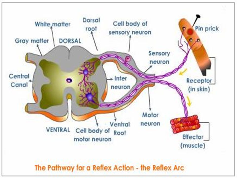
Normal Spinal Cord Reflex Arc Involuntary response to a stimulus
Where sensory and motor nerves arise from cord Sensory fibers enter posterior Synapse in the grey matter Motor fibers leave anterior Once outside cord join form spinal nerve reflex movement

9
Normal Spinal Cord Dermatones Skin innervated by sensory spinal nerves
Myotome- muscle group innervated by motor neurons

10
Nervous System and the Spinal Cord
ANS can be affected by SCI Sympathetic chains on both sides of the spinal column (T1-L2) Parasympathetic nervous system is the cranial-sacral branch (brainstem, S2-4)
 Parasympathetic nervous system is the cranial-sacral branch (brainstem, S2-4)")
11
Spinal Cord Protection
Bones- vertebral column 7 Cervical 12 Thoracic 5- Lumbar 5- Sacral Discs- between vertebra

12
Spinal Cord Protection
Internal and external ligaments Dura Meninges CSF in subarachnoid space allow for movement within spinal canal

13
Etiology of Traumatic SCI
MVA- most common cause Other: falls, violence, sport injuries SCI typically occurs from indirect injury from vertebral bones compressing cord SCI frequently occur with head injuries Cord injury may be caused by direct trauma from knives, bullets, etc

14
Etiology of Traumatic SCI
78% people with SCI are male Typically young men – 16-30 Number of older adults rising (>61 yr) Greater complications Life Expectancy 5 years less than same age without injury 90% go home
 Greater complications. Life Expectancy 5 years less than same age without injury. 90% go home.")
15
Spinal Cord Injury- SCI
Compression Interruption of blood supply Traction Penetrating Trauma

16
Spinal Cord Injury Primary Initial mechanism of injury Secondary
Ongoing progressive damage Ischemia Hypoxia Microhemorrhage Edema

17
Spinal Cord Injury Hemorrhage and edema occur in the cord post injury, causing more damage to cord Extension of the cord injury from cord edema can occur over the first few days watch the phrenic nerve! Initially SCI experience spinal shock depression of all cord & ANS function below injury. Lasts from few min to wks

18
Spinal and Neurogenic Shock
Spinal Shock Decreased reflexes and loss of sensation below the level of injury Motor loss- flaccid paralysis below level injury Sensory loss- loss touch, pressure, temperature pain and proprioception perception below injury Lasts days to months

19
Spinal and Neurogenic Shock
Due to loss of vasomotor tone SNS loss results in parasympathetic dominance with vasomotor failure Loss of SNS innervation causes peripheral pooling and decreased cardiac output Hypotension and Bradycardia Orthostatic hypotension and poor temperature control (poikilothermic)
")
20
How do you know spinal shock is over?
Clonus is one of the first signs Hyperreflexia of foot Test by flexing leg at knee & quickly dorsiflex the foot Rhythmic oscillations of foot against hand clonus

21
Classifications of SCI
Mechanism of Injury Skeletal and Neurologic Level Completeness (degree) of Injury Flexion Hyperextension Compression Flexion /Rotation
 of Injury. Flexion. Hyperextension. Compression. Flexion /Rotation.")
22
Classifications of SCI Mechanism of Injury
Flexion (hyperflexion) Most common because of natural protection position. Generally cause neck to be unstable because stretching of ligaments
 Most common because of natural protection position. Generally cause neck to be unstable because stretching of ligaments.")
23
Classifications of SCI Mechanism of Injury
Hyperextention Caused by chin hitting a surface area, such as dashboard or bathtub Usually causes central cord syndrome symptoms

24
Classifications of SCI Mechanism of Injury
Compression Caused by force from above, as hit on head Or from below as landing on butt Usually affects the lumbar region

25
Classifications of SCI Mechanism of Injury
Flexion/Roatation Most unstable Results in tearing of ligamentous structures that normally stabilize the spine Usually results in serious neurologic deficits

26
Levels of Function in Spinal Cord Injury
Skeletal level Vertebral level where the most damage to the bones Neurologic level The lowest segment of the spinal cord with normal sensory and motor function on both sides of the body Levels of Function in Spinal Cord Injury

27
Classification of SCI- Level of Injury
Spinal cord level When referring to spinal cord injury, it is the reflex arc level (neurologic)not the vertebral or bone level. the thoracic, lumbar & sacral reflex arcs are higher than where the spinal nerves actually leave through the opening of vertebral bone
not the vertebral or bone level. the thoracic, lumbar & sacral reflex arcs are higher than where the spinal nerves actually leave through the opening of vertebral bone.")
28
Classifications of SCI Completeness (Degree) of Injury
Incomplete Central cord syndrome Anterior Cord syndrome Brown-Sequard Syndrome Posterior Cord Syndrome Cauda Equina and Conus Medullaris

29
Classification of SCI Completeness (degree) of Injury
Complete (transection) After spinal shock: Motor deficits- spastic paralysis below level of injury Sensory- loss of all sensation perception Autonomic deficits- vasomotor failure and spastic bladder
 After spinal shock: Motor deficits- spastic paralysis below level of injury. Sensory- loss of all sensation perception. Autonomic deficits- vasomotor failure and spastic bladder.")
30
Classification of SCI Completeness (degree) of Injury
Incomplete Central Cord Syndrome Injury to the center of the cord by edema and hemorrhage Motor weakness and sensory loss in all extremities Upper extremities affected more

31
Classification of SCI Completeness (degree) of Injury
Incomplete Brown-Séquard Syndrome Hemisection of cord Ipsilateral paralysis Ipsilateral superficial sensation, vibration and proprioception loss Contralateral loss of pain and temperature perception

32
Classification of SCI Completeness (degree) of Injury
incomplete Anterior Cord Syndrome Injury to anterior cord Loss of voluntary motor, pain and temperature perception below injury Retains posterior column function (sensations of touch, position, vibration, motion)
")
33
Classification of SCI Completeness (degree) of Injury
incomplete Posterior Cord Syndrome Least frequent syndrome Injury to the posterior (dorsal) columns Loss of proprioception Pain, temperature, sensation and motor function below the level of the lesion remain intact
 columns. Loss of proprioception. Pain, temperature, sensation and motor function below the level of the lesion remain intact.")
34
Classification of SCI Completeness (degree) of Injury
incomplete Conus Medullaris Injury to the sacral cord (conus) and lumbar nerve roots Cauda Equina Injury to the lumbosacral nerve roots Result- areflexic (flaccid)bladder and bowel, flaccid lower limbs
 and lumbar nerve roots. Cauda Equina. Injury to the lumbosacral nerve roots. Result- areflexic (flaccid)bladder and bowel, flaccid lower limbs.")
36
Clinical Manifestations of SCI
Skin: pressure ulcers Neuro: pain sensory loss upper/lower motor deficits autonomic dysreflexia Cardio: dysrhythmias spinal shock loss of SNS control over blood vessels orthostatic hypotension, poikilothermic

37
Respiratory- GI GU Musculoskeletal
decrease chest expansion, cough reflex & vital capacity diaphragm function-phrenic nerve GI stress ulcers paralytic ileus bowel- impaction & incontinence GU upper/lower motor bladder Impotence sexual dysfunction Musculoskeletal joint contractures bone demineralization osteoporosis muscle spasms muscle atrophy pathologic fractures para/tetraplegia

38
Common Manifestation/Complications
Upper and Lower Motor Deficits Upper motor deficits result in spastic paralysis Lower motor deficits result in flaccid paralysis and muscle atrophy

39
Common Manifestations/Complications
Spinal cord injuries are described by the level of the injury– the cord segment or dermatome level Such as C6; L4 spinal cord injury Terms used to describe motor deficits Prefix: para- meaning two extremities tetra- or quadra- all four extremities Suffix : -paresis meaning weakness -plegia meaning paralysis Quadraparesis means what?

40
Common Manifestations/Complications
C1-3 usually fatal- Loss of phrenic innervation ventilator dependent No B/B control Spastic paralysis Electric w/c with chin/mouth control

41
Common Manifestations/Complications
C6- weak grasp Has shoulder/biceps to transfer & push w/c No bowel/bladder control. Considered level of independence

42
Common Manifestations/Complications
T1-6- full use of upper extremity Transfer Drive car with hand controls and do ADL’s No bowel/bladder control

43
Immediate Care Emergency Care at Scene, ER & ICU
Transport with cervical collar Assess ABC’s; O2; tracheotomy/vent IV for life line NG to suction Foley

44
Diagnostic Studies for SCI
X-ray of spinal column CT/MRI Blood gases

45
Therapeutic Interventions
Medications IV methylprednisolone (Solu-Medrol) within 8 hrs to decrease cord edema
 within 8 hrs to decrease cord edema.")
46
Therapeutic Interventions
Medications To control or to prevent complications of SCI and immobility: Vasopressors to maintain perfusion Histamine H2 blockers to prevent stress ulcers Anticoagulants Stool softeners Antispasmodics

47
Therapeutic Interventions
Stabilization/ Immobilization Traction- Gardner-wells tongs Halo Casts Splints Collars Braces

48
Therapeutic Interventions
Surgery for SCI Manipulation to correct dislocation or to unlock vertebrae Decompression laminectomy Spinal fusion Wiring or rods to hold vertebrae together

49
Nursing Management Assessment
HEALTH HISTOY Description of how and when injury occurred Other illnesses or disease processes Ability to move, breathe, and associated injury such as a head injury, fractures

50
Nursing Management Assessment
PHYSICAL EXAM LOC and pupils- may have indirect SCI from head injury Respiratory status- phrenic nerve (diaphragm) and intercostals; lung sounds Vital signs Motor Sensory Bowel and bladder function
 and intercostals; lung sounds. Vital signs. Motor. Sensory. Bowel and bladder function.")
51
Nursing Management Assessment
Motor Assessment Upper Extremity Movement, strength and symmetry Hand grips Flex and extend arm at elbow- with and without resistance

52
Nursing Management Assessment
Motor Assessment Lower Extremity Flex and extend leg at knee with and without resistance Planter and dorsi flexion of foot Assess for Clonus

53
Nursing Management Assessment
Sensory assessment With the sharp and dull ends of a paperclip have the individual, with their eyes closed identify Use the dermatome as reference to identify level C6 thumb; T4 nipple; T10 naval

54
Nursing Problems/Interventions
1.Impaired mobility 2.Impaired gas exchange 3. Impaired skin integrity 4. Constipation 5. Impaired urinary elimination 6. Risk for autonomic dysreflexia 7. Ineffective coping

55
1. Impaired Physical Mobility
Log roll as a single unit; provide assistance as needed to keep alignment; teach patient Care traction, collars, splints, braces, assistive devices for ADL’s Flaccid paralysis- use high top tennis shoes or splints to prevent contractures. Remove at least every 2 hrs for ROM (active ROM best)
")
56
1. Impaired Physical Mobility
Spastic Paralysis Prevent spasms by avoiding; sudden movements or jarring of the bed; internal stimulus (full bladder/skin breakdown; use of footboard; staying in one position too long; fatigue Treat spasms by decreasing causes; hot or cold packs; passive stretching; antispasmodic medications Assess skin break down thrombophlebitis; remove TED hose at least every shift

57
1. Impaired Physical Mobility
Prevent/treat orthostatic hypotension Abdominal binder, calf compressors, TED hose when individual gets up Assess BP, especially when rising Teach use of transfer board Assist Physical Therapy with tilt table as individual gradually gets use to being in an upright position

58
2. Impaired Gas Exchange Phrenic nerve (C3-5) controls the diaphragm bilaterally. If nerve is nonfunctioning then individual is ventilator dependent. Thoracic nerves control the intercostals muscles for breathing and abdominal muscles aide in breathing and coughing

59
2. Impaired Gas Exchange Respiratory rate, rhythm, depth, breath sounds, respiratory effort, ABG’s, O2 saturation Signs of impending extension of SCI up cord to phrenic nerve level (C3-5) Need for ventilatory assistance tracheotomy, ventilator Quad cough (assistive cough) as needed
 Need for ventilatory assistance tracheotomy, ventilator. Quad cough (assistive cough) as needed.")
60
3. Impaired Skin Integrity
Change position frequently Protection from extremes in temperature Inspect skin at least 2x/day especially over boney prominences Avoid shearing and friction to soft tissue with transfers Removal of TED hose every 8 hours Nutritional status

61
4. Constipation Bowels rely more on bulk than on nerves
Stimulate bowels at the same time each day. Best after a meal when normal peristalsis occurs Individual may progress from Dulcolax suppository to glycerin then to gloved finger for digital stimulation Assess bowel sounds prior to giving food for the first time– paralytic ileus!

62
5. Impaired Urinary Elimination
Flaccid bladder (lower motor neuron lesion) No reflex from S2,3,4 Automatic empting of bladder Urine fills the bladder and dribbles out Need Foley or freq intermittent self catheterization Spastic bladder (upper motor neuron lesion) Reflex arc but no connection to or from brain Reflex fires at will Bladder training- trigger points to stimulate empting; self catheterization
 No reflex from S2,3,4. Automatic empting of bladder. Urine fills the bladder and dribbles out. Need Foley or freq intermittent self catheterization. Spastic bladder (upper motor neuron lesion) Reflex arc but no connection to or from brain. Reflex fires at will. Bladder training- trigger points to stimulate empting; self catheterization.")
63
5. Impaired Urinary Elimination
Use bladder scan to see amount of urine in bladder Goal- residual <100ml/20% bladder capacity Some individuals may need suprapubic catheter Assess effectiveness of medication Urecholine to stimulate bladder contraction Urinary antiseptic

64
6. Risk for Autonomic Dysreflexia
SCI above T6 Results in loss of normal compensatory mechanisms when sympathetic nervous system is stimulated Life threatening- if goes unchecked BP can result in cerebral hemorrhage Vasodilatation symptoms above SCI Vasoconstriction symptoms below SCI The cause of SNS stimulation

66
6. Risk for Autonomic Dysreflexia
Elevate head of bed- causes orthostatic hypotension Identify cause/alleviate- if full bladder- cath; if skin- remove pressure, if full bowel- empty, etc Remove support hose/abdominal binder Monitor blood pressure- can get > 300 S Give PRN medication to lower BP If above not effective– call physician

67
7. Ineffective Coping/ Grief and Depression
Assess thoughts on ‘quality of life’; body image; role changes Physical and psychological support Most common SCI is yeas old and generally a risk taker– this greatly affects their perception of life and rehabilitation

68
7. Ineffective Coping/sexuality
Male Female UMN lesion reflexogenic (S2,3,4) erections LMN lesion psychogenic erections (psychological stimulation) Ejaculation/fertility may be affected hormones more than nerves regarding fertility. C-section because of chance for autonomic dysreflexia during labor. Lack of sensation/movement affects sexual performance
 erections. LMN lesion. psychogenic erections (psychological stimulation) Ejaculation/fertility may be affected. hormones more than nerves regarding fertility. C-section because of chance for autonomic dysreflexia during labor. Lack of sensation/movement affects sexual performance.")
69
7. Ineffective Coping/sexuality
Assess readiness/knowledge/your ability Use proper terminology Suggestions: empty bladder before sex withhold fluids and antispasmodics certain positions may increase spasms explore new erogenous zones penile implants Refer to specially trained counselor

70
Home Care Assess psychological, physiological resources
need for rehabilitation (in-house or out patient) need for community resources Home assessment
 need for community resources. Home assessment.")
71
What’s new in SCI treatment?
Superman breather YouTube - Superman breather – USA Kevin Everett hypothermia treatment for SCI Standing Tall Travis Roy- 11 Seconds Stem Cell treatment for SCI Lipitor for SCI

72
Case study- Jim Valdez 1. Why does Jim have flaccid paralysis on admission to ICU? 2. What symptoms indicate that he is in spinal shock? What was done about these symptoms? 3. How will we know when he is out of spinal shock? 4. How does progressive mobilization assist with orthostatic hypotension? What else can be done? 5. What are realistic functional goals for Jim?

73
Herniated Disc and Spinal Cord tumors

74
Spinal Cord Anatomy Function of disc is to allow for mobility of the spine and act as shock absorber spinal cord anatomy

75
Pathophysiology/Etiology
Located between vertebral bodies Composed of nucleus pulposus a gelatinous material surrounded by annulus fibrosis- a fibrous coil Spinal nerves come out between vertebra

76
Herniated Disc Herniated nucleus pulposus, (HNP) slipped disc, ruptured disc HNP- annulus becomes weakened/torn and the nucleus pulposus herniates through it. Risk Factors- Standing erect Aging changes Poor body mechanics Overweight Trauma

77
Common Manifestations/Complications
HNP compresses Spinal nerve (sensory or motor component) as it leaves the spinal cord Or the cord itself- the white tracts within the cord- rare
 as it leaves the spinal cord. Or the cord itself- the white tracts within the cord- rare.")
78
Common Manifestations/Complications
Sensory root or nerve usually affected pain, parenthesis, or loss of sensation Motor root or nerve may be affected paresis or paralysis Manifestations depend on what nerve root, spinal nerve is being compressed– which dermatomes Radiculopathy- pathology of the nerve root

79
Common Manifestations/Complications Lumbar HNP
Most common site for HNP L4-5 disc- the 5th lumbar nerve root posterior sensory nerve or root compressed Classic symptoms- low back sciatica pain pain increases with increase in intrathoracic pressure herniated disc L4-L5

80
Other Symptoms Lumbar HNP:
Postural changes Urinary/male sexual function changes Paresis or paralysis Foot drop Paresthesias Numbness Muscle spasms Absent cord reflexes

81
Common Manifestations/Complications Cervical HNP
C5-C6 disc- affects the 6th cervical nerve root Pain- neck, shoulder, anterior upper arm to thumb Absent/diminished reflexes to the arm Motor changes- paresis or paralysis Sensory- paresthesias or pain Muscle spasms

82
Therapeutic Interventions- Diagnostic Tests
X-ray identify deformities and narrowing of disk space CT/MRI Mylogram p1336 Nerve conduction studies (EMG) detect electrical activity of skeletal muscles
 detect electrical activity of skeletal muscles.")
83
Treatment- Conservative
Bed rest with firm mattress log roll side lying position with knees bent and pillow between legs to support legs Avoid flexion of the spine brace/corset, cervical collar to provide support Medications non-narcotic analgesics, anti-inflammatory, muscle relaxants, antispasmodics and tranquilizers

84
Treatment- Conservative
Heat/cold therapy to decrease muscle spasms Break the pain-spasm-pain cycle Ultrasound, massage, relaxation techniques Progressive mobilization with approved exercise program –includes abdominal/thigh strengthening Teaching good body mechanics Weight loss TENS unit

85
Treatment- Surgery Laminectomy-
removal of a portion of the lamina to relieve pressure and to get to the herniated nucleus pulposus that is protruding out herniated disc repair Foraminotomy Enlargement of the bony overgrowth at the opening which is compressing the nerve

86
Treatment- Surgery Microdiskectomy
Use of electron microscope through a small incision to remove a portion of the HNP that is displaced If cervical HNP, usually use the anterior approach in the neck anterior cervical fusion

87
Treatment- Surgery Spinal fusion spinal fusion Artificial Disc
removes most of the disc and replaces it with bone usually from the patient iliac crest Fusion also with rods, pins, synthetic protein Flexibility is lost at the site- requires longer hospital stay spinal fusion Artificial Disc Combination of metal and plastic Attached to vertebrae above and below

88
Prevention of HNP Back school approach-
Causes of HNP Learn how to prevent Good body mechanics Exercises to strengthen leg and abdominal muscles Change in life-style or occupation

89
Nursing Assessment Specific to HNP Health History
Assess for risk factors- The cumulative effect of standing erect and daily stress Aging changes in disc/ligaments Poor body mechanics Overweight Trauma Employment History of pain and other neuro changes

90
Nursing Assessment Specific to HNP Physical Exam
Use similar methods to assess as utilized SCI Muscle strength and coordination Sensation sharp/dull of paperclip using dermatome as reference Pain evaluation- pain scale Pre/Post-op assessment

91
Post-Op Assessment for HNP
Sensory/motor assessment- care not to injure op site Assess for CSF drainage or bleeding from op site Encourage turn (log roll, cough, deep breath) Assess for postural hypotension especially if client was on bed rest for several days/weeks prior to surgery
 Assess for postural hypotension. especially if client was on bed rest for several days/weeks prior to surgery.")
92
Post-op Assessment for HNP
If Anterior Cervical- Assess injury to the carotid, esophagus, trachea, laryngeal nerve (speech- hoarseness) Assess respiration, neck size, swallowing and speech If Post-Op Lumbar- Assess bowels sounds, voiding. Minimize stress of post-op site- flat with pillow between knees, log roll, etc
 Assess respiration, neck size, swallowing and speech. If Post-Op Lumbar- Assess bowels sounds, voiding. Minimize stress of post-op site- flat with pillow between knees, log roll, etc.")
93
Nursing Problems/Interventions 1. Acute Pain
Post surgery the individual may have similar pain as pre-op due to lack of resiliency of the spinal nerves to ‘bounce’ back quickly Donor site (illiac crest) may cause more pain than laminectomy Individual may be in a pain-spasm-pain cycle, therefore may need both antispasmodic as well as analgesic
 may cause more pain than laminectomy. Individual may be in a pain-spasm-pain cycle, therefore may need both antispasmodic as well as analgesic.")
94
2. Chronic Pain Surgery may not relieve pain
Nonpharmalogical methods to control pain Pain clinic

95
3. Constipation As a result of bed rest and decreased mobility and fear of pain with straining of stool Constipation prevention methods– fluids, diet, etc

96
4. Home Care When riding in a car, take frequent stops to move and stretch Prevention– Back school approach May have to deal with pain as a chronic condition May need to make life/job changes

97
Spinal Cord Tumors CNS is made up of neural tissue and support tissue
These tissues undergo changes and result in spinal cord tumors Blood vessels and bone also can be part of the tumor

98
Intramedullary- arise from neural tissues of the spinal cord
Extramedullary- arise from tissues outside the spinal cord may be benign or malignant Intradural-from the nerve roots or meninges in subarachnoid space Extradural- from the epidural tissue or vertebra

99
Classification by origin
Primary- originating in the spinal cord or meninges Secondary- metastases from other parts of the body Most spinal cord tumors are found in the thoracic region Spinal cord tumors can compress (benign), invade the neural tissue, or cause ischemia to the area because of vascular obstruction
, invade the neural tissue, or cause ischemia to the area because of vascular obstruction.")
100
Common Manifestations/Complications
Symptoms depend on the anatomical level of the spinal column, the anatomical location, the type of tumor and the spinal nerves affected Pain that is not relieved by bed rest is the most common presenting symptom Other symptoms are similar to those found with HNP or spinal cord injury- sensory or motor

101
Common Manifestations/Complications
Manifestations of thoracic cord tumor Paresis & spasticity of one leg then the other Pain back & chest, not relieved by bedrest Sensory changes Babinski reflex Bowel (ileus); bladder dysfunction (UMN in type)
; bladder dysfunction (UMN in type)")
102
Therapeutic Interventions
Diagnostic tests include: X-ray of the spinal column Myelogram Lumbar puncture with CSF analysis

103
Therapeutic Interventions
Medications spinal tumors Control pain- narcotic analgesics, epidural catheter, PCA, NSAID’s Reduce cord edema and tumor size- Steroids- high dose Dexamethasone

104
Therapeutic Interventions
Surgery for spinal cord tumors Laminectomy to remove or to decrease the size (decompression laminectomy) of the spinal cord tumor Spinal fusion or the insertion of rods if several vertebra involved and the column is unstable Radiation to reduce size and control pain
 of the spinal cord tumor. Spinal fusion or the insertion of rods if several vertebra involved and the column is unstable. Radiation to reduce size and control pain.")
105
Nursing Assessment Health history Physical exam
Pain, motor and sensory changes, bowel and bladder changes, Babinski reflex. Physical exam Similar to physical assessment for HNP

106
Nursing Problems/Interventions
1. Anxiety Metatastic tumor vs benign spinal cord tumor Education and support system 2. Risk for constipation From spinal cord compression, narcotics, bed rest Adjust fluid and diet

107
Nursing Problems/Interventions
3. Impaired physical mobility From bed rest and motor involvement Basic nursing- ROM, etc 4. Acute pain From compression or invasion of tumor Assess and treat 5. Sexual dysfunction Male sacral reflex arc (S 2,3,4) interference Similar care as discussed with SCI
 interference. Similar care as discussed with SCI.")
108
Nursing Problems/Interventions
6. Urinary retention Reflex arc (S2,3,4) interference can cause neurogenic bladder as discussed with SCI 7. Home care Rehabilitation Home evaluation Support groups case study
 interference can cause neurogenic bladder as discussed with SCI. 7. Home care. Rehabilitation. Home evaluation. Support groups. case study.")
109
A 30-year-old was admitted to the progressive care unit with a C5 fracture from a motorcycle accident. Which of the following assessments would take priority? Bladder distension Neurological deficit Pulse ox readings The client’s feelings about the injury

110
While in the ER, a client with C8 tetraplegia develops a blood pressure of 80/40, pulse 48, and RR of 18. The nurse suspects which of the following conditions? Autonomic dysreflexia Hemorrhagic shock Neurogenic shock Pulmonary embolism

111
A 22-year-old client with quadriplegia is apprehensive and flushed, with a blood pressure of 210/100 and a heart rate of 50 bpm. Which of the following nursing interventions should be done first? Place the client flat in bed Assess patency of the indwelling urinary catheter Give one SL nitroglycerin tablet Raise the head of the bed immediately to 90 degrees

112
The nurse is caring for an elderly client diagnosed with a herniated nucleus pulposus of L4-L5. Which scientific rationale explains the incidence of a ruptured disc in the elderly? The client did not use good body mechanics when lifting an object. There is an increased blood supply to the back as the body ages. Older clients develop atherosclerotic joint disease as a result of fat deposits. Clients develop intervertebral disc degeneration as they age.

113
A client is admitted with a spinal cord injury at the level of T12
A client is admitted with a spinal cord injury at the level of T12. He has limited movement of his upper extremities. Which of the following medications would be used to control edema of the spinal cord? Acetazolamide (Diamox) Furosemide (Lasix) Methylprednisolone (Solu-Medrol) Sodium bicarbonate
 Furosemide (Lasix) Methylprednisolone (Solu-Medrol) Sodium bicarbonate.")
114
A client with a cervical spine injury has Gardner-Wells tongs inserted for which of the following reasons? To hasten wound healing To immobilize the surgical spine To prevent autonomic dysreflexia To hold bony fragments of the skull together

115
Which of the following interventions describes an appropriate bladder program for a client in rehabilitation for spinal cord injury? Insert an indwelling urinary catheter to straight drainage Schedule intermittent catherization every 2 to 4 hours Perform a straight catherization every 8 hours while awake Perform Crede’s maneuver to the lower abdomen before the client voids.

116
A client has a cervical spine injury at the level of C5
A client has a cervical spine injury at the level of C5. Which of the following conditions would the nurse anticipate during the acute phase? Absent corneal reflex Decerebate posturing Movement of only the right or left half of the body The need for mechanical ventilation

117
The nurse is evaluating neurological signs of the male client in spinal shock following spinal cord injury. Which of the following observations by the nurse indicates that spinal shock persists? Positive reflexes Hyperreflexia Inability to elicit a Babinski’s reflex Reflex emptying of the bladder

118
Your T1 spinal cord injured patient complains of a headache. You should
Give him prn Tylenol Disimpact his bowels Call the doctor Take his blood pressure

119
What can the nurse do to best speed the patients recovery from a laminectomy of L5?
Keep patient flat in bed Teach the back school approach Medicate for pain q2 hours Ambulate as soon as orders permit

120
Your patient has a malignant metastatic lesion at T8 and is in for palliative radiation. What is your main goal with this patient? Teach patient self catheterization Ensure patient receives pain medication as needed Encourage patient to discuss fears Ambulate twice a shift

Similar presentations


 - Joints - Cartilages - Ligaments.>")


