Download presentation
Presentation is loading. Please wait.

1
GASTROINTESTINAL (G.I) BLEEDING
Fadi J. Zaben RN MSN

2
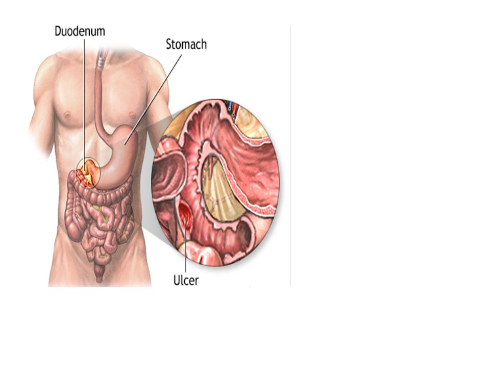
Normal Anatomy: The gastrointestinal tract starts at the mouth, which leads to the esophagus, stomach, small intestine, colon, and finally, the rectum and anus. The GI tract is a long, hollow, muscular tube through which food passes and nutrients are absorbed.

3
Definition: Gastrointestinal (GI) bleeding refers to any bleeding that starts in the gastrointestinal tract. GI bleeding is not just a gastroduodenal disorder but may occur anywhere along the alimentary tract. Bleeding from the GI tract is a common medical problem. Bleeding is a symptom of an upper or lower GI disorder. It may be obvious in emesis or stool, or it may be occult (hidden).
.")
4
Types of G.I Bleeding: Bleeding may come from any site along the GI tract, but is often divided into: Upper GI bleeding: The upper GI tract includes the esophagus (the tube from the mouth to the stomach), stomach, and first part of the small intestine. An upper source is characterized by hematemesis and melena. About half of cases are due to peptic ulcer disease. Esophagitis and erosive disease is the next most common causes.
, stomach, and first part of the small intestine. An upper source is characterized by hematemesis and melena. About half of cases are due to peptic ulcer disease. Esophagitis and erosive disease is the next most common causes.")
5
The most common cause is hemorrhoids.
Continue….. Lower GI Bleeding: The lower GI tract includes much of the small intestine, large intestine or bowels, rectum, and anus. It may be indicated by red blood per rectum, especially in the absence of hematemesis. The most common cause is hemorrhoids.

6
Incidence: Upper GI bleed 100/100,000. Lower GI Bleed 20/100,000.
Both are more common in males and elderly.

7
Etiology: Trauma anywhere along the GI tract. Erosions or ulcers.
Rupture of an enlarged vein such as a varicosity (esophageal or gastric varices). Inflammation, such as esophagitis (caused by acid or bile), gastritis, inflammatory bowel disease (chronic ulcerative colitis, Crohn's disease), and bacterial infection.
. Inflammation, such as esophagitis (caused by acid or bile), gastritis, inflammatory bowel disease (chronic ulcerative colitis, Crohn s disease), and bacterial infection.")
8
Anal disorders, such as hemorrhoids or fissures.
Continue…… Alcohol and drugs (aspirin-containing compounds, NSAIDs, anticoagulants, corticosteroids). Diverticular disease. Cancers. Vascular lesions or disorders, such as bowel ischemia, aortoenteric fistula. Mallory-Weiss tear. Anal disorders, such as hemorrhoids or fissures.
. Diverticular disease. Cancers. Vascular lesions or disorders, such as bowel ischemia, aortoenteric fistula. Mallory-Weiss tear. Anal disorders, such as hemorrhoids or fissures.")
10
Clinical Manifestations:
Characteristics of Blood: Bright red: vomited from high in esophagus (hematemesis): from rectum or distal colon (coating stool). Mixed with dark red: higher up in colon and small intestine: mixed with stool. Shades of black (coffee ground): esophagus, stomach, and duodenum; vomitus from these areas. Tarry stool (melena): occurs in patient who accumulates excessive blood in the stomach
: from rectum or distal colon (coating stool). Mixed with dark red: higher up in colon and small intestine: mixed with stool. Shades of black (coffee ground): esophagus, stomach, and duodenum; vomitus from these areas. Tarry stool (melena): occurs in patient who accumulates excessive blood in the stomach.")
11
Signs and Symptoms of Bleeding:
Massive bleeding: Acute, bright red hematemesis or large amount of melena with clots in the stool. Rapid pulse, drop in BP, hypovolemia, and shock. Subacute bleeding: Intermittent melena or coffee-ground emesis. Hypotension. Weakness, dizziness. Chronic bleeding: Intermittent appearance of blood. Increased weakness, paleness, or shortness of breath. Occult blood.

12
Diagnosis: It is not difficult to diagnose bleeding, but it may be difficult to locate the source of bleeding. History: change in bowel pattern, presence of pain or tenderness, recent intake of food and what kind, alcohol consumption, such drugs as aspirin or steroids. Complete blood count (CBC) (hemoglobin, hematocrit, platelets) and coagulation studies (partial thromboplastin time, prothrombin time with international normalized ratio) may show abnormalities.
 (hemoglobin, hematocrit, platelets) and coagulation studies (partial thromboplastin time, prothrombin time with international normalized ratio) may show abnormalities.")
13
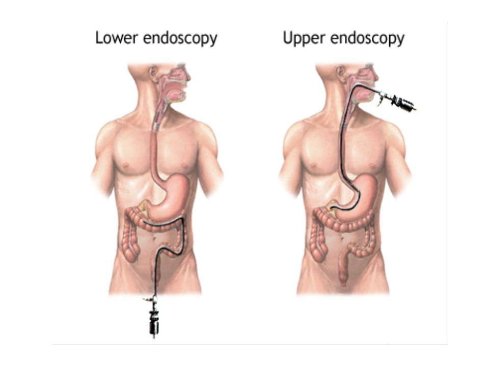
Continue….. Lower and Upper Endoscopy: identifies source of bleeding, determines risk of re-bleeding, and provides endoscopic therapy if needed. Imaging may detect etiology of bleeding. Test of stool for occult blood.

15
Management: Based on Etiology. Emergency Intervention.
Nasogastric Intubation. Other Measures.

16
Based on Etiology: If aspirin or NSAIDs are the cause, discontinue medication and treat bleeding. If ulcer is the cause, medications, dietary and lifestyle modifications. Therapeutic endoscopic procedure (cautery, injection). Surgery may be indicated for cancers, inflammatory diseases, and vascular disorders.
. Surgery may be indicated for cancers, inflammatory diseases, and vascular disorders.")
18
Emergency Intervention:
Patient remains on NPO status. I.V. lines and oxygen therapy initiated. If life-threatening bleeding occurs, treat shock, administer blood replacement, intra-arterial vasopressin or embolization. Surgical therapy, if indicated.

19
Nasogastric Intubation:
An NG tube should be in place for most patients with acute or upper GI bleeding. If the aspirate continues to be bloody after 2 to 3 L of tap water lavage, the patient may have an active bleed requiring more emergent intervention or endoscopic therapy.

20
Other Measures……… Electrocoagulation using a heater probe.
Injection of sclerosant or epinephrine. Endoscopy used in conjunction with management measures as well as in diagnostic evaluation. Pharmacotherapy depends on cause; can include histamine blockers as either continuous I.V. (preferred) or bolus infusion to block the acid-secreting action of histamine. Intra-arterial vasopressin can be used to slow or stop active bleeding from diverticulum or vascular ectasia. Surgery is indicated when more conservative measures fail.
 or bolus infusion to block the acid-secreting action of histamine. Intra-arterial vasopressin can be used to slow or stop active bleeding from diverticulum or vascular ectasia. Surgery is indicated when more conservative measures fail.")
21
Complications: Hemorrhage. Shock. Death.

22
Nursing Assessment: Obtain history regarding:
Change in bowel patterns or hemorrhoids. Change in color of stools (dark black, red, or streaked with blood). Alcohol consumption. Medications, such as aspirin, NSAIDs, antibiotics, anticoagulants, corticosteroids. Hematemesis. Other medical conditions. Evaluate for presence of abdominal pain or tenderness. Monitor vital signs and laboratory tests for changes that indicate bleeding (hemoglobin, hematocrit, platelet count, coagulation studies). Test for occult blood, if indicated.
. Alcohol consumption. Medications, such as aspirin, NSAIDs, antibiotics, anticoagulants, corticosteroids. Hematemesis. Other medical conditions. Evaluate for presence of abdominal pain or tenderness. Monitor vital signs and laboratory tests for changes that indicate bleeding (hemoglobin, hematocrit, platelet count, coagulation studies). Test for occult blood, if indicated.")
23
Nursing Diagnoses: Deficient Fluid Volume related to blood loss.
Imbalanced Nutrition: Less Than Body Requirements related to nausea, vomiting, diarrhea.

24
Nursing Interventions:
1. Attaining Normal Fluid Volume: Maintain NG tube and NPO status to rest GI tract and evaluate bleeding. Monitor intake and output as ordered to evaluate fluid status. Monitor vital signs as ordered. Observe for changes indicating shock, such as tachycardia, hypotension, increased respirations, decreased urine output, change in mental status. Administer I.V. fluids and blood products as ordered to maintain volume.

25
2. Attaining Balanced Nutritional Status:
Continue……… 2. Attaining Balanced Nutritional Status: Weigh daily to monitor caloric status. Administer I.V. fluids, TPN if ordered to promote hydration and nutrition while on oral restrictions. Begin liquids when patient is no longer NPO. Advance diet as tolerated. Diet should be high-calorie, high-protein. Frequent, small feedings may be indicated. Offer snacks; high-protein supplements.

26
Patient Education and Health Maintenance:
Discuss the cause and treatment of GI bleeding with patient. Instruct patient regarding signs and symptoms of GI bleeding: melena, emesis that is bright red or coffee ground color, rectal bleeding, weakness, fatigue, shortness of breath. Instruct patient on how to test stool or emesis for occult blood, if applicable.

27
QUESTIONS……..

Similar presentations


>")
 682-3793; (p) 413-3222.>")
 The McGraw-Hill Companies, Inc. Permission required for reproduction or display. 23-1 Chapter 23 Abdominal and Gastrointestinal Disorders.>")


