Download presentation
Presentation is loading. Please wait.

1
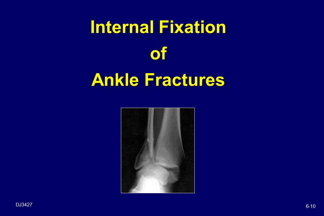
Internal Fixation of Ankle Fractures
6-10 1
2
Objectives Review ankle anatomy
Identify the indications & treatment goals for ORIF of ankle fractures Summarize the implant options

3
Anatomy Ankle Bones Formed by medial malleolus of tibia, and lateral malleolus (fibula) Talus sits in “mortise” (as in “mortise & tenon”) Fibula Tibia Talus
 Fibula. Tibia. Talus.")
4
Anatomy Ankle Soft Tissues
Ligaments connect ankle on medial & lateral sides Important for stability

5
Anatomy Ankle Soft Tissues
Fibula connected to tibia by fibrous band of tissue called syndesmosis Also important for stability

6
Ankle Fractures 1

7
Ankle Fractures History Physical examination Twisting injury
Immediate pain – lateral and/or medial Difficulty weight-bearing Physical examination Malleolar pain (posterior & anterior) Swelling Neurovascular involvement
 Swelling. Neurovascular involvement.")
8
Ankle Fractures Radiographs
Ankle Series: AP, mortise, lateral “Rule out” other injuries: Osteochondral injuries Lateral process fracture Anterior calcaneus fracture Base of 5th MT fracture AP Mortise Lateral

9
Ankle Fractures Classification
Weber / AO Classification based on level of fibula fracture A – Below syndesmosis B – At syndesmosis C – Above syndesmosis

10
Simple Classification: Stable & Unstable
Stable fractures Most commonly involve medial or lateral side only Talus remains anatomic relative to tibia

11
Simple Classification: Stable & Unstable
Unstable fractures Disruption of 2 or more aspects of the mortise -- bone and/or ligament Talus may sublux or be dislocated from tibia

12
Stable Examples

13
Unstable Examples

14
Indications for Surgery Ankle Fractures
Inability to obtain or maintain an anatomic mortise (unstable fracture pattern) Open fractures
 Open fractures.")
15
Basic Set-Up Ankle Fractures
Supine position most common Occasionally prone for direct approach to posterior malleolus Bump beneath ipsilateral buttocks (allows easier approach to fibula) Tourniquet Prep / drape to above knee Pre-op antibiotics Fluoroscopy or X-ray
 Tourniquet. Prep / drape to above knee. Pre-op antibiotics. Fluoroscopy or X-ray.")
16
General Considerations
Small size of ankle bones = dictates implant sizes Multiple complex 3-D articulations Weight bearing structure subject to high stresses (2 – 5x body weight)
")
17
General Considerations
Limited soft tissue coverage

18
Instrumentation Ankle Fractures
Small fragment set Cannulated screws K-wires Cerclage wire Power Have mini-frag available

19
Ankle Fracture Surgical Tx
Type One malleolus Bimalleolar Tri-malleolar Treatment Fix fibula with screw / TB wire / plate Plate fibula, lag screw tibia (medial malleolus) Plate fibula, lag screw tibia, fix posterior if > % articular surface involved
 Plate fibula, lag screw tibia, fix posterior if > % articular surface involved.")
20
Implant Considerations Lateral Malleolus
One-third tubular plate & mm cortex screws Lateral Posterior 3.5mm compression plate for unstable fractures

21
Implant Considerations Lateral Malleolus
Locking plates -- lateral or posterolateral Osteoporotic bone Unstable fractures Distal fractures

22
Implant Considerations Lateral Malleolus
Hook Plate Used to obtain purchase in very distal fibula fractures

23
Implant Considerations Posterior Malleolus
Posterior to anterior Anterior to posterior

24
Implant Considerations Medial Malleolus
Two partially threaded 4.0 mm cancellous screws K-wires Cerclage wire for tension band technique

25
Syndesmosis Fixation Indications
Syndesmotic instability after fixation of malleolus Consider if fibula fracture > 4 cm above joint line & Maisonneuve’s fracture Have bone hook on back table to check stability Have large frag screws & instruments available

26
Implant Considerations Syndesmosis
Surgeons choice of large or small fragment fully threaded screws, one or two Not inserted as lag screw, but as a positioning screw (threads engage all cortices) Secures position of fibula next to tibia allowing torn syndesmotic tissues to heal May be removed in weeks
 Secures position of fibula next to tibia allowing torn syndesmotic tissues to heal. May be removed in weeks.")
27
Implant Considerations Syndesmosis
Have pelvic forceps on back table May need longer plates than in small frag set: 1/3 tubular, compression or specialty fibula plate Bioresorbable screws

28
Case #1 Age: 81 Gender: Female Cause of Injury: Fall
Fixation: 3.5mm LCP Lateral Distal Fibula Plate

29
Case #2 Age: 64 Gender: Female Cause of Injury: Fall
Fixation: 3.5mm LCP Lateral Distal Fibula Plate

30
Summary Reviewed ankle anatomy
Identified the indications & treatment goals for ORIF of ankle fractures Summarized the implant options

31
Thank You

Similar presentations
>")


 Radiographic Evaluation of the Ankle>")


: Brian M. Fuller MD, Maine Medical Center.>")


