Download presentation
Presentation is loading. Please wait.

1
CHEST TUBES

2
Objectives: Discuss the anatomy of the chest and the mechanism of respirations Discuss the indications for a chest tube Discuss the structure and function of the chest bottle (drainage system) Troubleshoot the problems with/maintain chest tubes and chest tube bottles
 Troubleshoot the problems with/maintain chest tubes and chest tube bottles.")
3
Inspiration: Expiration: Lung expansion Ribs move outwards and upwards
Diaphragm moves downwards Expiration: Decreased lung volume Ribs move inwards and downwards Diaphragm moves upwards

4
Pleura: Lungs are surrounded by two thin layers (films) called the pleurae They function as lubricant, preventing the lungs from rubbing with the rib cage during inspiration and expiration Visceral: Outside the lungs Parietal: Inside the ribcage The area between the two pleurae is filled with a fluid called the pleural fluid (that fills a pleural space) The pleural fluid acts as a lubricant so the two films don’t rub each other
 called the pleurae They function as lubricant, preventing the lungs from rubbing with the rib cage during inspiration and expiration Visceral: Outside the lungs. Parietal: Inside the ribcage. The area between the two pleurae is filled with a fluid called the pleural fluid (that fills a pleural space) The pleural fluid acts as a lubricant so the two films don’t rub each other.")
5
THE PLEURAL SPACE HAS ALWAYS A NEGATIVE PRESSURE. OTHERWISE,
THE LUNGS WILL COLLAPSE

6
Pleural Pressure: The degree of negativity changes throughout the respiratory cycle Intrapleural pressure normally ranges from –(4) to –(10) cm of water Inspiration pressure drops to –(10) cm water Expiration pressure rises up to –(4) cm water It is the pressure variance that allows the air to move in and out easily
 to –(10) cm of water. Inspiration pressure drops to –(10) cm water. Expiration pressure rises up to –(4) cm water. It is the pressure variance that allows the air to move in and out easily.")
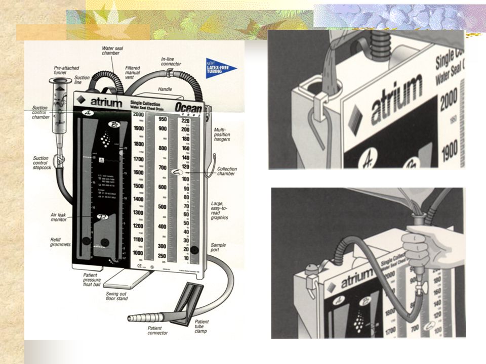
7
Definition of the chest drainage
Is the insertion of a tube into the pleural and/or mediastenal space and a physician order required for the type of evacuation force: a-normally 20cm for adult b-10-15cm for pediatric The drainage must be kept always lower than the patient to prevent backflow of fluid into the pleural or mediastinal space.

8
Indications of a Chest Tube:
Pneumothorax Hemothorax Hemopneumothorax Pleural Effusion Empyema Drainage

9
Chest tube location For evacuation of air: 2,3,4 intercostals space are commonly used sites. To drain fluid : 5,6 intercostals space are commonly used sites.

10
CHEST DRAINAGE SYSTEM UN DEUX TROIS

11
Fluid Collection Chamber:
Collects fluid as it drains from the pleural space or mediastinal space Water Seal Chamber: Acts as a one way valve, allowing air to escape from the patient and never return back. Always 2 cm of water. Suction Control Chamber: Controls the amount of suction applied directly to the patient. It improves the rate and flow of drainage.

13
Subcutaneous Emphysema:
When the lungs or the air passages are injured, air may enter the tissue planes and pass for some distance under the skin. The tissues give a crackling sensation when palpated, and the subcutaneous air produces an alarming appearance as body becomes misshapen. It is not a serious complication if the air is spontaneously absorbed or stopped, or if the leak is treated.

14
Chest Tube Assessment: STOP
S=SITE T=TUBING O=OUTPUT P=PATENCY

15
Site: Check for: Clean and dry dressing: change every 72 hrs ,use a prim pore dressing and clean with sterile technique. Subcutaneous emphysema 1. palpate contious monitoring 2.notify doctor

16
Tubing: Connections are secured No dependent loops
Straighten periodically Keep bottle below patient’s level Tape the connections if 2 suction tubes are used

17
Output: Amount, type and color Mark regularly Document
Use the white on column on the drainage chamber to mark drainage level

18
Patency: A-water seal chamber
Assess the Water seal with the suction off If water seal level is too high, it will be more difficult for air leave the chest If the water is too low ,it leaves the water seal chamber at risk for exposure to air can cause a pneumothorax

19
Patency: B-Bubbling: Bubbling means there is a leak in the system unless the patient has a pneumothorax.

20
How to Check for an Air Leak?
Clamp the tube below the dressing Bubbling continues? No Leak is between patient and dressing Yes Continue clamping

21
Patency: C-fluctuation
When inspiration the water seal level will reach -10cm of negative pressure normally When expiration the water seal level will reach -4cm of negative pressure normally When fluctuation stopped so the tubing may be obstructed If more than 2cm the drainage will decreased If less than 2cm there risk of pneumothorax

22
Patency :D-suction drainage units
Assess the suction control water level when the suction is off Excessive bubbling in the suction control chamber result in evaporation of the water which will decreased in applied suction and does not increase the pressure on the pleural or mediastinal cavity Normally 80 mmhg of wall suction is required to ensure consistent delivery of suction

23
Patient Activity A semi-fowler position is required
Position and turn the patient every 2 hrs3 Patent can be out of bed if there is no contraindication Do (ROM) exercises to the affected arm and shoulder site Encourage coughing and deep breathing
 exercises to the affected arm and shoulder site. Encourage coughing and deep breathing.")
24
Clamping: Clamp only when: Don’t clamp when: Changing the bottle
Checking for an air leak Tubing is disconnected and sterile water is out of reach If changing the position and when elevating the tube system above the patient level is required Don’t clamp when: : Ambulating a patient

25
Milking: Milking the tube is not recommended.
It creates a pressure of –(400) to – (100) cm of water that can cause a tension pneumothorax
 to – (100) cm of water that can cause a tension pneumothorax.")
26
Problem solving...

Similar presentations
, The process through which the respiratory system moves air into and out of the lungs. In contrast, Respiration.>")


>")
