Download presentation
Presentation is loading. Please wait.

1
1 st Lecture Biome II Dr.Manal Radwan Salim Lecturer of Physical Therapy Pharos University Fall 2013-2014 28-9-2013

2
I. Types of fractures: A) Common types of fractures. B) According to site of injury. C) According to nature of loading. D) According to loading modes.
 According to site of injury. C) According to nature of loading. D) According to loading modes..")
3
A) Common types of fractures: A)Stable fracture. The broken ends of the bone line up and are barely out of place. B) Open, compound fracture. The skin may be pierced by the bone or by a blow that breaks the skin at the time of the fracture. The bone may or may not be visible in the wound. C) Comminuted fracture. In this type of fracture, the bone shatters into three or more pieces.
 Open, compound fracture. The skin may be pierced by the bone or by a blow that breaks the skin at the time of the fracture. The bone may or may not be visible in the wound. C) Comminuted fracture. In this type of fracture, the bone shatters into three or more pieces..")
4
B) According to site of injury: 1-Direct Injury: fracture occur at site of force application. 2- Indirect Injury: resulting from force transmission through other tissues e.g. avulsion fracture.

5
C) According to nature of loading : 1- High energy loading (acute loading): in response to a single large-magnitude loading as in violent collision. A fall, a motor vehicle accident, or a tackle during a football gamecan all result in fractures.

6
Example 1: Radiograph of a high- energy fracture shows marked comminution resulting from trauma sustained in an automobile accident.

7
Example 2: Radiograph of a very high energy fracture produced by a bullet fired from a gun at high muzzle velocity. The energy of the projectile imparted to the soft tissue and bone upon impact results in extensive tissue destruction and very fine bony comminution. associated exclusively with gunshot injuries produced by missiles having high muzzle velocity.

8
2- Low energy acute loading: occur in osteoporosis. This disorder weakens bones and makes them more likely to break.

9
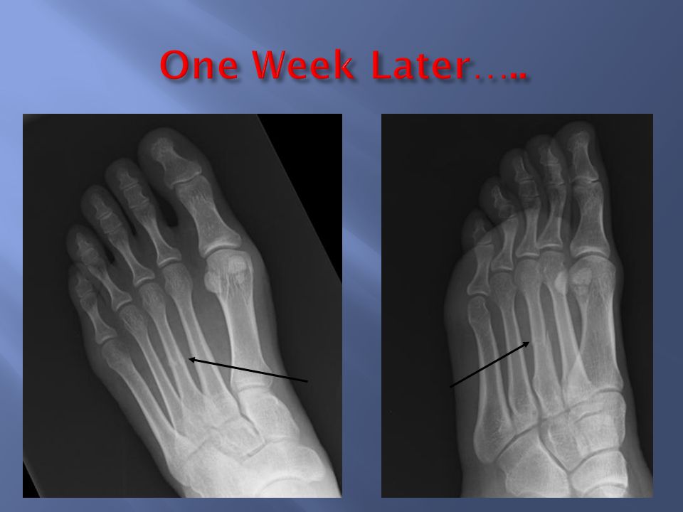
5th metatarsal stress fracture 3- Chronic Loading: resulting from application of lower magnitude forces e.g. stress fracture. Small number of repetitions with large load Large number of repetitions with usual load Intermediate combination of increased load and repetition

10
Stress fracture: imbalance between bone resorption and formation As there is abrupt increase in duration, intensity, frequency without adequate rest (re- modeling). Microfracture -> continued load -> stress fracture

13
1- Fracture due to Tension. 2- Fracture due to compression. 3-Fracture due to torsional loads. 4- Fracture due to bending. 5-Fracture due to combined loading.

14
1- Fracture due to Tension: Fracture due to pure tensile loading is uncommon in the long bone diaphysis. Fracture pattern is transverse fracture as the maximum tensile stress is generated at a transverse plane

15
Example : Radiograph of an injury produced by tensile forces imposed on the tibial tuberosity by the quadriceps musculature. (Courtesy of RB Hohn, DVM) in tension as a function of stress orientation.
 in tension as a function of stress orientation..")
16
2- Fracture due to compression: Fracture due to compression may occur at vertebra or long bones. a)In the vertebrae: A shortening of the vertebral bodies, In the long bone: cause the shear stresses to be high on the planes oblique to the longitudinal axis. Fracture occurs along these oblique angles where shear stresses are greatest.
In the vertebrae: A shortening of the vertebral bodies, In the long bone: cause the shear stresses to be high on the planes oblique to the longitudinal axis. Fracture occurs along these oblique angles where shear stresses are greatest..")
17
3- fracture due to bending: Due to bending load, the bone is subjected to two types of stresses (tensile and compressive loads). - At the tensile side (transverse fracture). - At the compressive side a small butterfly fracture results which is formed by propagation of two cracks on two oblique fracture planes.
. - At the compressive side a small butterfly fracture results which is formed by propagation of two cracks on two oblique fracture planes..")
18
Example: Radiograph of a short oblique long- bone fracture probably produced by bending forces imposed on the midshaft humerus.

19
4-Fracture due to torsional loads: Cross section of the cylinder loaded in torsion shows the shear stress distribution about the neutral axis. Shear stress increases as a function of distance from the neutral axis. Maximum shear stresses on the surface of a cylinder subjected to torsional forces occur on planes perpendicular and parallel to the neutral axis.

20
Fracture pattern: The fracture initiates as a small crack and then run in a spiral manner (spiral fracture) to maintain an angle of 45° as fracture occurs along the oblique plane of high tensile stress. Schematic illustration of a two- piece spiral fracture of the femur drawn from a radiograph of a clinical case.

21
Example: Radiograph of a spiral fracture of the humerus shows the spiral fracture surface and a fissure extending proximally along the line of maximum tensile stress.

22
5- fracture due to combined loading: Bone fracture seen clinically are usually caused by a complex loading situation. Many loading situations may combine together forming a fracture pattern. 1- example, combined bending and axial compressive loading: This will cause compressive stress at one side to be bigger than tensile stress at the opposite side.

23
due to combination between bending and axial compressive loads. Fracture pattern will be as in bending load but the butterfly fragments will be bigger in this case 2- example; combined torsional, bending and compressive forces : will cause spiral fracture at one side associated with transverse fracture at the opposite side and also combined with small oblique lesions.

24
a) In nature: Healing by callus. b) With operative intervention: (reduction + compression). During this operation, the bone fragments are first repositioned (reduced) in their normal alignment Primary bone healing. c) With operative intervention (nailing or external fixation). Healing by callus.
 in their normal alignment Primary bone healing. c) With operative intervention (nailing or external fixation). Healing by callus..")
25
According to the principle ‘’bone formed when needed and resorbed when not needed’’ So bones are formed in areas of higher stresses will form and those of lower stresses, bone density will decrease. Fracture callus bridges a gap between fractured bone ends, so the diameter will increase more than normal. As healing occurs, the callus becomes more mineralized and increases the material strength of the bone. When stress distribution returns to normal, the bone resorbs and resumes its normal geometry.

26
Relation between mechanical stresses and metabolic activity of connective tissue: Pure tensile and pure compressive stresses causes connective tissue to form bone and enhance fracture healing. Shear stresses causes connective tissue to form fibrous tissue. If the shear stress is limited, fracture callus will be formed. If it is severe, non union may result. Stresses from all directions causes connective tissues to form cartilage.

27
Clinically Upper limb Lower limb Adult 6-8 weeks 12-16 weeks Child3-4 weeks 6-8 weeks Radiologically Bridging callus formation Remodelling

Similar presentations

>")
.>")


 Bone fractures are classified by: –The position of the bone ends after fracture –The completeness.>")
>")


 fracture: involve.>")