Download presentation
Presentation is loading. Please wait.

1
Dr. Sufia Husain MBBS, MD, FRCPath.
The Uterine Corpus Dr. Sufia Husain MBBS, MD, FRCPath.

7
Acute endometritis Is most often related to intrautreine trauma e.g. after an abortion either spontaneous or induced, complications of pregnancy, medical instrumentation or intrauterine contraceptive devices. Is most often caused by Staphylococci, Streptococci. Others like N. gonorrhoeae, gram-negative bacilli and occasionally fungi and viruses can also cause infection.

8
Chronic endometritis Chronic endometritis is associated with Intrauterine contraceptive device use, pelvic inflammatory disease, and retained products of conception following an abortion or delivery. The etiologic agent is often not apparent and the patient is said to have non- specific chronic endometritis.

9
Chronic endometritis Patients present with irregular bleeding.
Histologically, the presence of plasma cells in the endometrium is diagnostic. The stromal cells become spindled and swirl around the glands. Sometimes granulomatous endometritis is noted in patients with tuberculosis.

10
Endometriosis This is the presence of ectopic endometrial glands and stroma outside the uterus. The lesions are usually found on the peritoneal surfaces of the reproductive organs and adjacent pelvic organs. The most frequent location is the ovary (approx. 50%) followed by the pouch of Douglas, uterine ligaments. Occasional sites include the cervix, vagina, perineum, bladder, large bowel and the umbilicus. Rare lesions are seen as far as small bowel, kidneys, lungs and brain. It has been reported in men. The sites involved have been the bladder, scrotum and prostate
 followed by the pouch of Douglas, uterine ligaments. Occasional sites include the cervix, vagina, perineum, bladder, large bowel and the umbilicus. Rare lesions are seen as far as small bowel, kidneys, lungs and brain. It has been reported in men. The sites involved have been the bladder, scrotum and prostate.")
11
Endometriosis is non-neoplastic.
Like the uterine endometrium it is responsive to the hormonal variations of the menstrual cycle. It is characterized by menstrual type bleeding at the site of the ectopic endometrium, resulting in blood filled areas (e.g.chocolate cysts).
.")
12
Endometriosis :Clinical appearances
Clinical presentation depends on the site of endometriosis. Dysmenorrhea, cyclic abdominal pain and dyspareunia are common symptoms. Usually there is severe menstrual-related pain. Often results in infertility. Endometriosis usually appears as multiple red or brown (due to hemosiderin) 1mm to 5mm nodules (some may form larger masses or cysts). Dense fibrous adhesions may surround the foci.
 1mm to 5mm nodules (some may form larger masses or cysts). Dense fibrous adhesions may surround the foci.")
13
Endometriosis Repeated hemorrhage into foci in the ovary with each menstrual cycle produces cysts, which contain inspissated, chocolate-brown material, called "chocolate cysts". Clinical behavior Benign with no malignant potential. May recur after surgical excision but the risk is low.

14
Adenomyosis This is defined as the presence of endometrial glands and stroma in the myometrium. The condition involves the posterior wall more often than the anterior wall. Clinical appearances It is associated with menorrhagia and severe dysmenorrhea. In about a third of patients there are no symptoms and the lesions are discovered accidentally.

15
Adenomyosis When extensive the lesions produce myometrial thickening with small yellow or brown cystic spaces containing fluid or blood. Clinical behavior. This is a benign condition with no known malignant potential that regresses after the menopause

16
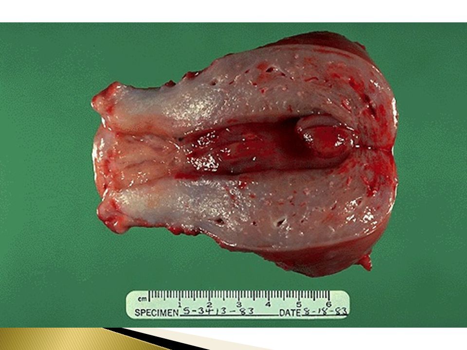
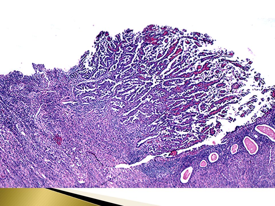
Endometrial polyp Is a localized benign overgrowth of endometrial tissue covered by epithelium. Endometrial polyps are most common in women between 40 and 50 years. The polyp may cause irregular bleeding. It may be broad-based and sessile, pedunculated or attached to the endometrium by a slender stalk. The size is variable from 1mm to a mass that fills the endometrial cavity. Occasionally a polyp may protrude through the external os.

17
Endometrial polyp Histology
Composed of glands of variable size and shape, fibrotic stroma and thick-walled blood vessels. Clinical behavior Endometrial polyps are benign with no malignant potential but sometimes malignant tumors may be found in them.

19
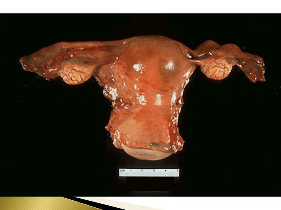
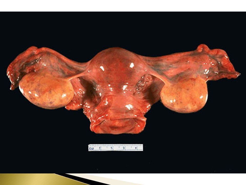
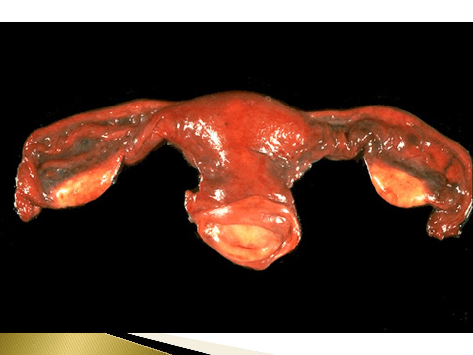
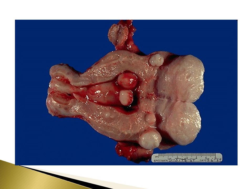
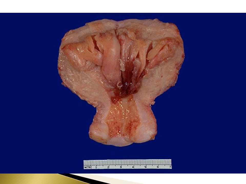
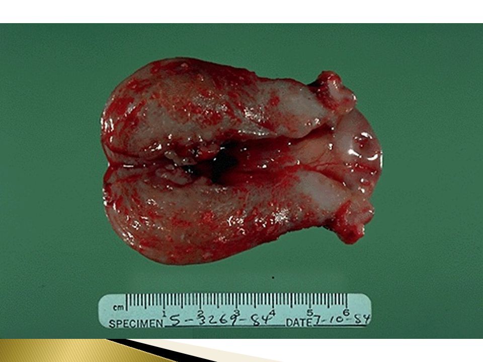
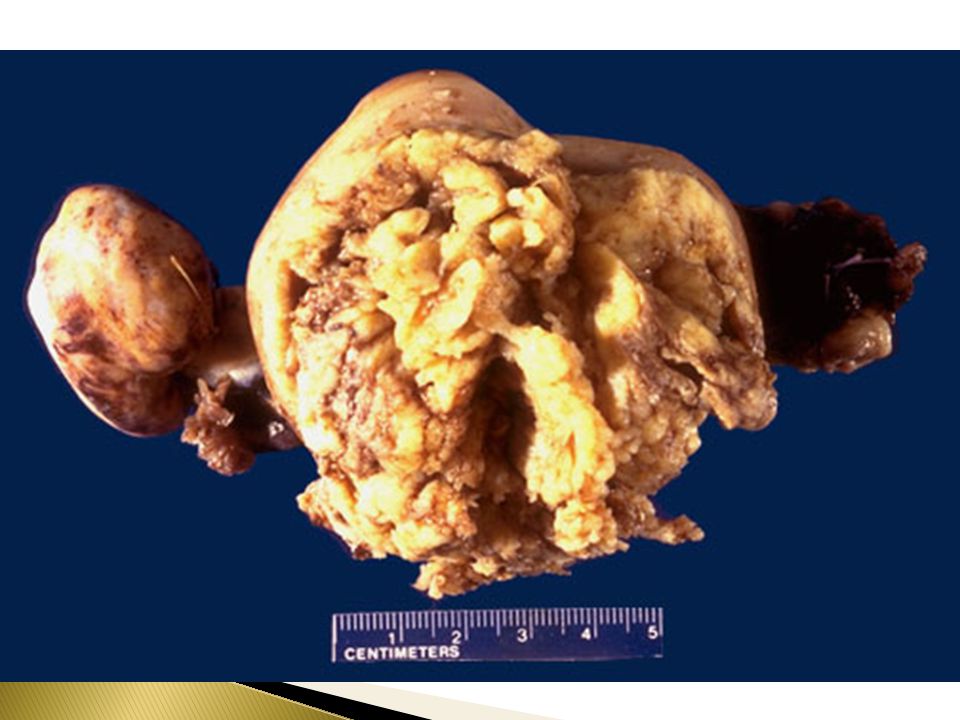
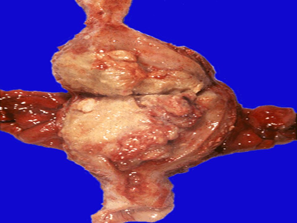
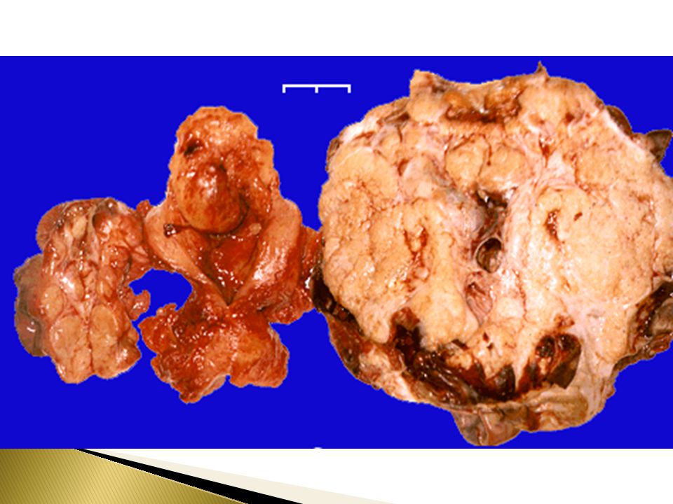
Leiomyoma (Fibroid) Leiomyoma is a benign tumor of smooth muscle origin. It is the most common neoplasm of the female genital tract and probably the most common neoplasm in women. Is more common in women of African lineage. Clinical and gross appearances Patients may present with irregular bleeding, pelvic pain, pelvic mass, infertility. The tumor is estrogen responsive and often increases in size during pregnancy and decreases in size during menopause. It can be single or multiple. Mostly it is multiple.

20
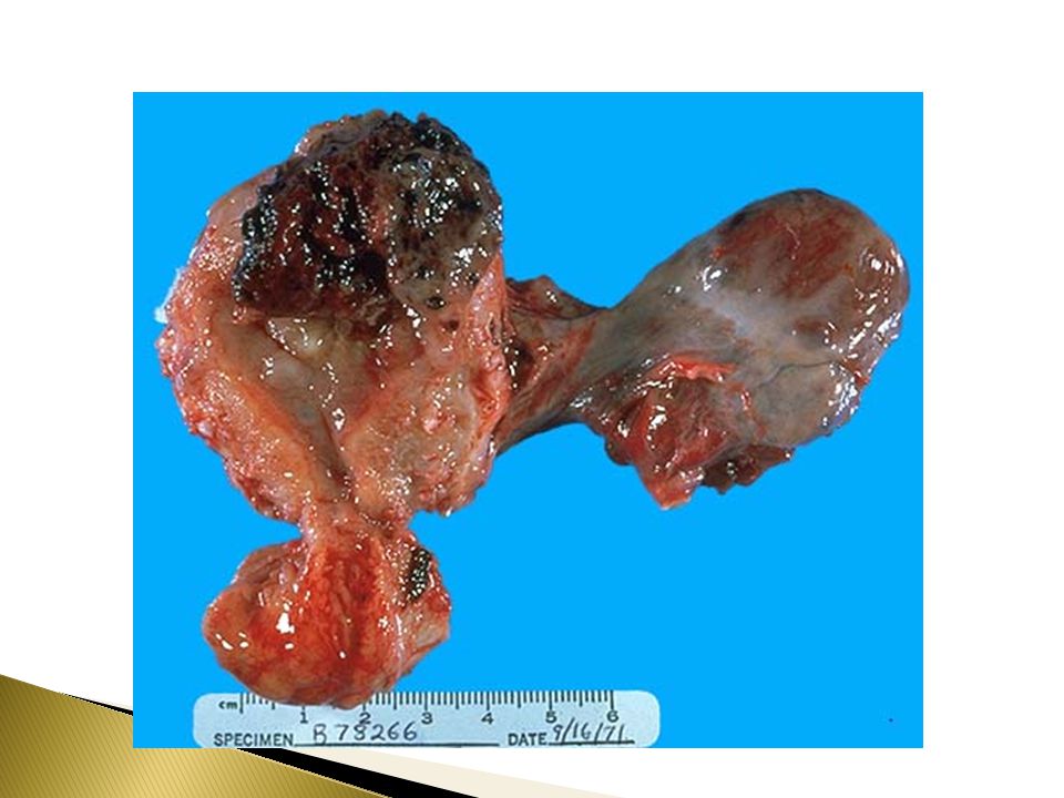
Leiomyoma (Fibroid) Leiomyoma may be located anywhere in the myometrium. Submucosal tumors are present immediately below the endometrium, may be pedunculated and occasionally protrude though the cervix. Intramural tumors, the most common, lie within the myometrium. Subserosal fibroids lie beneath the serosal covering of the uterus or are pedunculated and attached to the serosa. Pedunculated ones may undergo torsion and infarction or loose their connection to the uterus and become attached to another pelvic organ forming a "parasitic leiomyoma". Grossly : the tumors appear as well circumscribed, spherical, dense and firm-to-hard masses with whorled, tan-white cut surfaces.

21
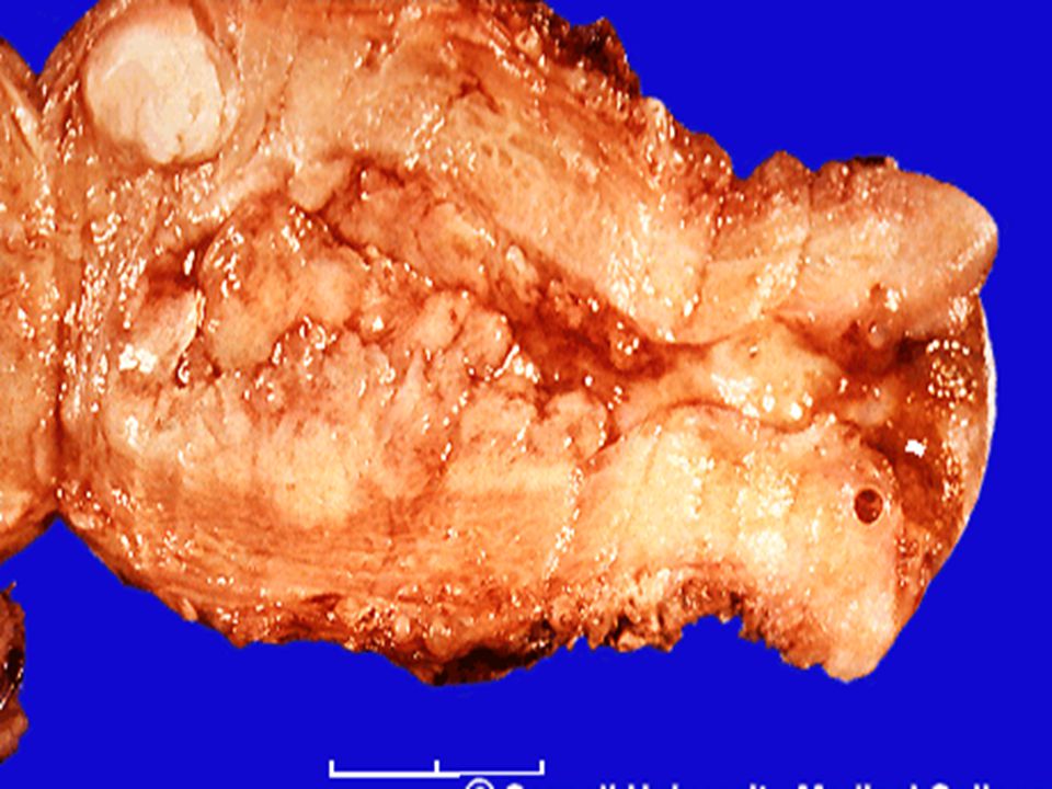
Leiomyoma. Note whorled cut surface

23
Histology Microscopically, there are interlacing bundles of smooth muscle cells with collagenous stroma between bundles

24
Leiomyoma composed of interlacing fibers of bland smooth muscle

25
Leiomyoma (Fibroid) Degenerative changes may be seen. These take the form of: Atrophy — the tumor reduces in size at menopause or after pregnancy following drop in estrogen level. Hyaline change (hyalinization) — Usually occurs as the tumor "ages". Myxoid and cystic change. Calcification — common in menopausal women. Septic with necrosis of the center due to circulatory inadequacy. Red degeneration— venous thrombosis and congestion with interstitial hemorrhage may occur, most commonly in pregnancy. This is usually accompanied by pain, which may produce a clinical picture of acute abdomen.
 — Usually occurs as the tumor ages . Myxoid and cystic change. Calcification — common in menopausal women. Septic with necrosis of the center due to circulatory inadequacy. Red degeneration— venous thrombosis and congestion with interstitial hemorrhage may occur, most commonly in pregnancy. This is usually accompanied by pain, which may produce a clinical picture of acute abdomen.")
26
Leiomyoma (Fibroid) Clinical behavior
This is a benign tumor with no appreciable malignant potential (incidence of malignant transformation is %). It may cause anemia from heavy bleeding, or urinary or bowel obstruction (subserosal or parasitic tumors) In pregnant women it may cause spontaneous abortion, precipitate labor, obstructed labor ,post partum hemorrhage (due to interference with uterine contraction), and red degeneration.
. It may cause anemia from heavy bleeding, or urinary or bowel obstruction (subserosal or parasitic tumors) In pregnant women it may cause spontaneous abortion, precipitate labor, obstructed labor ,post partum hemorrhage (due to interference with uterine contraction), and red degeneration.")
27
Endometrial Hyperplasia

28
Endometrial Hyperplasia
Endometrial hyperplasia refers to a process in which there is a proliferation of endometrial glands of irregular size and shape with an increase in gland/stroma ration compared to proliferative endometrium. Induced by persistent, prolonged estrogenic stimulation of the endometrium.

29
Endometrial Hyperplasia
The endometrial hyperplasia may progress to endometrial carcinoma. The development of cancer is based on the level and duration of the estrogen excess. The risk is depending on the severity of the hyperplastic changes and associated cellular atypia.

30
Endometrial Hyperplasia, causes
Causes of Endometrial Hyperplasia: A common cause is a succession of anovulatory cycles (failure of ovulation ). It may also be caused by excessive endogenously produced estrogen in -polycystic ovary syndrome including Stein- Leventhal syndrome, -granulosa cell tumors of the ovary -excessive ovarian cortical function (cortical stromal hyperplasia) Prolonged exogenous administration of estrogenic steroids without counter balancing progestins
. It may also be caused by excessive endogenously produced estrogen in. -polycystic ovary syndrome including Stein- Leventhal syndrome, -granulosa cell tumors of the ovary. -excessive ovarian cortical function (cortical stromal hyperplasia) Prolonged exogenous administration of estrogenic steroids without counter balancing progestins.")
31
Endometrial Hyperplasia ,Clinical
Milder forms of hyperplasia tends to occur in younger patients The great majority of mild hyperplasia regress , either spontaneously or after treatment . The more severe forms ,occur predominantly in peri- and postmenopausal women .This form has a significant premalignant potential. Patients usually present with abnormal uterine bleeding .

32
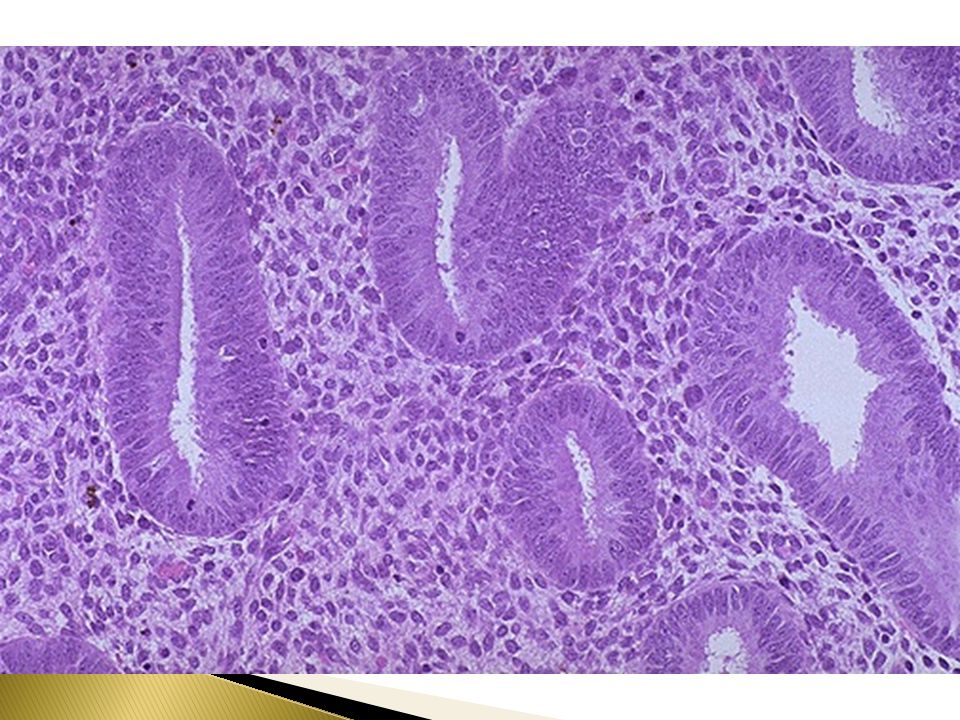
Endometrial Hyperplasia: classification
simple hyperplasia complex hyperplasia Both are classified as with or without atypia.

33
Endometrial Hyperplasia: microscopy
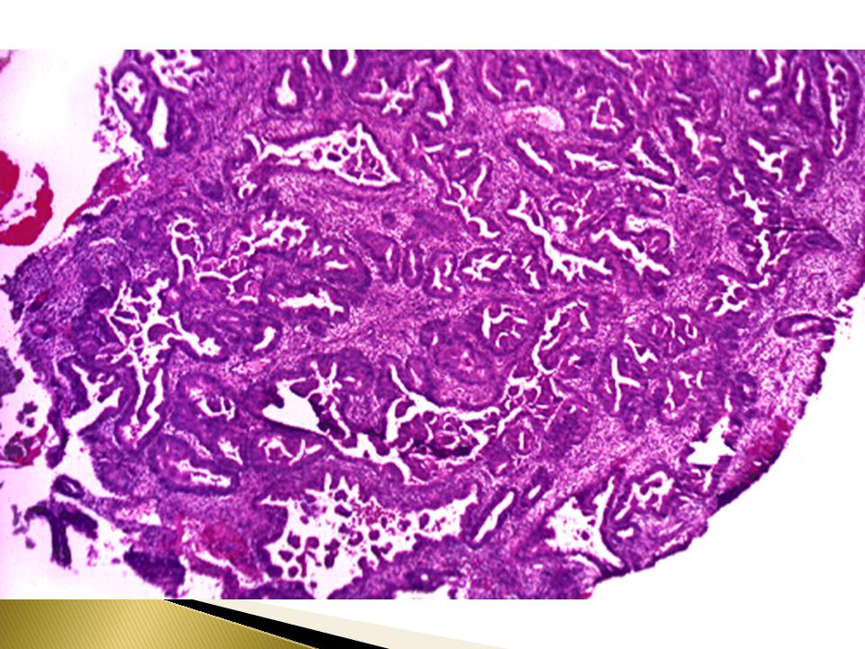
In general, the condition is characterized by proliferation of both glands and stroma. In spite of proliferation of both components, glandular overcrowding occurs. Endometrial hyperplasia is histologically classified according to: 1)Architecture as: simple or complex depending on the degree of glandular complexity and crowding, and 2)Cytologic features as: with or without atypia.
Architecture as: simple or complex depending on the degree of glandular complexity and crowding, and. 2)Cytologic features as: with or without atypia.")
34
Endometrial Hyperplasia: microscopy
Simple hyperplasia (cystic hyperplasia) — glands are cystically dilated and dispersed within abundant cellular stroma and give a "Swiss Cheese" appearance. Complex hyperplasia —characterized by complex crowded glands with papillary infoldings and irregular shapes.The crowded glands are back-to-back with very little intervening stroma. Both simple and complex hyperplasia can be with or without atypia.
 — glands are cystically dilated and dispersed within abundant cellular stroma and give a Swiss Cheese appearance. Complex hyperplasia —characterized by complex crowded glands with papillary infoldings and irregular shapes.The crowded glands are back-to-back with very little intervening stroma. Both simple and complex hyperplasia can be with or without atypia.")
35
Endometrial Hyperplasia: Clinical behavior and premalignant potential
Some endometrial hyperplasias revert to normal spontaneously or with medical treatment, others persist as hyperplasia, and a few progresses to endometrial adenocarcinoma. Generally, patients who have hyperplasia with atypia are more likely to develop carcinoma than those without atypia. The risks for developing adenocarcinoma in each are as follows: Complex atypical — 30% Simple atypical — 10% Complex — 3% Simple — 1% Atypical hyperplasia in postmenopausal women appears to have a higher rate of progression to adenocarcinoma.

36
Endometrial Hyperplasia, Risk Factors
Obesity Western diet Nulliparity Diabetes Mellitus Hypertension Hyperestrinism

38
Simple hyperplasia with dilated glands

39
Simple hyperplasia with dilated glands

40
Simple hyperplasia with dilated glands

41
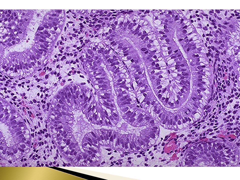
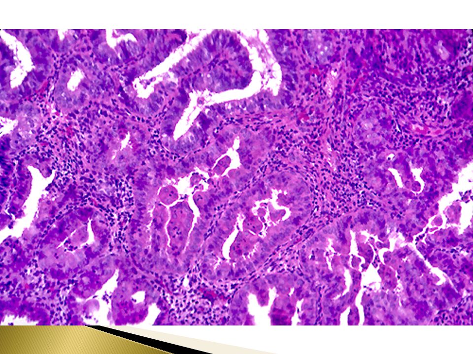
Complex atypical hyperplasia with back-to-back arrangement of glands and papillary tufting

45
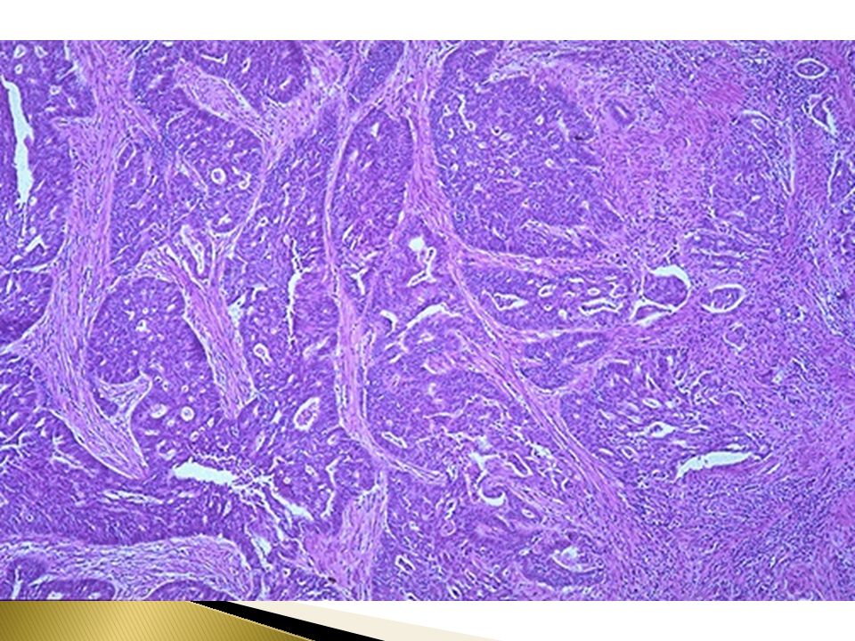
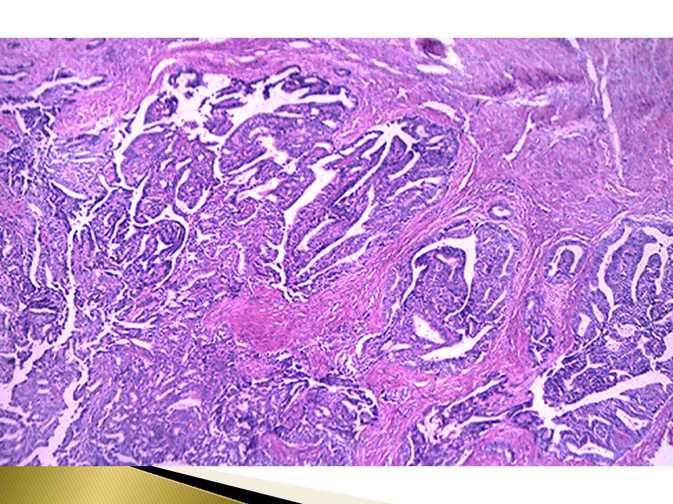
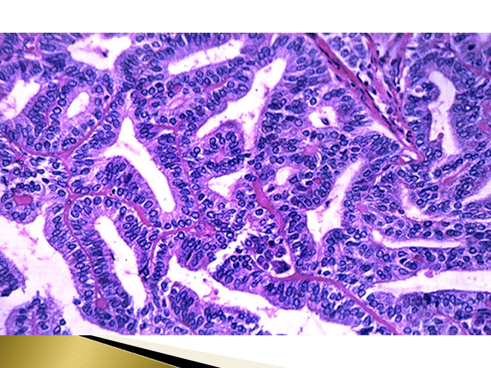
Endometrial adenocarcinoma
Epidemiology This is a common neoplasm in women. The incidence varies widely throughout the world. It is the most common invasive tumor of the female genital tract in the U.S. Worldwide, it is the fifth commonest cancer in women. Risk factors include obesity (women with upper body fat have 3 times the risk of women with lower body fat), estrogen therapy, nulliparity (as a result of infertility due to chronic anovulation), chronic anovulation, late menopause, hypertension, diabetes, tamoxifen therapy, and high socioeconomic status. The disease may follow atypical hyperplasia but may occur independently of it especially in older patients.
, estrogen therapy, nulliparity (as a result of infertility due to chronic anovulation), chronic anovulation, late menopause, hypertension, diabetes, tamoxifen therapy, and high socioeconomic status. The disease may follow atypical hyperplasia but may occur independently of it especially in older patients.")
46
Endometrial adenocarcinoma
Clinical presentation Most patients are between 50 and 59 years. Approximately 5% of affected women are under 40 years of age. Nearly 50% of these women who are under 40 years are nulliparous and more than 75% of them are obese. Endometrial adenocarcinoma manifests as abnormal vaginal bleeding. The tumor may grow in a diffuse or polypoid pattern. It often involves multiple areas of the endometrium

47
Endometrial Adenocarcinoma,morphology:
May closely resemble normal endometrium May be exophytic May be Infiltrative May be polypoid

48
Endometrial adenocarcinoma, histology:
The tumors are composed of glandular cells. The commonest type is Endometrioid adenocarcinoma. Other types include clear cell, adenosquamous, and papillary serous carcinoma. Endometrioid carcinoma may show areas of benign looking squamous epithelium. A tumor with such features is referred to as adenoacanthoma. In adenosquamous carcinoma both glandular and squamous components appear malignant.

49
Endometrial adenocarcinoma,prognosis:
Clinical behavior of endometrial adenocarcinoma depends on the histologic type, the grade (degree of differentiation) and the stage (extent of spread). Endometrioid carcinoma has a better prognosis than the other histologic types, which tend to occur at a higher stage. Staging is based on degree of myometrial invasion, cervical, adnexal and adjacent pelvic organ invasion, result of peritoneal fluid cytology and distant organ metastasis. Lymph node status is an important prognostic factor.
 and the stage (extent of spread). Endometrioid carcinoma has a better prognosis than the other histologic types, which tend to occur at a higher stage. Staging is based on degree of myometrial invasion, cervical, adnexal and adjacent pelvic organ invasion, result of peritoneal fluid cytology and distant organ metastasis. Lymph node status is an important prognostic factor.")
50
Endometrial Carcinoma,prognosis:
75% of patients present with stage I disease and these have 95% 5-year survival. The tumors associated with unopposed estrogen tend to have low histologic grade and clinical stage, hence tend to have better prognosis. These usually occur in young women. 20% of endometrial carcinoma there is no association with hyperestrinism or preexisting hyperplasia ,these cancers tend to occur late in life and have a poor prognosis.

51
Endometrial Carcinoma , Grading and staging
Grading is from 1 to 3 Staging is from 1 to 4 Stage 1 : Confined to uterus corpus Stage 2 : Cervix involvement Stage 3 : beyond the uterus ,but within the true pelvis Stage 4 : Distant metastasis/extrapelvic extension.

62
Gestational Trophoblastic Disease
Includes disorders charecterized by degenerative or neoplastic changes of trophoblastic tissue which is normally seen in the placental tissue. Gestational trophoblastic disease (GTD) can be benign or malignant. Majority of GTD cases are noncancerous.
 can be benign or malignant. Majority of GTD cases are noncancerous.")
63
Most forms of gestational trophoblastic disease can be cured with prompt management. Surgery and chemotherapy are the most common forms of treatment. Most women who have had gestational trophoblastic disease can have normal pregnancies later.

64
Gestational Trophoblastic Disease
Diagnosis: A blood test for the human chorionic gonadotropin (HCG) hormone is most commonly used to diagnose GTD. Serum HCG is markedly increased is GTD. Serum HCG is also elevated in normal and ectopic pregnancy, choricarcinoma and germ cell tumor. Serum hCG levels continue to rise after 14th week as opposed to drop in normal gestation
 hormone is most commonly used to diagnose GTD. Serum HCG is markedly increased is GTD. Serum HCG is also elevated in normal and ectopic pregnancy, choricarcinoma and germ cell tumor. Serum hCG levels continue to rise after 14th week as opposed to drop in normal gestation.")
65
Gestational Trophoblastic Disease
Histologically, it is classified into Hydatidiform mole (complete and incomplete) Invasive mole (chorioadenoma destruens) Choriocarcinoma Placental site trophoblastic tumor (PSTT).
 Invasive mole (chorioadenoma destruens) Choriocarcinoma. Placental site trophoblastic tumor (PSTT).")
66
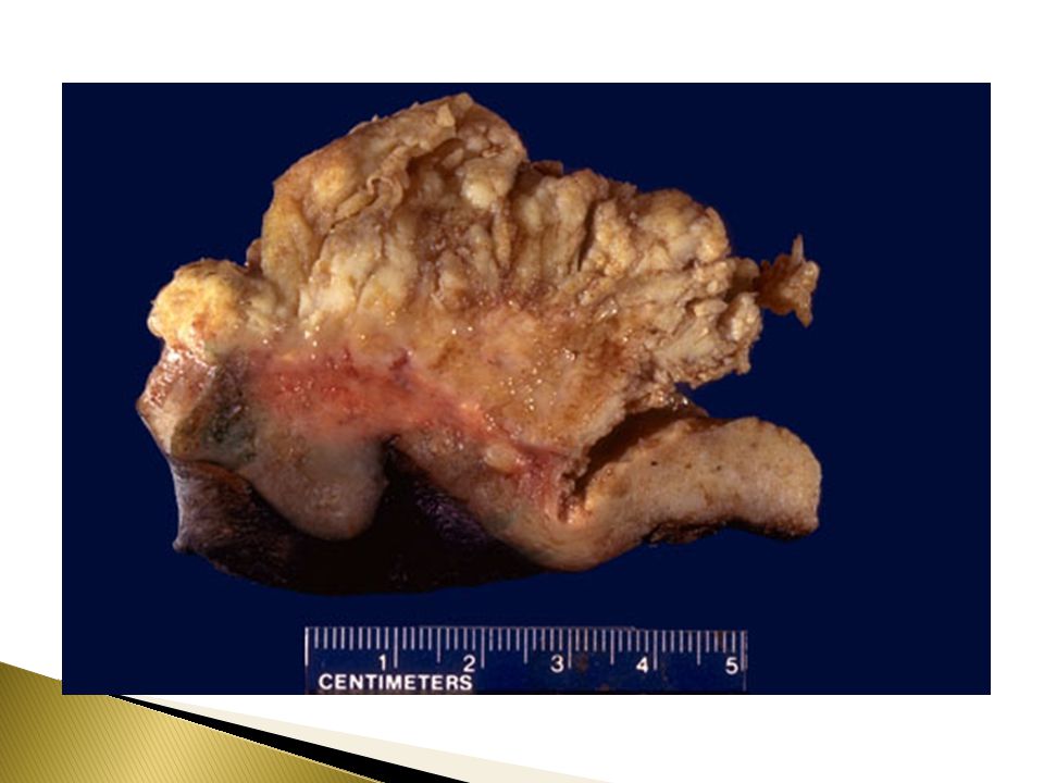
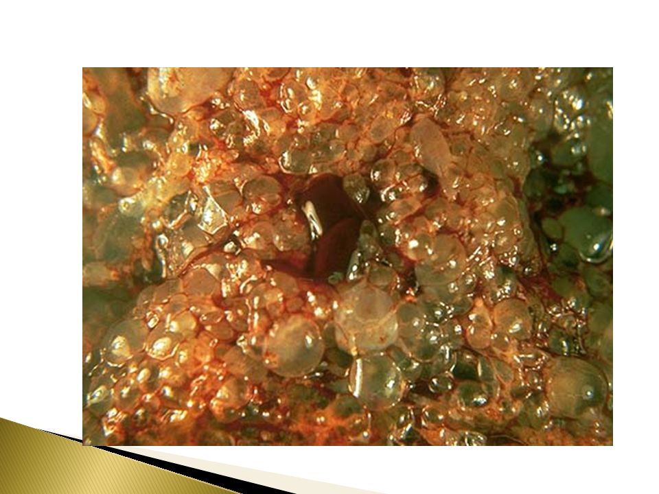
Hydatidiform Mole In it there is enlarged and odematous placental villi filling the lumen of the uterus. Grossly it looks like a bunch of grapes Symptoms: Patients present with increased abdominal swelling (rapid increase in uterine size) mistaken for normal pregnancy but the uterus is disproportionately large for stage of pregnancy. In addition patient has some vaginal bleeding, severe nausea and vomiting.
 mistaken for normal pregnancy but the uterus is disproportionately large for stage of pregnancy. In addition patient has some vaginal bleeding, severe nausea and vomiting.")
67
Hydatidiform Mole Caused by abnormal gametogenesis and fertilization.
There are 2 types of hydatidiform mole (HM). Complete HM Partial HM
. Complete HM. Partial HM.")
68
Complete HM Defintion: Genetically abnormal placenta with hyperplastic trophoblast, without fetus or embryo. Uterus is distended and filled with swollen/large villi. No embryo, or fetal tissue is present. There is elevated HCG levels. On chromosomal analysis there are 46 chromosome (2 haploid sets), 46XX karyotype and all the chromosomes come from the male/paternal side.
, 46XX karyotype and all the chromosomes come from the male/paternal side.")
69
Complete HM Treatment : Evacuation of uterus by curettage, chemotherapy. With appropriate therapy cure rate is very high spontaneous regression in 81% 17% developed an invasive mole 2% developed choriocarcinoma

71
Partial Mole (PM) Definition: PM is a genetically abnormal placenta with a mixture of large and small villi with slight hyperplasia of the trophoblast, filling the uterus. Embryonal/fetal tissue is present. Grossly large vesicular chorionic villi mixed with normal-appearing villi. .

72
Partial Mole (PM) It makes up 15–35% of all moles
Uterine size usually small or appropriate for gestational age Serum hCG levels elevated but not as high as complete mole. Chromosomal analysis of partial moles shows 69 chromosomes (i.e. 3 haploid sets also called as triploidy), XXY in which 2 haploids are paternal (from the male) and one is maternal
, XXY in which 2 haploids are paternal (from the male) and one is maternal.")
73
Partial Mole (PM) Prognosis
Risk for development of choriocarcinoma very low. Only 2-3% become malignant. Follow-up is mandatory.

74
Invasive Mole Definition: Hydatidiform mole, generally of the complete type, in which villi penetrate deeply in the myometrium and/or its blood vessels. It occurs in about 15% of complete moles and rarely in partial mole. Can cause hemorrahge and uterine perforation.

75
Choriocarcinoma Definition: Malignant tumor derived from normal or abnormal placental tissue, composed of a proliferation of malignant cytotrophoblast and syncytiotrophoblast, without villi formation. It is an aggressive malignant neoplasm. It is characterized by highly increased serum concentration of HCG. Choriocarcinomas are aneuploid.

76
Choriocarcinoma It spreads early via blood to the lungs and other organs. Responds well to chemotherapy About half the choriocarcinoma are preceded by complete hydatidiform mole . Others are preceded by partial mole (very unusual), abortion,ectopic pregnancy and occasionally normal term pregnancy.
, abortion,ectopic pregnancy and occasionally normal term pregnancy.")
77
Choriocarcinoma

Similar presentations


 ”.>")


.>")

