Download presentation
Presentation is loading. Please wait.

1
Structure Stains (Spore & Capsule Stains) Abdelraheem BA
 Abdelraheem BA")
3
Some bacteria have two forms of life: ◦ Active vegetative cells. Under normal conditions. ◦ Inactive cells called spores. When environmental conditions become unfavorable. Examples of such bacteria: ◦ Anaerobic genus Clostridium. ◦ Aerobic genus Bacillus.

4
Under unfavorable conditions cells undergo sporogenesis; ◦ Giving rise to an extra intracellular structure (Endospore). ◦ Endospore is surrounded be three layers: Outermost spore coat. Intermediate spore wall. Innermost spore membrane. ◦ Endospore surrounds the matrix (DNA, RNA, ribosomes… etc.)
.")
5
As conditions worsen; ◦ Endospore is released, becoming independent spore. Such spores are resistant to the damaging effect of: ◦ Excessive heat. ◦ Freezing. ◦ Radiations. ◦ Chemical agents (Including stains).
..")
6
When environmental conditions become favorable; ◦ The free spore germinates to a vegetative cell by the rupture of spores. ◦ This process is called germination. NOTE: ◦ Germination and sporogenesis are not means of reproduction. ◦ They are means of survival.

8
Shape: ◦ Spherical Vs Oval. ◦ Swollen. Location: ◦ Terminal, sub-terminal or central. Figure: (a) Oval & Terminal. (b) Spherical & Sub-terminal. (c) Oval & Central. (d) Spherical & Terminal (Swollen). (e) Oval & Terminal (Swollen). (f) Oval & Central (Swollen).
 Oval & Terminal. (b) Spherical & Sub-terminal. (c) Oval & Central. (d) Spherical & Terminal (Swollen). (e) Oval & Terminal (Swollen). (f) Oval & Central (Swollen)..")
9
C. botulinum : ◦ Swollen oval sub-terminal endospore. C. perfringens : ◦ Large oval central or sub-terminal endospore. C. tetani : ◦ Swollen spherical terminal “drumstick” endospore. Bacillus spp. ◦ Including B. anthracis, B. subtilis, B. cereus). ◦ All have non-swollen oval central endospores.
. ◦ All have non-swollen oval central endospores..")
10
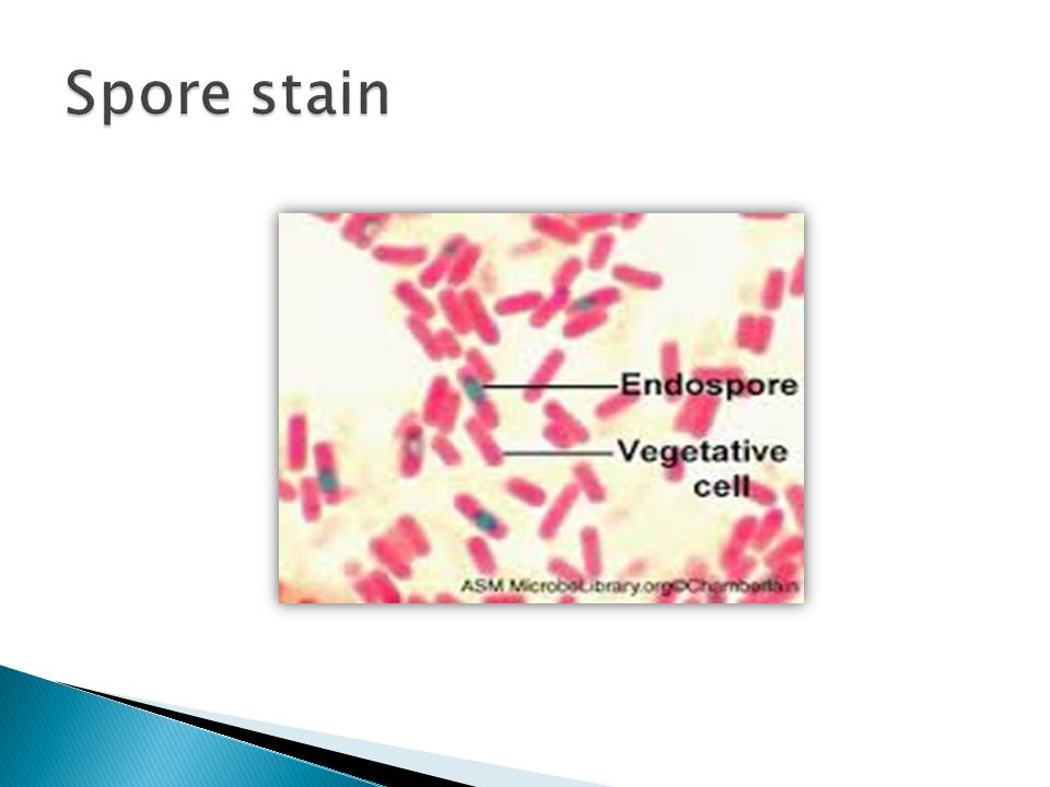
Primary stain: ◦ Malachite green. ◦ Due to their impervious coats, heat must be applied. Don’t allow the stain to evaporate, so add stain as needed. ◦ Now: Both, spores and vegetative cells are green. Decolorizer: ◦ Tap water. ◦ Spores cannot be decolorized by tap water (remain green). ◦ Vegetative cells become colorless. Counter stain: ◦ Safranin. ◦ Spores remain green. ◦ Vegetative cells become red.
. ◦ Vegetative cells become colorless. Counter stain: ◦ Safranin. ◦ Spores remain green. ◦ Vegetative cells become red..")
12
Definition: ◦ A gelatinous outer layer secreted by the cell, surrounding and adhering to cell wall. Function: ◦ Protection of bacteria against phagocytic activity of host cells. Chemical composition: ◦ Polysaccharide. ◦ Glycoprotein, or ◦ Polypeptide.

13
Principle: ◦ Indirect stain. ◦ Using an acidic stain; like Eosin or Nigrosin. Procedure: Place a small drop of eosin close to one end of a glass slide. Using a sterile technique, place a loopful of bacteria into the drop of eosin, mix well. With the edge of a second slide (held at 30º), push the mixture to form a bacterial smear. Air dry (Don’t heat). Examine under microscope.
, push the mixture to form a bacterial smear. Air dry (Don’t heat). Examine under microscope..")
Similar presentations


 Prokaryotic 2) Single celled (some say multicellular) 3) Microscopic in size 4) Cell Wall usually present (varies, but contains.>")


 the spore is formed inside the parent vegetative cell – hence the name „endospores“>")


>")


 Abdelraheem BA>")


