Download presentation
Presentation is loading. Please wait.

1
Sorting Through the Tangles: Alzheimer’s Disease
SHP – Neurobiology of Development and Disease

2
Alzheimer’s Disease Early symptoms feature memory loss (amnesia) beginning with minor forgetfulness which intensifies with progression of the disease. Deficits over time spread to processes including: Coordinated motor function (apraxia) Language (aphasia) Recognition of familiar people (agnosia) Variety of loss in prefrontal lobe processes
 beginning with minor forgetfulness which intensifies with progression of the disease. Deficits over time spread to processes including: Coordinated motor function (apraxia) Language (aphasia) Recognition of familiar people (agnosia) Variety of loss in prefrontal lobe processes.")
3
Discovery: Symptoms of the disease were initially described by Emil Kraepelin. In 1907 Alois Alzheimer (Kraepelin’s student) characterizes the clinical case of a middle-aged woman suffering from memory loss and cognitive defects. She was unreasonably suspicious of her husband, memory declined, hid objects in her home, and felt as if someone were trying to kill her. She was interned in a psychiatric hospital where she died 5 years later. Autopsy of patient reveals classic AD neuropathology: neurofibrillary tangles and senile plaques in the neocortex and hippocampus.
 characterizes the clinical case of a middle-aged woman suffering from memory loss and cognitive defects. She was unreasonably suspicious of her husband, memory declined, hid objects in her home, and felt as if someone were trying to kill her. She was interned in a psychiatric hospital where she died 5 years later. Autopsy of patient reveals classic AD neuropathology: neurofibrillary tangles and senile plaques in the neocortex and hippocampus.")
4
Epidemiology: Most common cause of dementia in the elderly.
Effects 7% of people older than 65 and ~40% above 80. As baby boomer generation become seniors, the incident of this disease is expected to triple

5
Progression of the AD

6
The Ensuing Damage Damage seems to be selective to certain parts of the brain and some cell types are more vulnerable than others. Most obvious ultrastructural changes are a shrinking of hippocampus, expansion of the ventricles and sulcus enlargement (or gyrus shrinkage).
.")
7
Cellular and Local Death

8
Areas most susceptible
Neocortex and enterrhinal cortex are the most severely damaged – primary a loss of excitatory large glutaminergic pyramidal neurons and interneurons. Hippocampus – pyramidal neurons are more vulnerable and damage focuses on CA1 and CA2 region. Cholinergic neurons in nucleus basalis, medial spetal nucleus, and diagonal band of Broca are destroyed.

9
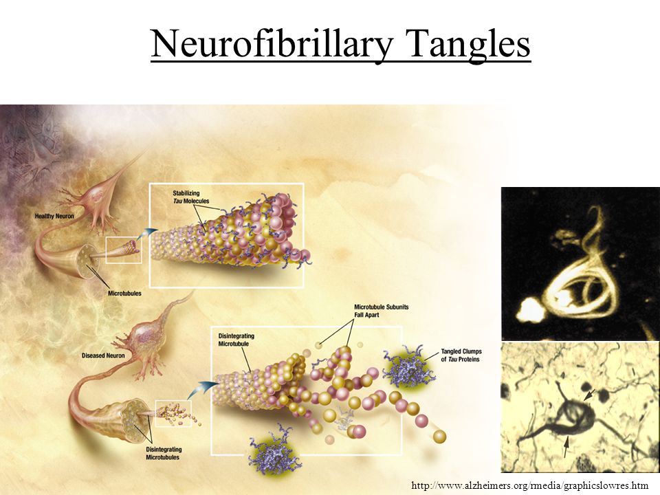
Neurofibrillary Tangles
10
5 Principle Genetic Risks:
Mutation in the amyloid precursor protein (App) (chromosome 21) Mutation sin the presenilin 1 gene (chromosome 14) Mutation in the presenilin 2 gene (chromosome 1) Alleles for the ApoE gene (chromosome 19) Potential mutation or polymorphism in gene on chromosome 12 that encodes alpha-2 macroglobulin
 (chromosome 21) Mutation sin the presenilin 1 gene (chromosome 14) Mutation in the presenilin 2 gene (chromosome 1) Alleles for the ApoE gene (chromosome 19) Potential mutation or polymorphism in gene on chromosome 12 that encodes alpha-2 macroglobulin.")
11
Amyloid Precursor Protein (APP)
Transmembrane glycoprotein that is 695, 751, or 770 amino acids long Localized to dendrites, soma, and axons of neurons (neuronal APP is thought to be the source of most of the amyloid-beta. It is internalized and processed by a number of proteases, releasing a number of species of peptide fragments 1-40, 1-42, and 1-43. A-beta 1-41/43 have been shown to be more likely to oligomerize into amyloid plaques. 1-42 fragment appears to be neurotoxic for unknown reasons

12
Amyloid Theory Amyloid: histological name for fibrillar peptides arranged as beta-sheets in aggregates that are refractive in polarlized light and Congo Red stain Extracellular APP fragments can associate into plaques around neurons and cause degeneration and death in surrounding cells.

13
APP undergoes extensive Proteolytic Processing
Primary cleavage by Beta-secretase appears to Be required for amyloid-forming peptide fragments

14
APP Alleles Various missense mutations lie in the APP gene in different populations and are inherited ~10% of individuals with these mutations develop clinical AD symptoms by age 50 Inherited in autosomal dominant manner mutation in the 717 position increase 1-42 and 1-43 levels and is especially toxic and amyloid forming

15
Human APP expressed in mice
APP-mut Wt Expression of these human mutants in mice replicate the neuropathological features, degeneration and death. Astrogliosis can be seen (in d) by GFAP staining Amyloid formation is highlighted by thioflavin S (Congo Red) These mice also have impaired memory and learning
 by GFAP staining. Amyloid formation is highlighted by thioflavin S (Congo Red) These mice also have impaired memory and learning.")
16
AD Genes in Development
Presenilin is homologous to a C elegans gene called sel-1 that is required for cell lineage decisions in neural development The physiological role of APP is still unclear. Presenilins have been shown to be required for Notch signaling in Drosophila for transmembrane proteolytic cleavage

17
Notch Pathway Notch is a transmembrane receptor that binds a transmembrane ligand (Delta/Serrate) This binding causes proteolytic cleavage of Notch, which diffuses to the nucleus and regulates a cell transcription program that blocks neural fate.

18
APP Destroys Synaptic Integrity
PSD95 postsynaptic adaptor protein is dramatically diminished in APP mutant neurons Blockade of APP cleavage by gamma-secretase inhibitor DAPT blocks this effect.

19
Mutant APP Blocks Integration of Glutamate Receptors
In neurons expressing the mutant form of APP, the glutamate receptor (GluR1) cannot make it to the cell membrane Inhibition of gamma-secretase (DAPT) blocks this effect
 cannot make it to the cell membrane. Inhibition of gamma-secretase (DAPT) blocks this effect.")
20
Treatment Clinical treatment for AD only focus on the symptoms but can do nothing to the etiology of the disease. The cholinergic signaling defects can be overcome by acetylcholinesterase inhibitors Glutamate excitotoxicity seem to be involved to come extent so treatment with NMDA antagonists are often used. The latest approach as been developing vaccines and immunotreatments to target the most likely cause of the degeneration: APP amyloid fragments.

21
Three Approaches of Immunotherapy

22
Mechanism of Antibody function
Small percentage of antibodies can cross the blood-brain barrer and bind to extracellular A-beta monomers, oligomers, and amyloid plaques These antibodies then recruit immune cells to clear the “wreckage” of damaged tissue and proteins themselves or block protein-protein interaction directly.

23
Synopsis of Immunotherapies for AD

Similar presentations
 A 42 deposits subtle.>")


>")


 observations/experiences.>")


