Download presentation
Presentation is loading. Please wait.

1
BONE

2
Support. Bone is hard and rigid; cartilage is flexible yet strong. Cartilage in nose, external ear, thoracic cage and trachea. Ligaments- bone to bone Protection. Skull around brain; ribs, sternum, vertebrae protect organs of thoracic cavity Movement. Produced by muscles on bones, via tendons. Ligaments allow some movement between bones but prevent excessive movement Storage. Ca and P. Stored then released as needed. Fat stored in marrow cavities Blood cell production. Bone marrow that gives rise to blood cells and platelets Skeletal System Functions

3
6-3 Bone Histology Bone matrix. – Organic: collagen and proteoglycans – Inorganic: hydroxyapetite. CaPO 4 crystals Bone cells (see following slides for particulars) – Osteoblasts – Osteocytes – Osteoclasts – Stem cells or osteochondral progenitor cells Woven bone: collagen fibers randomly oriented Lamellar bone: mature bone in sheets Cancellous bone: trabeculae Compact bone: dense
 – Osteoblasts – Osteocytes – Osteoclasts – Stem cells or osteochondral progenitor cells Woven bone: collagen fibers randomly oriented Lamellar bone: mature bone in sheets Cancellous bone: trabeculae Compact bone: dense.")
4
Osteoblasts Synthesize organic components of matrix (collagen type I, proteoglycans, glycoproteins.) Collagen forms osteoids: strands of spiral fibers that form matrix Influence deposit of Ca++, PO4.
 Collagen forms osteoids: strands of spiral fibers that form matrix Influence deposit of Ca++, PO4.")
5
Osteoblasts

6
Osteocytes Mature bone cells that sit in lacunae Gap junctions between osteocytes provide nutrition (15 cells in a row) Maintain bony matrix; long lived cells Stimulated by calcitonin; inhibited by PTH
 Maintain bony matrix; long lived cells Stimulated by calcitonin; inhibited by PTH")
7
Osteocyte with Cytoplasmic Extensions

8
Osteocytes with Canaliculi Photomicrograph of dried bone ground very thin. The lacunae and canaliculi filled with air deflect the light and appear dark, showing the communication between these structures through which nutrients derived from blood vessels flow. Medium magnification.

9
Osteoclasts Derived from monocytes; engulf bony material Active osteoblasts stimulate osteoclast activity Large, branched, motile cells Secrete enzymes that digest matrix

10
Osteoclasts

11
Bone Resorption

12
Bone Anatomy Diaphysis Metaphysis Epiphysis – Prox/Dist Epiphyseal line Periosteum Compact cortical bone Spongy bone Articular Cartilage Medullary cavity Marrow Nutrient artery

13
Types of Bone tissue: Woven bone – Irregular – immature – fetus Lamellar bone – regular – mature. – Circumferential – Concentric – Interstitial – Trabecular Compact & Spongy - lamellar bone Enchondral / intramembranous formation.

14
6-14 Woven and Lamellar Bone Woven bone. Collagen fibers randomly oriented. – Formed During fetal development During fracture repair Remodeling – Removing old bone and adding new – Woven bone is remodeled into lamellar bone Lamellar bone – Mature bone in sheets called lamellae. Fibers are oriented in one direction in each layer, but in different directions in different layers for strength.

15
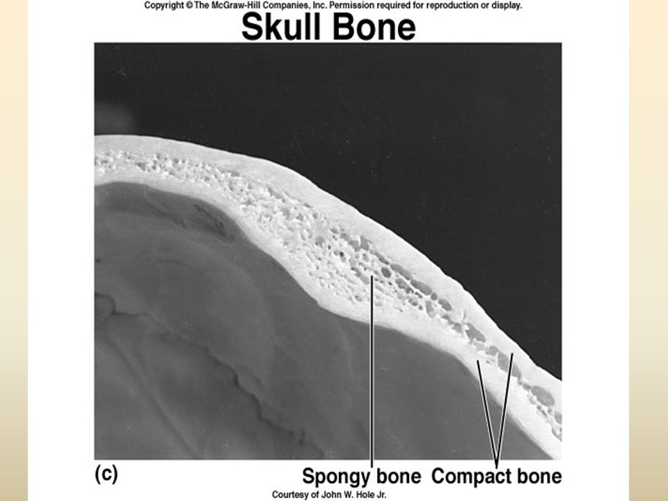
6-15 Cancellous (Spongy) Bone Trabeculae: interconnecting rods or plates of bone. Like scaffolding. – Spaces filled with marrow. – Covered with endosteum. – Oriented along stress lines

16
6-16 Compact Bone Central or Haversian canals: parallel to long axis Lamellae: concentric, circumferential, interstitial Osteon or Haversian system: central canal, contents, associated concentric lamellae and osteocytes Perforating or Volkmann’s canal: perpendicular to long axis. Both perforating and central canals contain blood vessels. Direct flow of nutrients from vessels through cell processes of osteoblasts and from one cell to the next.

17
Compact Bone:

19
6-19 Bone Development: Two Types Intramembranous ossification – Takes place in connective tissue membrane Endochondral ossification – Takes place in cartilage Both methods of ossification – Produce woven bone that is then remodeled – After remodeling, formation cannot be distinguished as one or other

20
6-20 Intramembranous Ossification Takes place in connective tissue membrane formed from embryonic mesenchyme Forms many skull bones, part of mandible, diaphyses of clavicles When remodeled, indistinguishable from endochondral bone. Centers of ossification: locations in membrane where ossification begins Fontanels: large membrane-covered spaces between developing skull bones; unossified

21
6-21 Intramembranous Ossification

22
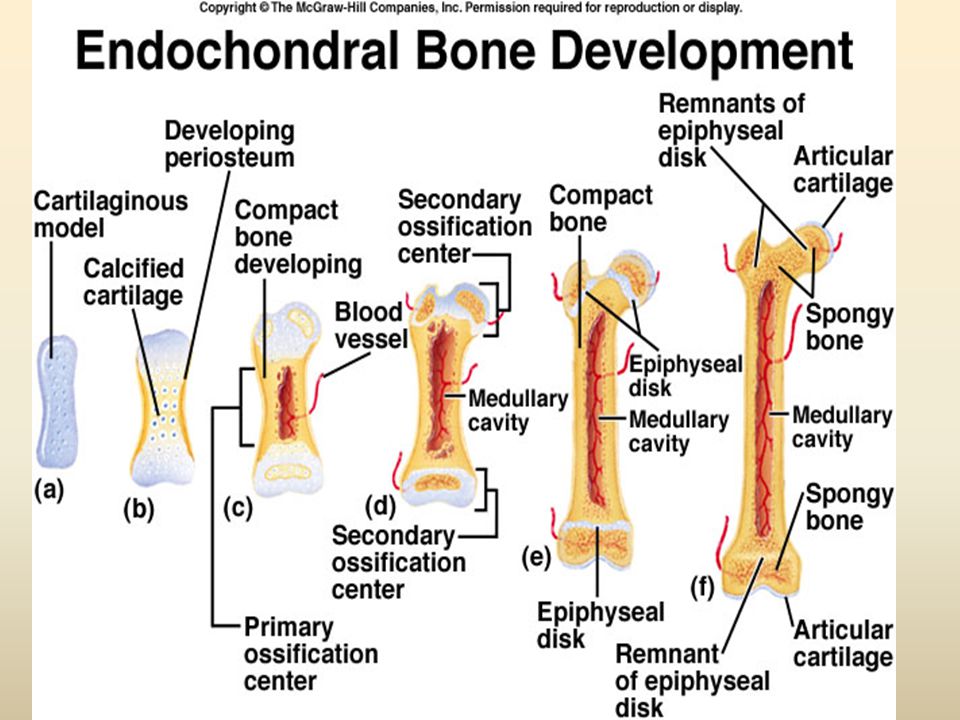
6-22 Endochondral Ossification Bones of the base of the skull, part of the mandible, epiphyses of the clavicles, and most of remaining bones of skeletal system Cartilage formation begins at end of fourth week of development Some ossification beginning at about week eight; some does not begin until 18-20 years of age

24
6-24 Endochondral Ossification

25
6-25 Endochondral Ossification

26
6-26 Growth in Bone Length

27
6-27 Factors Affecting Bone Growth Size and shape of a bone determined genetically but can be modified and influenced by nutrition and hormones Nutrition – Lack of calcium, protein and other nutrients during growth and development can cause bones to be small – Vitamin D Necessary for absorption of calcium from intestines Can be eaten or manufactured in the body Rickets: lack of vitamin D during childhood Osteomalacia: lack of vitamin D during adulthood leading to softening of bones – Vitamin C Necessary for collagen synthesis by osteoblasts Scurvy: deficiency of vitamin C Lack of vitamin C also causes wounds not to heal, teeth to fall out

28
6-28 Factors Affecting Bone Growth, cont. Hormones – Growth hormone from anterior pituitary. Stimulates interstitial cartilage growth and appositional bone growth – Thyroid hormone required for growth of all tissues – Sex hormones such as estrogen and testosterone Cause growth at puberty, but also cause closure of the epiphyseal plates and the cessation of growth

29
6-29 Calcium Homeostasis Bone is major storage site for calcium The level of calcium in the blood depends upon movement of calcium into or out of bone. – Calcium enters bone when osteoblasts create new bone; calcium leaves bone when osteoclasts break down bone – Two hormones control blood calcium levels- parathyroid hormone and calcitonin.

30
6-30 Calcium Homeostasis

31
Bone Repair

Similar presentations


 Nestor T. Hilvano, M.D., M.P.H.>")

