Download presentation
Presentation is loading. Please wait.

1
Tissues Cells work together in functionally related groups called tissues How is this done? Attachments Communication Types of tissues: 1. Epithelial – lining and covering 2. Connective – support 3. Muscle – movement 4. Nervous – control

2
Epithelial Tissue – General Characteristics & Functions Covers a body surface or lines a body cavity & forms most glands Functions of epithelium: Protection- skin Absorption, secretion, and ion transport- pancreatic cells Filtration- stomach, intestine Forms slippery surfaces- lungs

3
Special Characteristics of Epithelia Cellularity Mostly cells that are in close contact (tightly packed)… thus they form effective barriers Specialized contacts Specialized cell contacts bind adjacent cells together (helps w/ communication) Location- body surfaces, lining of hollow organs, forms glands Outside surface of the body Lining of digestive, respiratory and urogenital systems Heart and blood vessels Linings of many body cavities
… thus they form effective barriers Specialized contacts Specialized cell contacts bind adjacent cells together (helps w/ communication) Location- body surfaces, lining of hollow organs, forms glands Outside surface of the body Lining of digestive, respiratory and urogenital systems Heart and blood vessels Linings of many body cavities")
4
Special Characteristics of Epithelia Surfaces Basal, apical and lateral Supported by connective tissue At the basal surface, epithelial tissue and connective tissue form the basement membrane

5
Special Characteristics of Epithelia Avascular No blood vessels; nutrients must diffuse Regenerative epithelial tissues have a high capacity for regeneration (mitosis!)
")
6
Recap. Identify two special characteristics about epithelial tissue? Identify two places epithelial tissue can be found.

7
Bell Work What surface of an epithelial cell opens up to the outside of the opening of an internal space? What surface connects to the side of another cell?

8
Basal Surface What is it? Where is it? Non-cellular, non-living supporting sheet two layers (basal lamina & reticular lamina) Composed of: proteins secreted by the epithelial cells Function: Selective filter selectively permeable to molecules from capillaries Point of attachment and support for overlying epithelial tissues (regenerating cells migrate from this point)
 Composed of: proteins secreted by the epithelial cells Function: Selective filter selectively permeable to molecules from capillaries Point of attachment and support for overlying epithelial tissues (regenerating cells migrate from this point).")
9
Apical Surface What is it? Where is it? Surface that is exposed to the outside or internally to an open space Located above the Basal Lamina Composed of: Microvilli – finger-like extensions of plasma membrane Found in the small intestine and kidney Maximize SA across which small molecules enter or leave Cilia – whip-like, highly motile extensions Found in the lungs Movement is coordinated waves

10
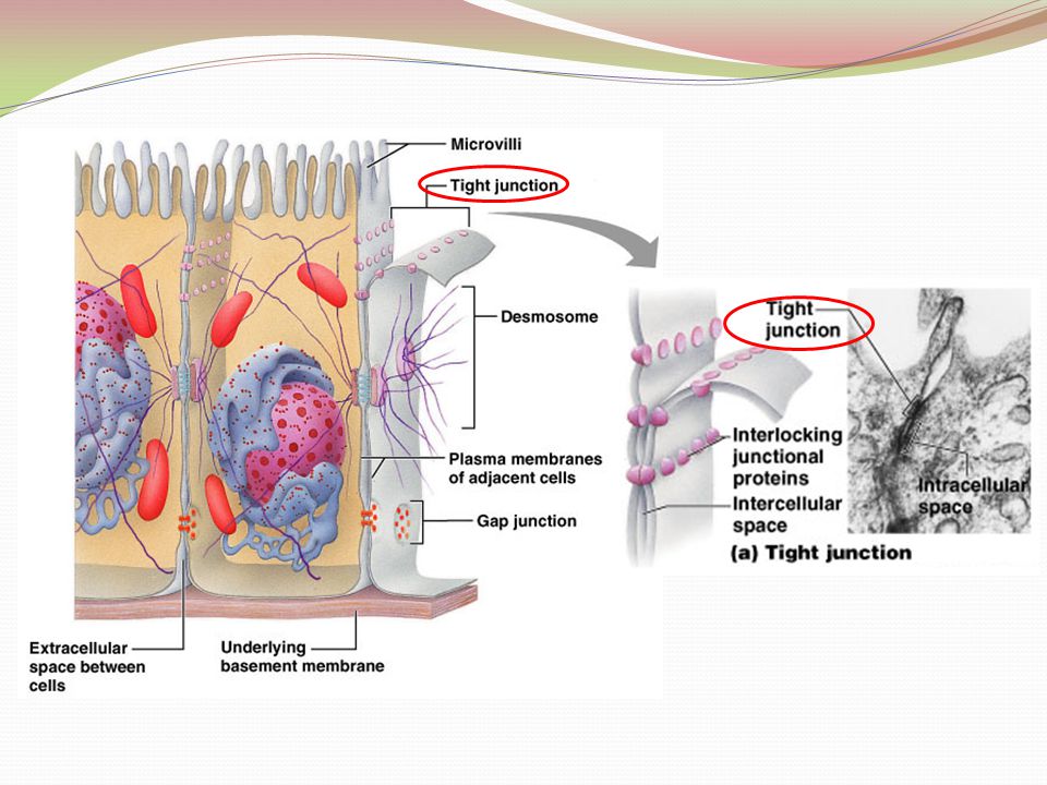
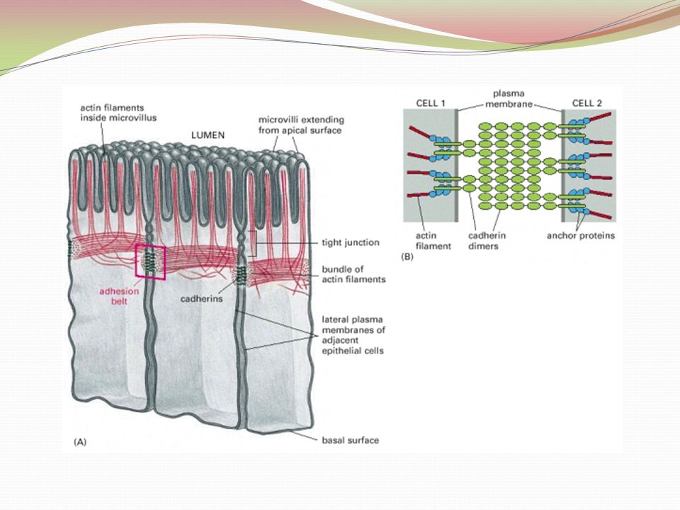
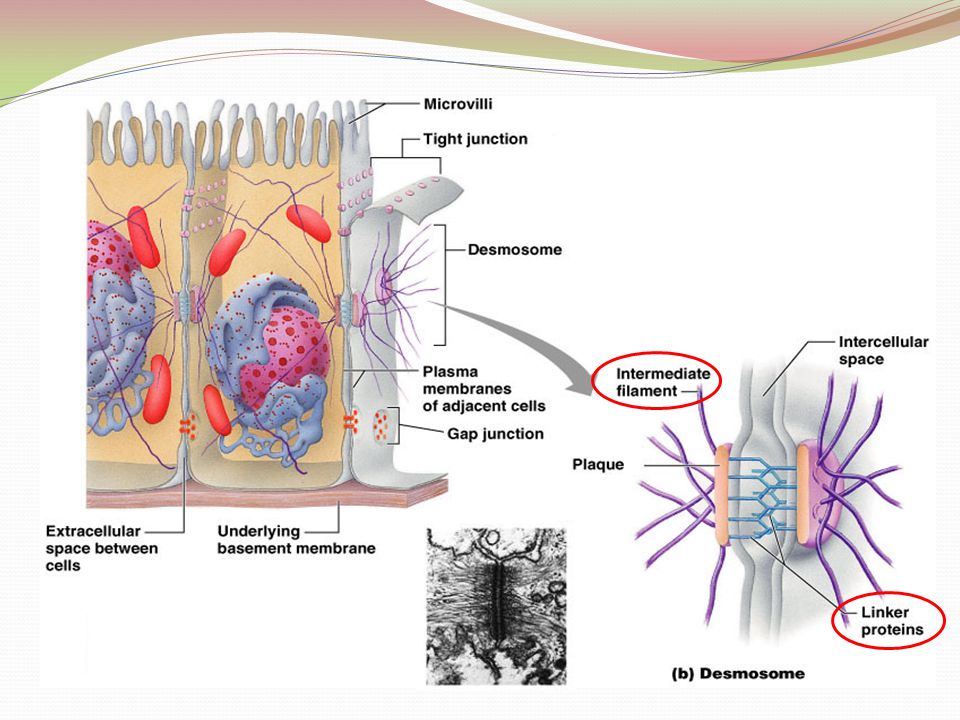
Lateral Surface Features What is it? sides of epithelial cells that face adjacent cells on either side Factors holding epithelial cells together: Adhesion proteins link plasma membranes of adjacent cells Special cell junctions Tight Junctions Adherens Junctions Desmosomes Gap Junctions

11
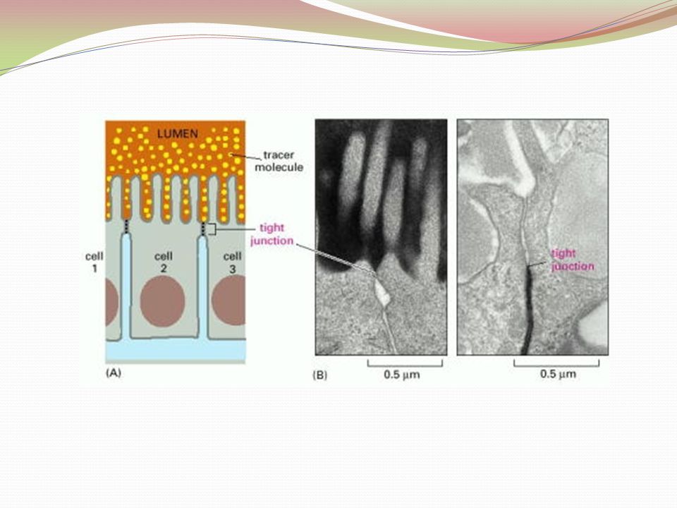
Tight Junctions Tight junctions– closes off intercellular space Location: near apical region Purpose: forms an impermeable junction; prevents molecules from passing between cells Formation: transmembrane proteins in the plasma membrane of adjacent cells fuse together Ie.: epithelial tissue lining the stomach, intestines & urinary bladder prevent contents of these organs from leaking

14
Adherens Junction Adherens junctions – anchoring junction Location- apical lateral borders Purpose: helps form the tight junction around apical lateral borders Formation: A dense layer of proteins on inside of plasma membrane (plaque) attaches to the cytoskeleton. Transmembrane linker proteins (cadherins) are anchored into the cell’s plaque and they bind to cadherins of another cell thus joining the two cells. Ie.: help epithelial surfaces resist separation during contractile activities (food moving through the intestine)
 are anchored into the cell’s plaque and they bind to cadherins of another cell thus joining the two cells. Ie.: help epithelial surfaces resist separation during contractile activities (food moving through the intestine).")
16
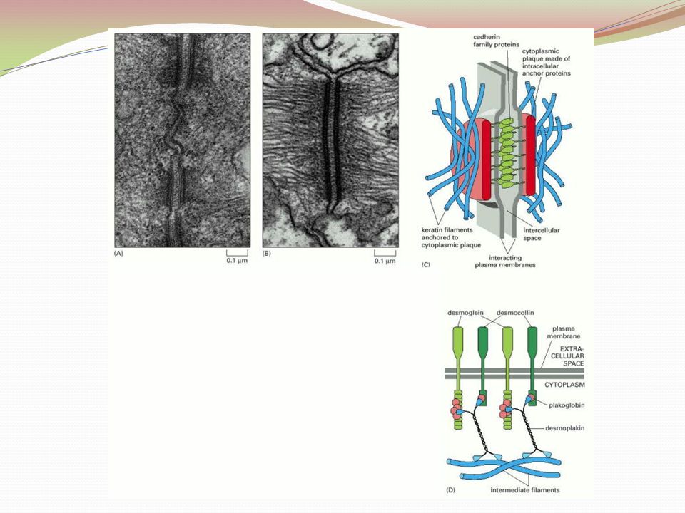
Desmosomes Desmosomes – 2 disc-like plaques connected across intercellular space Location: found in superficial layers of skin Purpose: reduces tearing, twisting, stretching Formation: Plaques of adjoining cells are joined by proteins called cadherins Desomosomes on one side of the cell are imbedded with intermediate filaments (keratin protein) that extends across the cytosol of a cell to desmosomes on the other side of the same cell
 that extends across the cytosol of a cell to desmosomes on the other side of the same cell")
19
Gap Junctions Gap junctions – passageway between two adjacent cells Location: Present in electrically excitable tissues (heart, smooth muscle) Purpose: Let small molecules move directly between neighboring cells Formation: Cells are connected by a protein called connexins that form hollow cylinders called connexons Ie.: lens and the cornea of the eye; enable nerve or muscle impulses to spread rapidly among cells
 Purpose: Let small molecules move directly between neighboring cells Formation: Cells are connected by a protein called connexins that form hollow cylinders called connexons Ie.: lens and the cornea of the eye; enable nerve or muscle impulses to spread rapidly among cells")
21
Bell Work What type of junction is a passage way between two cells? What type of junction resists contractile activity?

22
Create a flip book on the following: Draw & label the surfaces of an epithelial cell (Basal, apical and lateral) Draw & label each of the following (include the cell junction’s purpose, where it is found and an analogy to remember it) Tight junction Adherens junction Desmosome Gap junction
 Draw & label each of the following (include the cell junction’s purpose, where it is found and an analogy to remember it) Tight junction Adherens junction Desmosome Gap junction")
23
Bingo! tissue Epithelial Connective Muscle nerve apical basal lateral surface tight junction adherens junction desmosome gap junction Connexons Connexins Cadherins plaque microvilli cilia avascular impermeabe regenerative intermediate filament linker proteins

Similar presentations


 Bram Welch-Horan (tbw5) October 11, 2005.>")


