Download presentation
Presentation is loading. Please wait.

2
Systemic injection of Mecp2Bnull/y mice with scAAV9/MeCP2 virus results in MeCP2 expression in different cell types in brain. Systemic injection of Mecp2Bnull/y mice with scAAV9/MeCP2 virus results in MeCP2 expression in different cell types in brain. A, Schematic of scAAV9/MeCP2 vector. Mouse Mecp2-e1 is cloned downstream of 730 bp fragment of Mecp2 promoter. Other abbreviations are as noted in the legend to Figure 1. B, Experimental paradigm. C, Immunostaining of MeCP2 expression in different brain regions. Cell counts are relative to total DAPI-positive cells (36 sections, n = 2 mice). D, Expression of MeCP2 in neurons and non-neuronal cells varies with brain region. Cell counts are relative to indicated cell-specific marker (12 sections, n = 2 mice). *p < 0.05, **p < 0.01, ***p < 0.001, NS by one-way ANOVA (Newman–Keuls multiple-comparison test). Data are means ± SEM. E, Western blot of MeCP2 protein in different brain regions. Lane 1, Mecp2+/y; Lane 2, Mecp2Bnull/y; and Lane 3, Mecp2Bnull/y-AAV9/MeCP2. Garg S K et al. J. Neurosci. 2013;33: ©2013 by Society for Neuroscience
. D, Expression of MeCP2 in neurons and non-neuronal cells varies with brain region. Cell counts are relative to indicated cell-specific marker (12 sections, n = 2 mice). *p < 0.05, **p < 0.01, ***p < 0.001, NS by one-way ANOVA (Newman–Keuls multiple-comparison test). Data are means ± SEM. E, Western blot of MeCP2 protein in different brain regions. Lane 1, Mecp2+/y; Lane 2, Mecp2Bnull/y; and Lane 3, Mecp2Bnull/y-AAV9/MeCP2. Garg S K et al. J. Neurosci. 2013;33: ©2013 by Society for Neuroscience.")
3
MeCP2 expressed from virus binds to DNA, restores normal neuronal somal size, and improves survival.
MeCP2 expressed from virus binds to DNA, restores normal neuronal somal size, and improves survival. A, Ectopic MeCP2 localizes to DAPI+ heterochromatin puncta in Mecp2Bnull/y-scAAV9/MeCP2–injected mice. Shown is colocalization of DAPI and MeCP2 in olfactory neuron (top), CA3 pyramidal neuron (middle), and dentate gyrus astrocyte (bottom). Pearson's correlation coefficient = 0.943, 0.932, and 0.985, respectively. Scale bar, 5 μm. B, Image (left) shows representative MeCP2-positive (arrow) and MeCP2-negative (arrowhead) CA3 pyramidal neurons. Scale bar, 10 μm. Average somal diameters (right) of MeCP2-positive (purple) and MeCP2-negative (red) CA3 pyramidal neurons and olfactory bulb mitral cells from Mecp2Bnull/y-scAAV9/MeCP2–injected mice (n = 2). Also shown are measurements from WT mice (black bars; n = 2). The number of cells analyzed is indicated above each bar. ***p < by one-way ANOVA (Newman–Keuls multiple-comparison test). Data are means ± SEM. C, Kaplan-Meier survival curve. D, Observational scores. Mecp2Bnull/y-scAAV9/MeCP2 (n = 5), Mecp2Bnull/y-AAV9/Control (n = 6), and Mecp2+/y (n = 6). Data are means ± SEM. E, Field pixel intensities of MeCP2-Cy3 immunofluorescence measured from brainstem sections of Mecp2+/y and Mecp2Bnull/y-scAAV9/MeCP2 mice. Traces represent pixel intensities from individual fields and each field is indicated by a differently colored trace, n = 2 mice per genotype, 5 fields per mouse. Garg S K et al. J. Neurosci. 2013;33: ©2013 by Society for Neuroscience
, CA3 pyramidal neuron (middle), and dentate gyrus astrocyte (bottom). Pearson s correlation coefficient = 0.943, 0.932, and 0.985, respectively. Scale bar, 5 μm. B, Image (left) shows representative MeCP2-positive (arrow) and MeCP2-negative (arrowhead) CA3 pyramidal neurons. Scale bar, 10 μm. Average somal diameters (right) of MeCP2-positive (purple) and MeCP2-negative (red) CA3 pyramidal neurons and olfactory bulb mitral cells from Mecp2Bnull/y-scAAV9/MeCP2–injected mice (n = 2). Also shown are measurements from WT mice (black bars; n = 2). The number of cells analyzed is indicated above each bar. ***p < by one-way ANOVA (Newman–Keuls multiple-comparison test). Data are means ± SEM. C, Kaplan-Meier survival curve. D, Observational scores. Mecp2Bnull/y-scAAV9/MeCP2 (n = 5), Mecp2Bnull/y-AAV9/Control (n = 6), and Mecp2+/y (n = 6). Data are means ± SEM. E, Field pixel intensities of MeCP2-Cy3 immunofluorescence measured from brainstem sections of Mecp2+/y and Mecp2Bnull/y-scAAV9/MeCP2 mice. Traces represent pixel intensities from individual fields and each field is indicated by a differently colored trace, n = 2 mice per genotype, 5 fields per mouse. Garg S K et al. J. Neurosci. 2013;33: ©2013 by Society for Neuroscience.")
5
Inappropriate Silencing of Genes
Fragile-X Syndrome

6
Completely methylated
Fragile-X Syndrome Length Methylation Females Males Stable 6 to ~45 Unmethylated Not affected Gray zone ~45 to ~55 Premutation ~55 to ~200 Usually not affected Full mutation >200 Completely methylated ~50% affected All affected

7
11_05.jpg 11_05.jpg

8
11_05_2.jpg 11_05_2.jpg

10
Skewed X-Chromosome inactivation in a family with Fragile X

11
Southern Blot Analysis
Blood sample Digest genomic DNA with EcoRI and EagI Electrophoresis and transfer to membrane Hybridize with FMR1 specific probe “A normal female will show an unmethylated 2.8-kb band and a 5.2-kb methylated band that correspond to the normal FMR1 gene present in the active and inactive X chromosome, respectively.”

12
DNA Methylation Beckwith-Wiedemann syndrome

13
DNA Methylation Beckwith-Wiedemann syndrome
Above average birth weight Increase growth after birth (>95% growth curve) Enlarged organs Hypoglycemic following birth Increase risk of cancers Imprinting defect located at 11p15.5
 Enlarged organs. Hypoglycemic following birth. Increase risk of cancers. Imprinting defect located at 11p15.5.")
14
Beckwith-Wiedemann syndrome
Genetic causes of BWS: Maternal DMR hypermethylation UPD Remainder unknown

16
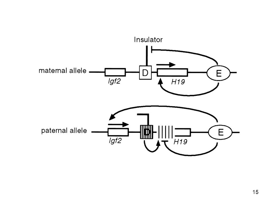
Reading of imprinted gene marks and regulation of imprinted gene expression. (A) A scheme showing the function of CTCF binding and barrier function at the IGF2/H19 locus. Transcriptional enhancer elements are indicated by gray circles. The imprinting control region (ICR) is indicated by a checkered box. CTCF bound to the non‐methylated maternal ICR is shown to block enhancer access to the IGF2 promoter. (B) A scheme of the KCNQ1 locus showing maternally and paternally expressed genes. The untranslated RNA transcript on the paternal allele is indicated by a dotted line. Black arrows indicate the absence of transcription, while white arrows indicate active transcription. Red dots indicate sites of allele‐specific ICR methylation. The genes indicated by abbreviations are given in full below each part of the figure © IF THIS IMAGE HAS BEEN PROVIDED BY OR IS OWNED BY A THIRD PARTY, AS INDICATED IN THE CAPTION LINE, THEN FURTHER PERMISSION MAY BE NEEDED BEFORE ANY FURTHER USE. PLEASE CONTACT WILEY'S PERMISSIONS DEPARTMENT ON OR USE THE RIGHTSLINK SERVICE BY CLICKING ON THE 'REQUEST PERMISSION' LINK ACCOMPANYING THIS ARTICLE. WILEY OR AUTHOR OWNED IMAGES MAY BE USED FOR NON-COMMERCIAL PURPOSES, SUBJECT TO PROPER CITATION OF THE ARTICLE, AUTHOR, AND PUBLISHER. The Journal of Pathology Volume 211, Issue 3, pages , 18 DEC 2006 DOI: /path

17
Model for the establishment of male germline imprinting marks
Model for the establishment of male germline imprinting marks. A proposal for male germline imprinting establishment is presented. The proposal derives from Jelinic et al. (2006). Individual histones are indicated by green spheres. The zinc finger region of CTCFL is indicated by small projections which recognize specific sequences within the H19 ICR. Methylation of adjacent histones is indicated by red stars. Methylation of cytosines within the dinucleotide sequence CpG is indicated by red stars within the indicated ICR. The DNA strand is indicated in blue. SDMA = symmetrical dimethylated arginine; PRMT7 = protein arginine methyltransferase 7; CTCFL = CTCF like; Dnmt3s = denovo methyltransferases 3a, b, and L. The proposed scheme of events is described in greater detail in the text © IF THIS IMAGE HAS BEEN PROVIDED BY OR IS OWNED BY A THIRD PARTY, AS INDICATED IN THE CAPTION LINE, THEN FURTHER PERMISSION MAY BE NEEDED BEFORE ANY FURTHER USE. PLEASE CONTACT WILEY'S PERMISSIONS DEPARTMENT ON OR USE THE RIGHTSLINK SERVICE BY CLICKING ON THE 'REQUEST PERMISSION' LINK ACCOMPANYING THIS ARTICLE. WILEY OR AUTHOR OWNED IMAGES MAY BE USED FOR NON-COMMERCIAL PURPOSES, SUBJECT TO PROPER CITATION OF THE ARTICLE, AUTHOR, AND PUBLISHER. The Journal of Pathology Volume 211, Issue 3, pages , 18 DEC 2006 DOI: /path
. Individual histones are indicated by green spheres. The zinc finger region of CTCFL is indicated by small projections which recognize specific sequences within the H19 ICR. Methylation of adjacent histones is indicated by red stars. Methylation of cytosines within the dinucleotide sequence CpG is indicated by red stars within the indicated ICR. The DNA strand is indicated in blue. SDMA = symmetrical dimethylated arginine; PRMT7 = protein arginine methyltransferase 7; CTCFL = CTCF like; Dnmt3s = denovo methyltransferases 3a, b, and L. The proposed scheme of events is described in greater detail in the text. © IF THIS IMAGE HAS BEEN PROVIDED BY OR IS OWNED BY A THIRD PARTY, AS INDICATED IN THE CAPTION LINE, THEN FURTHER PERMISSION MAY BE NEEDED BEFORE ANY FURTHER USE. PLEASE CONTACT WILEY S PERMISSIONS DEPARTMENT ON OR USE THE RIGHTSLINK SERVICE BY CLICKING ON THE REQUEST PERMISSION LINK ACCOMPANYING THIS ARTICLE. WILEY OR AUTHOR OWNED IMAGES MAY BE USED FOR NON-COMMERCIAL PURPOSES, SUBJECT TO PROPER CITATION OF THE ARTICLE, AUTHOR, AND PUBLISHER. The Journal of Pathology Volume 211, Issue 3, pages , 18 DEC 2006 DOI: /path")
18
Genes Dev. Vol. 11, No. 23, pp. 3128-3142, December 1, 1997
Mouse mutant embryos overexpressing IGF-II exhibit phenotypic features of the Beckwith-Wiedemann and Simpson-Golabi-Behmel syndromes Jonathan Eggenschwiler,1 Thomas Ludwig,2 Peter Fisher,3 Philip A. Leighton,4,5 Shirley M. Tilghman,4 and Argiris Efstratiadis1,6

23
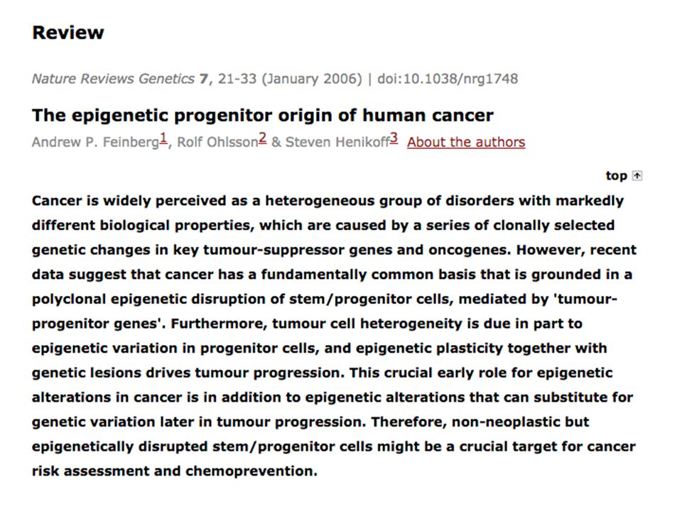
Nat Rev gene. 7: 21–33 doi:10.1038/nri1748
Figure 2 The epigenetic progenitor model of cancer. Feinberg AP et al. (2005) The epigenetic progenitor origin of human cancer Nat Rev gene. 7: 21–33 doi: /nri1748
 The epigenetic progenitor origin of human cancer. Nat Rev gene. 7: 21–33 doi: /nri1748.")
24
Prader-Willi and Angelman Syndrome
Mild mental retardation Severe impairment and loss of speech endocrine abnormalities seizures and ataxia temper tantrums unprovoked laughter Obesity hyperactivity 1 in 15,000

25
Prader-Willi and Angelman Syndrome
Genes/proteins involved Prader-Willi syndrome 15q11-13

26
Prader-Willi and Angelman Syndrome
UBE3A is paternally silenced This primarily occurs in brain, other tissues show biallelic expression

27
Prader-Willi and Angelman Syndrome
What happens in each pathologies? If the maternal copy of chromosome 15 is missing, then genes normally expressed from this parental origin are not expressed Consequences…

28
Prader-Willi and Angelman Syndrome
If paternal chromosome 15 is missing, then only the maternally expressed proteins are made Consequence: UBE3A is ok, but other genes in the region are not expressed…Prader-Willi syndrome

29
Prader-Willi and Angelman Syndrome
Thus, two different diseases based on the cells “memory” of methylation – alter the memory, alter the phenotype

30
Prader-Willi and Angelman Syndrome
How do you “lose” chromosome 15? Microdeletion of 15q11-13 on one chromsome – 70% Single gene mutation – 15% of AS Defect in imprinting centre (IC) – 5% Uniparental Disomy – 30% of PWS, 5% AS
 – 5% Uniparental Disomy – 30% of PWS, 5% AS.")
31
Prader-Willi and Angelman Syndrome

32
Uniparental Disomy Receive two chromosomes from one parent
Eg. Paternal disomy – both of chromosome 15 are from father, thus both have silenced UBE3A

33
Uniparental Disomy How does it happen? Trisomic Rescue - majority
Monosomic Duplication

34
Meiosis I

35
Meiosis II

36
Meiosis I Non-disjunction

37
Meiosis I Non-disjunction
Fertilization Meiosis I non-disjunction always creates a problem Trisomy

38
Meiosis II Non-disjunction
2/3 gametes following Meiosis II non-disjunction are normal Fertilization Fertilization Trisomy Normal

39
Trisomy Trisomy for most autosomal chromosomes is lethal
BIG exception: Trisomy 21, smallest autosomal chromosome, fewest genes, not lethal Under rare conditions, some autosomal trisomies can escape – Trisomic Rescue

40
Trisomic Rescue following Meiosis I Non-disjunction
Two copies of homologous, but not identical, chromosomes + Maternal Anaphase lag Maternal Paternal M,M 1/3 M,P 1/3 M,P 1/3

41
Trisomic Rescue following Meiosis II Non-disjunction
Two copies of identical chromosomes + Anaphase lag M,M 1/3 M,P 2/3

42
Uniparental Disomy How does it happen? Trisomic Rescue - majority
Monosomic Duplication

43
Monosomic Duplication
+ Two identical copies of paternal chromosome - isodisomy P,P

44
Parental Origin Determines Phenotype
M,M { 15 Prader-Willi Syndrome M,M Prader-Willi Syndrome P,P Angelman Syndrome

45
Prader-Willi and Angelman syndrome
Non-disjunction is more common in Meiosis I in females In human females, Meiosis I starts before birth but is arrested at diplotene stage (late prophase I) Oocytes sit like this for decades Complete meiosis II once each month While arrested at the diplotene stage, the tetrad chromosomes are held together by chiasmata (formed during recombination) If a pair of chromosomes don’t undergo recombination, the lack of chiasmata can contribute to non-disjunction Uniparental Disomy – 30% of PWS, 5% AS Maternal non-disjunction and trisomic rescue leading to the pair of maternal chromosomes
 Oocytes sit like this for decades. Complete meiosis II once each month. While arrested at the diplotene stage, the tetrad chromosomes are held together by chiasmata (formed during recombination) If a pair of chromosomes don’t undergo recombination, the lack of chiasmata can contribute to non-disjunction. Uniparental Disomy – 30% of PWS, 5% AS. Maternal non-disjunction and trisomic rescue leading to the pair of maternal chromosomes.")
46
Uniparental Disomy and Human Disease
Eric Engel. Some lessons from uniparental disomy (UPD) in the framework of contemporary cytogenetics and molecular biology. Atlas Genet Cytogenet Oncol Haematol. December 2003.
 in the framework of contemporary cytogenetics and molecular biology. Atlas Genet Cytogenet Oncol Haematol. December")
47
Trisomic rescue of Meiosis II non-disjunction can have other problems
Anaphase lag { Pair of identical chromosomes (isochromosomes)
")
48
Harboring CFTR mutation
Uniparental disomy as a mechanism for human genetic disease. Spence JE, Perciaccante RG, Greig GM, Willard HF, Ledbetter DH, Hejtmancik JF, Pollack MS, O'Brien WE, Beaudet AL. Am J Hum Genet Feb;42(2): CFTR-/+ CFTR+/+ CFTR-/- Uniparental isodisomy and reduction to homozygosity { Pair of identical Chromosome 7 Harboring CFTR mutation
: CFTR-/+ CFTR+/+ CFTR-/- Uniparental isodisomy and reduction to homozygosity. { Pair of identical. Chromosome 7. Harboring CFTR mutation.")
Similar presentations


 W2015 Prerequisite:>")


 imprinting Regulation of gene expression Developmental programming.>")


 of the Igf2 gene correlates with colon cancer With family.>")

 Thomas Morgan - fruit fly.>")

