Download presentation
Presentation is loading. Please wait.

2
References 1-Gray’s anatomy 2-Human nervous system Noback 3- Basic clinical neuroanatomy Young 4- Head and neck anatomy Berkovitz 5- clinical neuroanatomy Snell

3
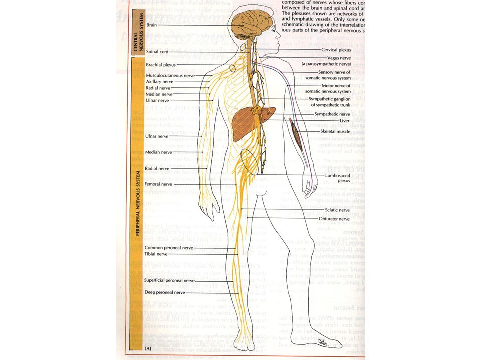
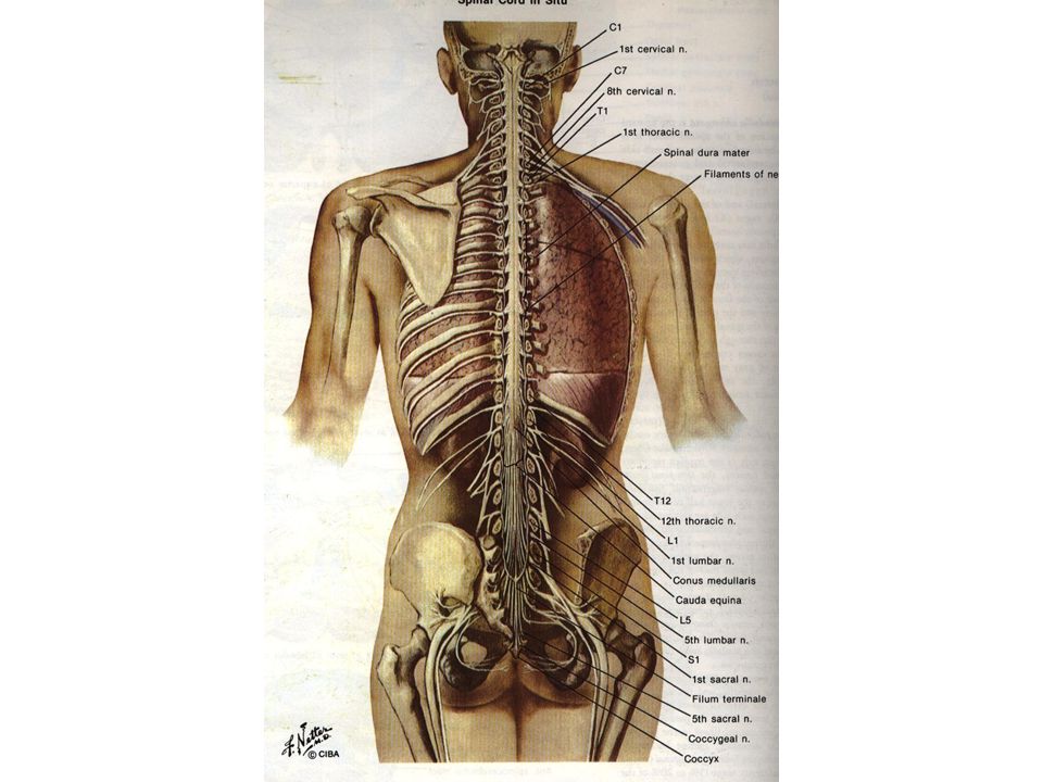
Anatomical division of nervous system 1- CNS brain and spinal cord 2- PNS peripheral nerves ganglions ganglions receptors receptors

6
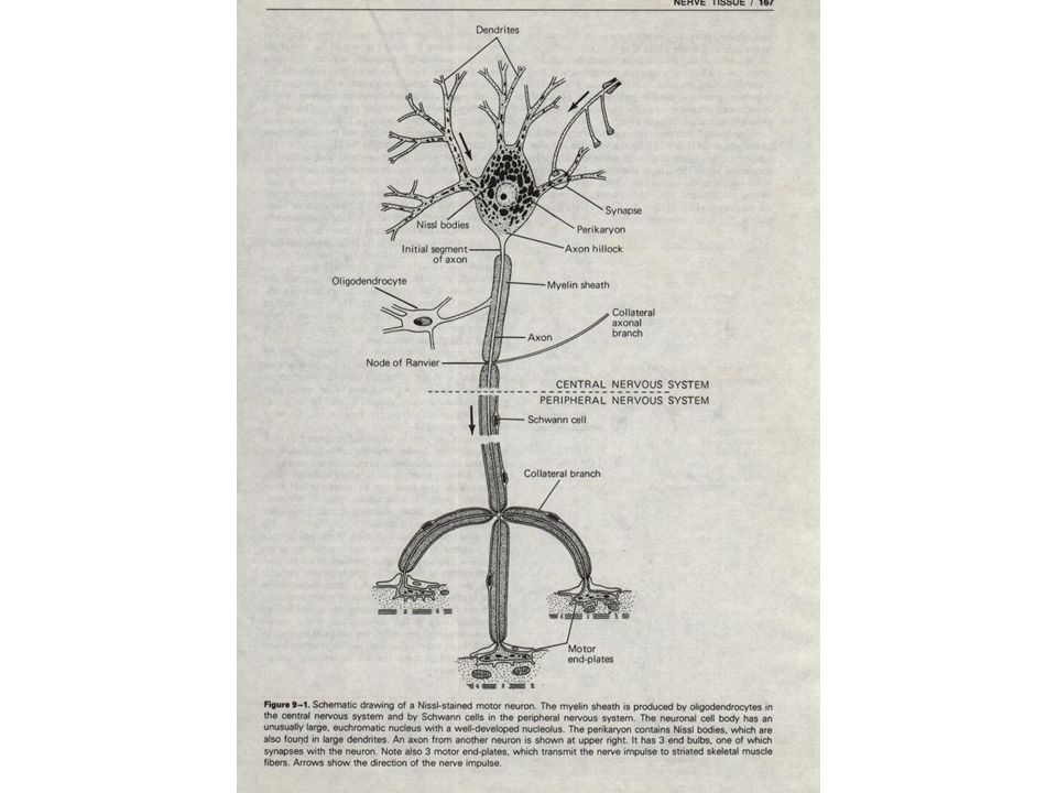
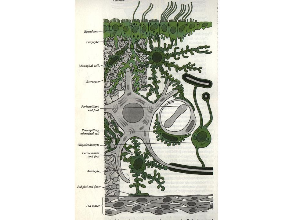
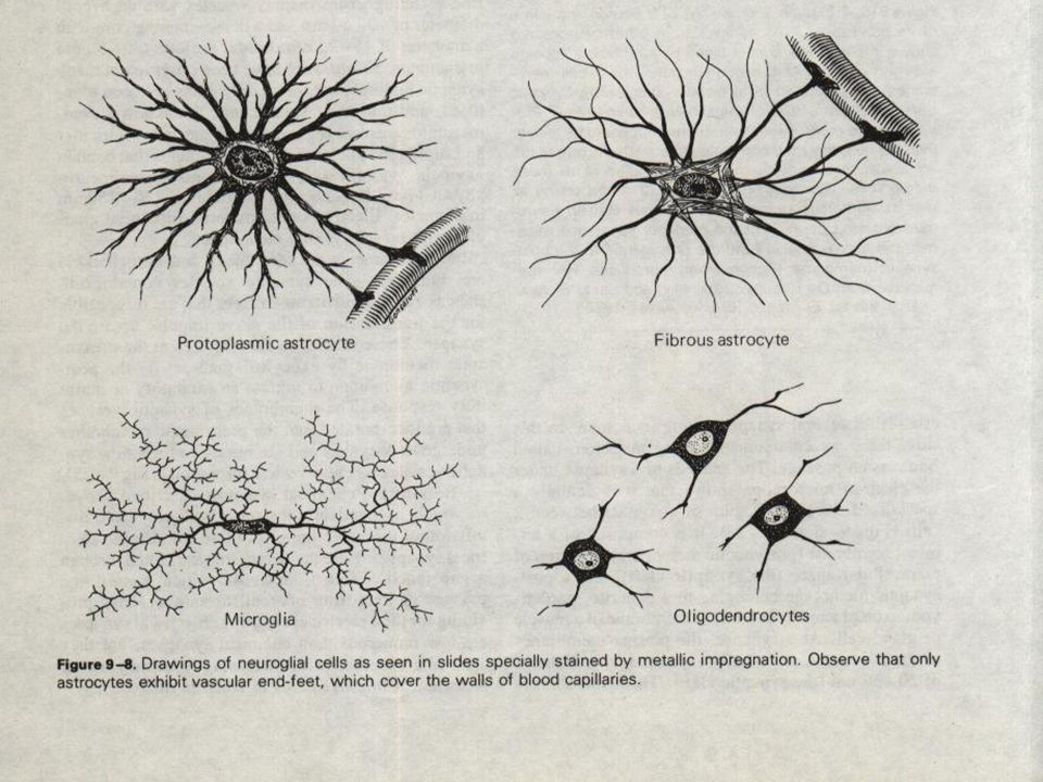
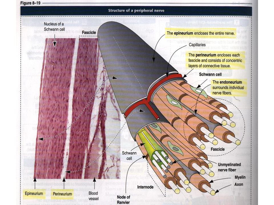
Cells in this system 1- neuron 2-neuroglia A- Central neuroglia Macroglia (astrocyte,oligodendrocyte) Microglia Ependymal cells B- Peripheral neuroglia Schwann cells Satellite cells
 Microglia Ependymal cells B- Peripheral neuroglia Schwann cells Satellite cells")
8
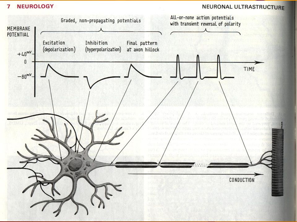
Specialist cell 1- produce action potential 2- convey

10
central neuroglia Macroglia (astrocyte,oligodendrocyte) Microglia Ependymal cells
 Microglia Ependymal cells")
14
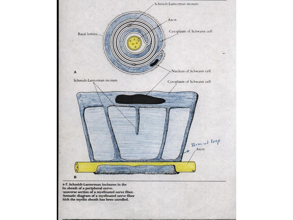
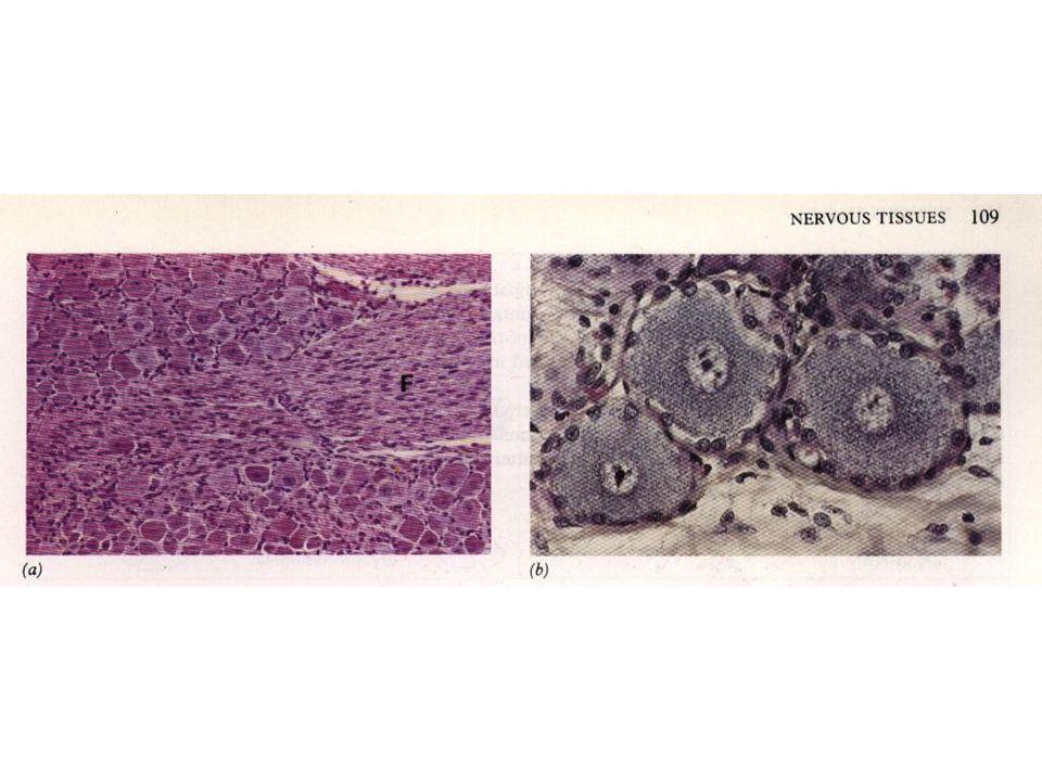
Peripheral neuroglia 1- schwann cell 2- satellite cell

19
Function of this system

20
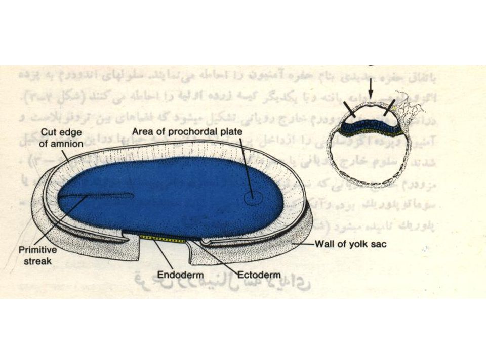
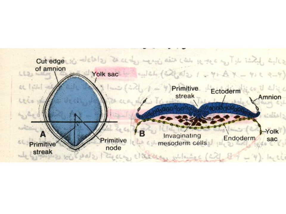
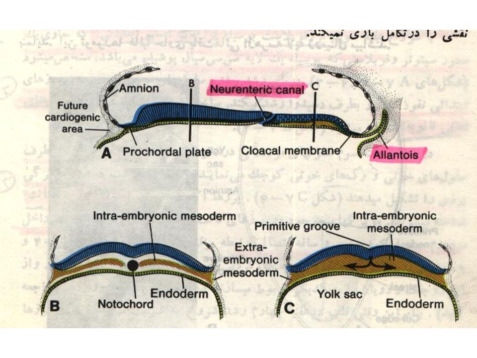
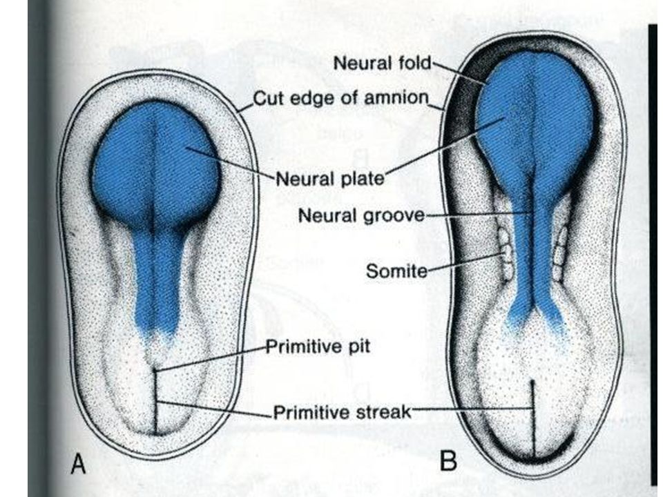
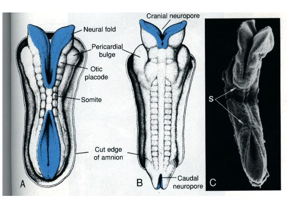
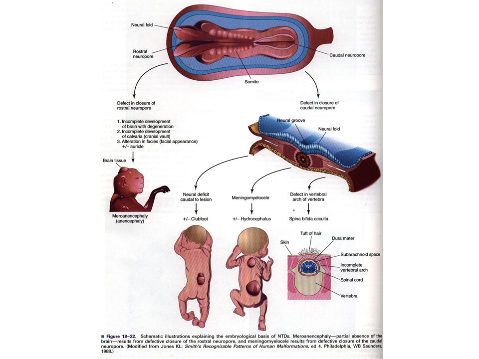
Embryology of the nervous system

28
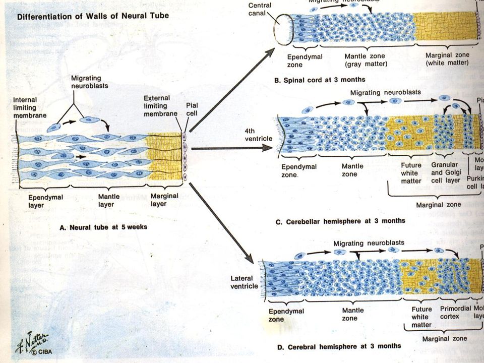
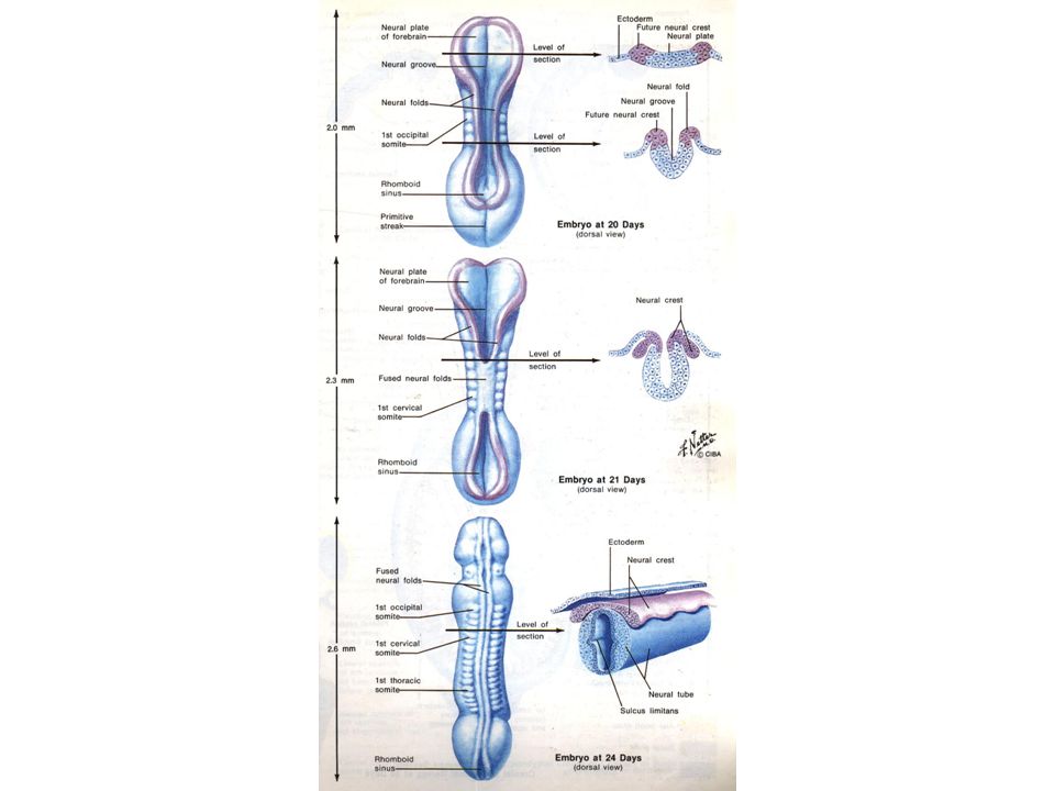
Neural Tube Histogenesis

31
What is the identity of these two cells?

32
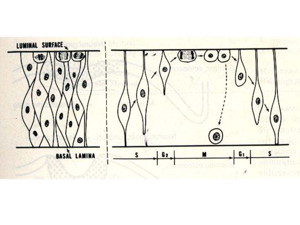
Three points about division of neural tube cells 1- Synchronise formation of neuronal and glial cells. 2- Continuous of glial division after stopping of neuronal cell division. 3- Early formation of large neuron in comparison with small neuron.

33
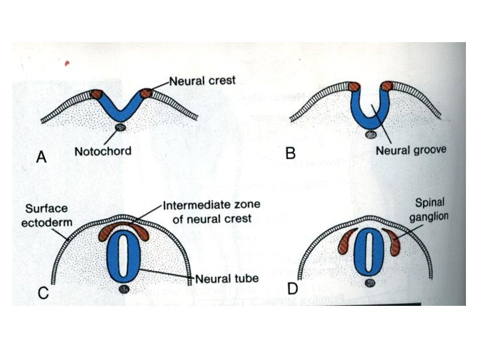
لایه پوشاننده لایه حاشیه ایی لایه اپاندیمی Formation of three layers in the thickness of NEURAL TUBE NEURAL TUBE

35
Neuronal cell migration 1- Gene activation 2- Epigenetic signals (mechanical or contact guidance)
")
36
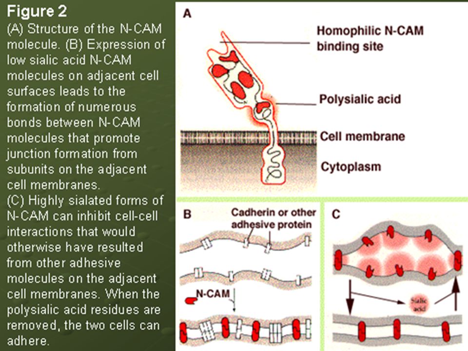
Neuronal cell aggregation Morphological appearance Morphological appearance NCAMs (nerve cell adhesion molecules) N cadherin
 N cadherin")
38
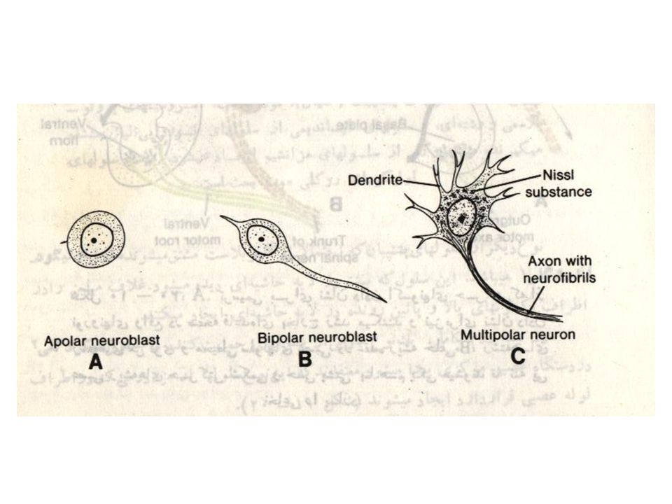
Neuroblast differentiation Apolar neuroblast Bipolar neuroblast Unipolar neuroblast Multipolar neuroblast

41
Neuroblast differentiation Neurotrophic Factors Neurotrophic Factors 1- NGFs ( Nerve Growth Factors) 2- Outgrowth Promoting proteins ( Laminin, Netrin, Fibronectin) ( Laminin, Netrin, Fibronectin) 3-NCAMs 3-NCAMs
 2- Outgrowth Promoting proteins ( Laminin, Netrin, Fibronectin) ( Laminin, Netrin, Fibronectin) 3-NCAMs 3-NCAMs")
42
Cell Programmed Death Apoptosis Apoptosis 30 to 60 percent of cells survive 30 to 60 percent of cells survive

43
Effects of Experience and Activation on neurons Structural Plasticity Structural Plasticity Axonal sprouting Axonal sprouting Changes in the number of Dendritic Spines Changes in the number of Dendritic Spines Amblyopia Disease Amblyopia Disease

44
Reveiw

45
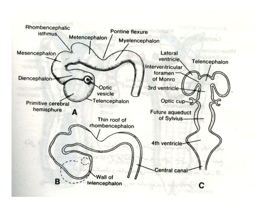
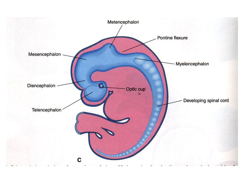
Formation of brain and spinal cord 1- Primary vesicles 2- Flexures

46
Midbrain or

47
Formation of brain and spinal cord 1- secondary vesicles

50
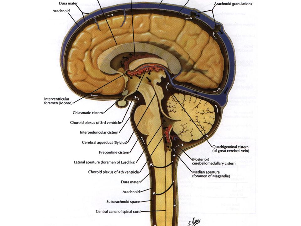
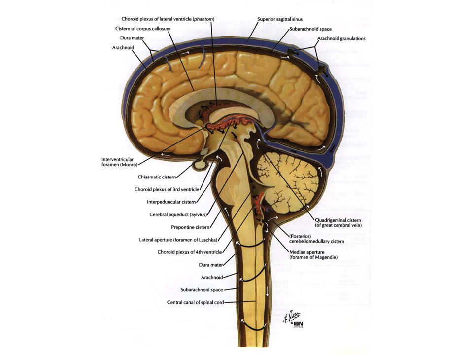
Ventricular spaces and central canal 1-Telencephalon------ lateral ventricals1&2 2-Diencephalon-------third ventricle 3-Mesencephalon----cerebral aqueduct 4- Rhombencephalon--- forth ventricle 5-Spinal cord----- central canal 6- in the end of spinal cord----- terminal ventricle Monro Foramen(interventricular F)
")
53
Neural crest derivates

56
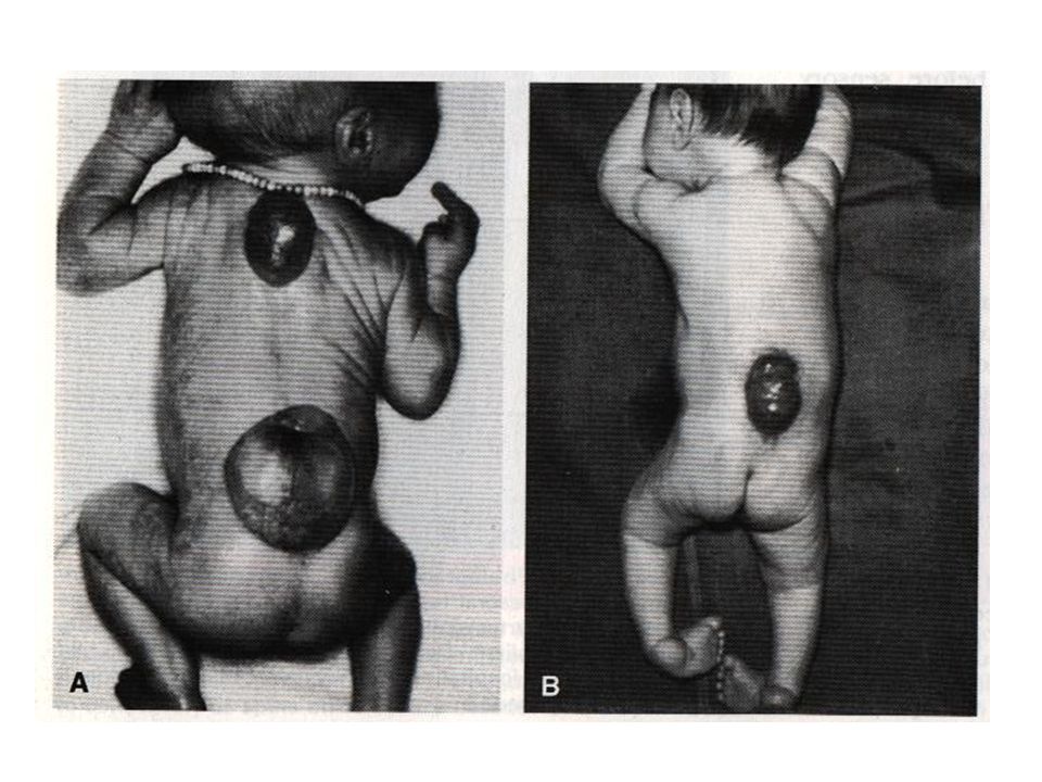
Neural Tube Defects (NTDs) 1- Spina Bifida Oculta Meningocele Meningomyelocle cystica Rachischhisis
 1- Spina Bifida Oculta Meningocele Meningomyelocle cystica Rachischhisis")
58
occulta meningocele meningomyelocelemyeloschesis

59
10 %of normal people L5 or S1

61
Neural Tube Defects (NTDs) 1- Cranial Bifida Cranial Meningocele Meningoencephalocele Meningohydroencephalocele Anencephaly
 1- Cranial Bifida Cranial Meningocele Meningoencephalocele Meningohydroencephalocele Anencephaly")
62
Cranial Meningocele Meningoencephalocele Meningohydroencephalocele Cranial Bifida

Similar presentations


 3. Name this muscle 4. Meg tore her semitendinosus muscle. What movement will be.>")
 both within.>")


 3. Name this muscle 4. Meg tore her semitendinosus muscle. What movement will.>")


