Download presentation
Presentation is loading. Please wait.

2
Metastatic involvement (M) M0 - No metastases M1 - Metastases present
 M0 - No metastases M1 - Metastases present")
3
Metastases (M) M0: No distant metastasis M1: Distant metastasis present; or Separate tumor nodules in the ipsilateral nonprimary-tumor lobes of the lung. Separate tumor nodules in the contralateral lung are considered M1 if they are of the same histologic cell type as the primary lesion. A contralateral lung tumor with a different cell type is considered a synchronous primary lesion and should be staged independently

13
StageTumorNodesMetastases Stage 0 TIS- Carcinoma in situ IA IB T1 T2 N0 M0 IIA IIB T1 T2 T3 N1 N1 N0 M0 M0 M0 IIIA T1 or T2 T3 N2 N1 or N2 M0 IIIB Any T T4 N3 Any N M0 IV Any T Any NM1

14
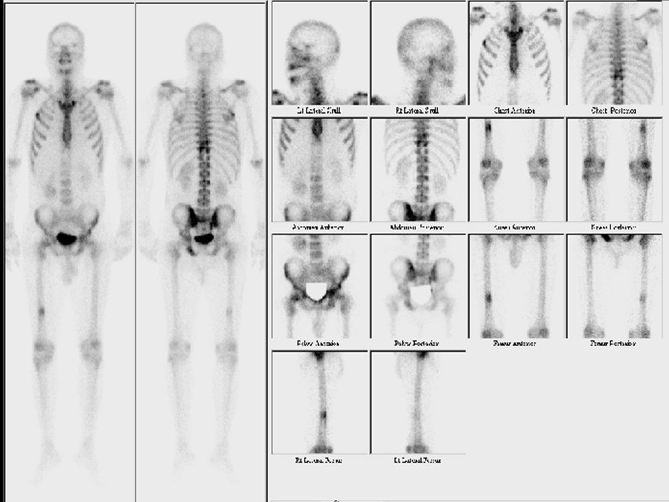
Advantages MRI has over CT in Tumor assessment Mediastinal and chest wall invasion and involvement of the diaphragm. MRI is most useful when evaluating spinal cord compression and brain metastasis. In Pancoast tumours, invasion into the brachial plexus, subclavian artery or vertebral body by MRI has been found to be 94% accurate as opposed to 63% for CT.

15
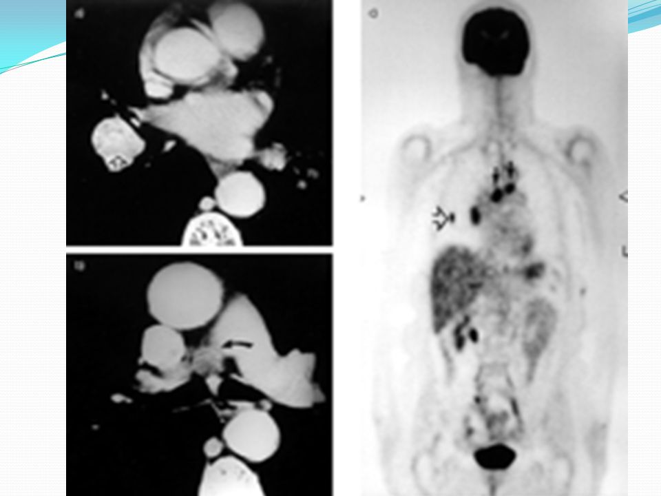
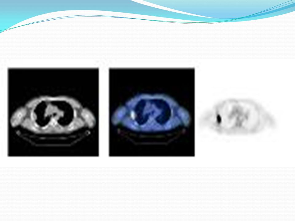
Positron Emission Tomography PET scans appear to be more sensitive, specific, and accurate than CT scans for staging mediastinal disease. PET is more accurate than conventional studies in detecting recurrent lung cancer. False-positive studies do occur secondary to postirradiation inflammatory change and delaying the examination until 4 or 5 weeks postirradiation is recommended

16
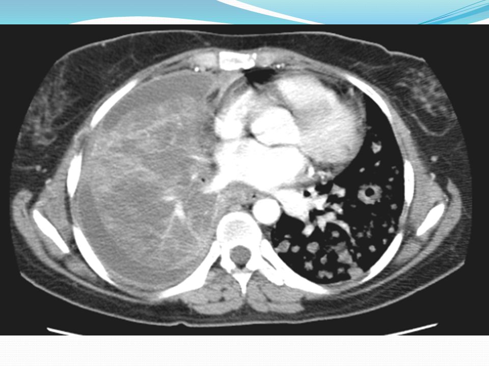
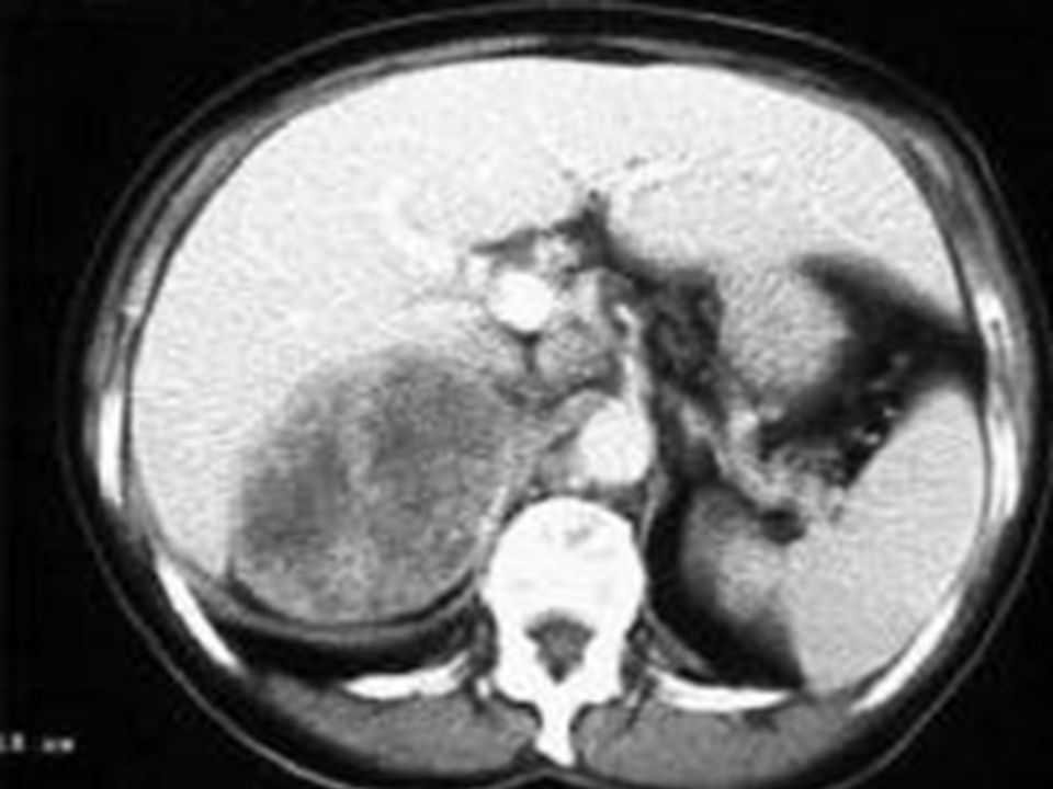
The solitary pulmonary nodule A common incidental CXR finding. CT detects many more lung nodules than CXR. Numerous differential diagnoses. 50% are malignant: 40% are primary CA, 10% are solitary metastases. Prompt diagnosis and management of early lung cancer manifesting as SPN may be the only chance for cure. No significant mortality reduction with screening.

18
Calcification in SPN CT scanning can further refine the detection of calcification and fat within nodules. A total 22–38% of noncalcified nodules on chest radiographs appear calcified on CT. Eccentric or stippled calcification is seen in 10% of lung cancers.

19
Features of SPN suggesting benignity Clinical history, especially of T.B. Compared with old films, no growth over a 2-yr period. Age <35 yrs, No history of cigarette smoking. No history of extrathoracic malignancy.

Similar presentations


; FRCS (Ireland); MMed (Wits); FCS (SA) Urology 38 th BMA CONGRESS.>")


