Download presentation
Presentation is loading. Please wait.

1
Chapter 4 Neural Conduction and Synaptic Transmission
How Neurons Send and Receive Signals This multimedia product and its contents are protected under copyright law. The following are prohibited by law: any public performance or display, including transmission of any image over a network; preparation of any derivative work, including the extraction, in whole or in part, of any images; any rental, lease, or lending of the program.

2
The Neuron’s Resting Membrane Potential
Inside of the neuron is negative with respect to the outside Resting membrane potential is about -70mV Membrane is polarized, it carries a charge Why?

3
Ionic Basis of the Resting Potential
Ions, charged particles, are unevenly distributed Factors influencing ion distribution Homogenizing Factors contributing to uneven distribution

4
Ionic Basis of the Resting Potential
Homogenizing Random motion – particles tend to move down their concentration gradient Electrostatic pressure – like repels like, opposites attract Factors contributing to uneven distribution Membrane is selectively permeable Sodium-potassium pumps

5
Ions Contributing to Resting Potential
Sodium (Na+) Chloride (Cl-) Potassium (K+) Negatively charged proteins (A-) synthesized within the neuron found primarily within the neuron
 Chloride (Cl-) Potassium (K+) Negatively charged proteins (A-) synthesized within the neuron. found primarily within the neuron.")
6
The Neuron at Rest Ions move in and out through ion-specific channels
K+ and Cl- pass readily Little movement of Na+ A- don’t move at all, trapped inside

7
Equilibrium Potential
The potential at which there is no net movement of an ion – the potential it will move to achieve when allowed to move freely Na+ = 120mV K+ = -90mV Cl- = -70mV (same as resting potential)
")
8
The Neuron at Rest Na+ is driven in by both electrostatic forces and its concentration gradient K+ is driven in by electrostatic forces and out by its concentration gradient Cl- is at equilibrium Sodium-potassium pump – active force that exchanges 3 Na+ inside for 2 K+ outside

9
Something to think about
What would happen if the membrane’s permeability to Na+ were increased? What would happen if the membrane’s permeability to K+ were increased?

10
Generation and Conduction of Postsynaptic Potentials (PSPs)
Neurotransmitters bind at postsynaptic receptors These chemical messengers bind and cause electrical changes Depolarizations (making the membrane potential less negative) Hyperpolarizations (making the membrane potential more negative)
 Hyperpolarizations (making the membrane potential more negative)")
11
Generation and Conduction of Postsynaptic Potentials (PSPs)
Postsynaptic depolarizations = Excitatory PSPs (EPSPs) Postsynaptic hyperpolarizations = Inhibitory PSPs (IPSPs) EPSPs make it more likely a neuron will fire, IPSPs make it less likely PSPs are graded potentials – their size varies
 Postsynaptic hyperpolarizations = Inhibitory PSPs (IPSPs) EPSPs make it more likely a neuron will fire, IPSPs make it less likely. PSPs are graded potentials – their size varies.")
12
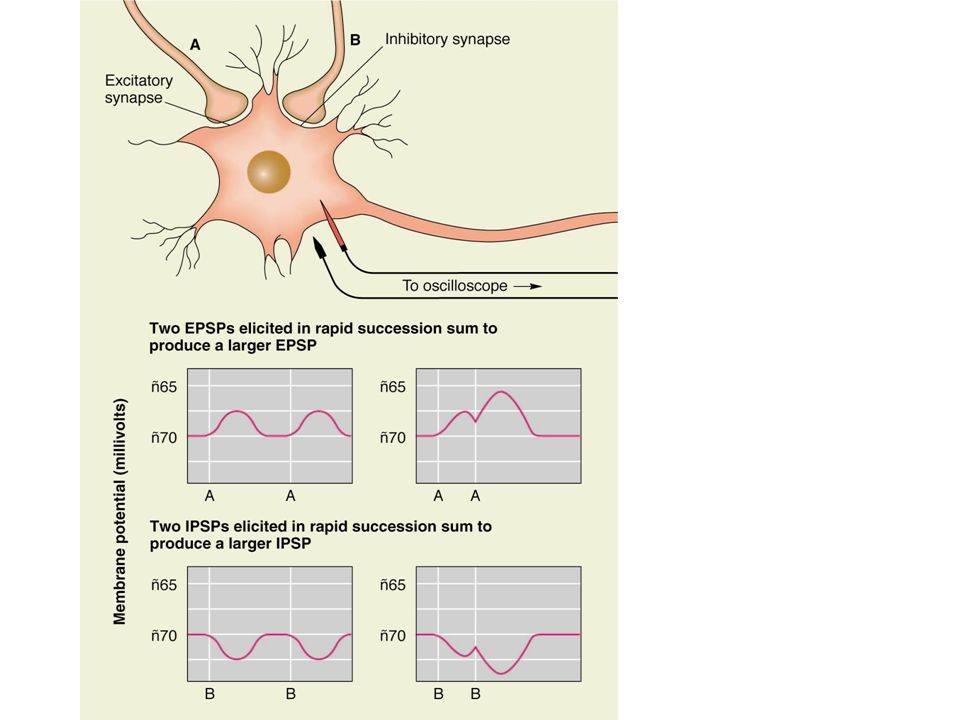
EPSPs and IPSPs Travel passively from their site of origination
Decremental – they get smaller as they travel 1 EPSP typically will not suffice to cause a neuron to “fire” and release neurotransmitter – summation is needed

13
Integration of PSPs and Generation of Action Potentials (APs)
In order to generate an AP (or “fire”), the threshold of activation must be reached at the axon hillock Integration of IPSPs and EPSPs must result in a potential of about -65mV in order to generate an AP
, the threshold of activation must be reached at the axon hillock. Integration of IPSPs and EPSPs must result in a potential of about -65mV in order to generate an AP.")
14
Integration Adding or combining a number of individual signals into one overall signal Temporal summation – integration of events happening at different times Spatial - integration of events happening at different places

15
What type of summation occurs when:
One neuron fires rapidly? Multiple neurons fire at the same time? Several neurons fire repeatedly? Both temporal and spatial summation occur simultaneously

18
The Action Potential All-or-none, when threshold is reached the neuron “fires” and the action potential either occurs or it does not. When threshold is reached, voltage-activated ion channels are opened.

19
The Ionic Basis of Action Potentials
When summation at the axon hillock results in the threshold of excitation (-65mV) being reached, voltage-activated Na+ channels open and sodium rushes in. Remember, all forces were acting to move Na+ into the cell. Membrane potential moves from -70 to +50mV.
 being reached, voltage-activated Na+ channels open and sodium rushes in. Remember, all forces were acting to move Na+ into the cell. Membrane potential moves from -70 to +50mV.")
21
The Ionic Basis of Action Potentials
Rising phase: Na+ moves membrane potential from -70 to +50mV. End of rising phase: After about 1 millisec, Na+ channels close. Change in membrane potential opens voltage-activated K+ channels. Repolarization: Concentration gradient and change in charge leads to efflux of K+. Hyperpolaization: Channels close slowly - K+ efflux leads to membrane potential <-70mV.

22
Refractory Periods Absolute – impossible to initiate another action potential Relative – harder to initiate another action potential Prevent the backwards movement of APs and limit the rate of firing

23
The action potential in action
24
PSPs Vs Action Potentials (APs)
EPSPs/IPSPs Decremental Fast Passive (energy is not used) Action Potentials Nondecremental Conducted more slowly than PSPs Passive and active
 Action Potentials. Nondecremental. Conducted more slowly than PSPs. Passive and active.")
25
Conduction in Myelinated Axons
Passive movement of AP within myelinated portions occurs instantly Nodes of Ranvier (unmyelinated) Where ion channels are found Where full AP is seen AP appears to jump from node to node Saltatory conduction
 Where ion channels are found. Where full AP is seen. AP appears to jump from node to node. Saltatory conduction.")
26
Structure of Synapses Most common
Axodendritic – axons on dendrites Axosomatic – axons on cell bodies Dendrodendritic – capable of transmission in either direction Axoaxonal – may be involved in presynaptic inhibition

27
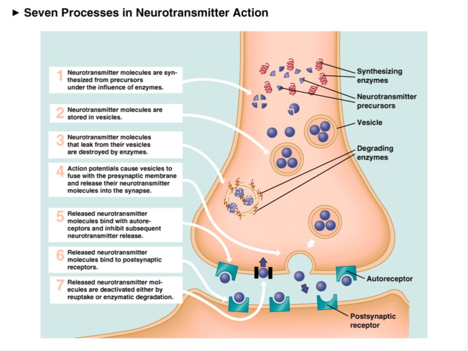
Synthesis, Packaging, and Transport of Neurotransmitter (NT)
NT molecules Small Synthesized in the terminal button and packaged in synaptic vesicles Large Assembled in the cell body, packaged in vesicles, and then transported to the axon terminal

28
Release of NT Molecules
Exocytosis – the process of NT release The arrival of an AP at the terminal opens voltage-activated Ca++ channels. The entry of Ca++ causes vesicles to fuse with the terminal membrane and release their contents

29
Activation of Receptors by NT
Released NT produces signals in postsynaptic neurons by binding to receptors. Receptors are specific for a given NT. Ligand – a molecule that binds to another. A NT is a ligand of its receptor.

30
Receptors There are multiple receptor types for a given NT.
Ionotropic receptors – associated with ligand-activated ion channels. Metabotropic receptors – associated with signal proteins and G proteins.

31
Ionotropic Receptors NT binds and an associated ion channel opens or closes, causing a PSP. If Na+ channels are opened, for example, an EPSP occurs. If K+ channels are opened, for example, an IPSP occurs.

32
Metabotropic Receptors
Effects are slower, longer-lasting, more diffuse, and more varied. NT (1st messenger) binds > G protein subunit breaks away > ion channel opened/closed OR a 2nd messenger is synthesized > 2nd messengers may have a wide variety of effects
 binds > G protein subunit breaks away > ion channel opened/closed OR a 2nd messenger is synthesized > 2nd messengers may have a wide variety of effects.")
34
Reuptake, Enzymatic Degradation, and Recycling
As long as NT is in the synapse, it is active – activity must somehow be turned off. Reuptake – scoop up and recycle NT. Enzymatic degradation – a NT is broken down by enzymes.

35
Small-molecule Neurotransmitters
Amino acids – the building blocks of proteins Monoamines – all synthesized from a single amino acid Soluble gases Acetylcholine (ACh) – activity terminated by enzymatic degradation
 – activity terminated by enzymatic degradation.")
36
Amino Acid Neurotransmitters
Usually found at fast-acting directed synapses in the CNS Glutamate – Most prevalent excitatory neurotransmitter in the CNS GABA – synthesized from glutamate Most prevalent inhibitory NT in the CNS Aspartate and glycine

37
Monoamines Effects tend to be diffuse
Catecholamines – synthesized from tyrosine Dopamine Norepinephrine Epinephrine Indolamines – synthesized from tryptophan Serotonin

38
Soluble-Gases and ACh Soluble gases – exist only briefly
Nitric oxide and carbon monoxide Retrograde transmission – backwards communication Acetylcholine (Ach) Acetyl group + choline Neuromuscular junction
 Acetyl group + choline. Neuromuscular junction.")
39
Neuropeptides Large molecules Example – endorphins
“Endogenous opiates” Produce analgesia (pain suppression) Receptors were identified before the natural ligand was
 Receptors were identified before the natural ligand was.")
40
Pharmacology of Synaptic Transmission
Many drugs act to alter neurotransmitter activity Agonists – increase or facilitate activity Antagonists – decrease or inhibit activity A drug may act to alter neurotransmitter activity at any point in its “life cycle”

42
Agonists – 2 examples Cocaine - catecholamine agonist
Blocks reuptake – preventing the activity of the neurotransmitter from being “turned off” Benzodiazepines - GABA agonists Binds to the GABA molecule and increases the binding of GABA

44
Antagonists – 2 examples
Atropine – ACh antagonist Binds and blocks muscarinic receptors Many of these metabotropic receptors are in the brain High doses disrupt memory Curare - ACh antagonist Bind and blocks nicotinic receptors, the ionotropic receptors at the neuromuscular junction Causes paralysis

Similar presentations


>")
 hyperpolarization.>")


 Nervous system functions Structure of a neuron Sensory, motor, inter- neurons Membrane potential Sodium.>")