Download presentation
Presentation is loading. Please wait.

1
Radiology Package 23 OCD

2
16-month old Labrador Retriever “Nipper” Hx: 2 week history of lameness in the left hind. The tarsus is swollen and there is crepitation and pain on palpation of the joint.

4
16-month old Labrador Retriever “Nipper” RF –The right tarsus is normal and is provided for comparison. –In the left stifle there is evidence of capsular distension of the tibiotarsal joint. –There is widening of the medial aspect of the tibiotarsal joint and the medial trochlear ridge appears shorter than that in the right tarsus. –In the lateral view careful evaluation shows subtle flattening of mid-to plantar surface of one of the trochlear ridges. –The articular surface of the distal tibia is subtly flattened, an indication of early osteoarthritis. RD –Osteochondrosis of the medial trochlear ridge of the talus

5
14-month old Old English Sheepdog Hx: Presented with left forelimb lameness of one-month’s duration. Initially the dog was lam after rest but now the lameness is almost constant. The lameness is worse after exercise. PE reveals pain when the left shoulder is manipulated.

6
14-month old Old English Sheepdog RF –There is an irregular radiolucent defect in the subchondral bone on the caudal aspect of the left humeral head. –Faint sclerosis is preset surrounding the defect. –Immediately adjacent to the defect is a thin, linear radiopacity within the joint space. –The articular margins of the caudal aspect of the humeral head and glenoid cavity of the scapula are irregular. –The right shoulder has a radiolucent defect in the subchondral bone on the caudal aspect of the humeral head. RD –Bilateral osteochondritis dissecans of the shoulders –Cartilaginous flap is visible in the left shoulder

7
10-month old Kuvasz “Mariuska” Hx: Bilateral forelimb lameness and shoulder pain.

9
10-month old Kuvasz “Mariuska” RF –Flattening of the subchondral bone of the caudal surface of both humeral head is present. RD –Osteochondrosis of both humeral heads

10
7-month old Newfoundland Hx: Presented for evaluation of an OCD lesion in the right shoulder. On PE it is noted that there is mild joint effusion in both stifles.

11
7-month old Newfoundland RF –The infrapatellar fat pad is displaced cranially and decreased in size. –Increase in size of the joint capsule has caused displacement of the fascial plane of the gastrocnemius muscle. –In the lateral biew there is an ill-defined area of lucency in one of the femoral condyles. The lucent area is dome-shaped with the base of the dome at the articular surface of the bone. –In the CC view the area of lucency is visible in the lateral femoral condyle. In this view it appears round and is surrounded by a rim of sclerotic bone. RD –Capsular distension (joint effusion and/or synovial thickening) –Osteochondrosis of the lateral femoral condyle Subchondral bone cyst
 –Osteochondrosis of the lateral femoral condyle Subchondral bone cyst.")
12
4-month old German Shepherd Hx: Lame in the left hind limb for 4 days.

14
4-month old German Shepherd RF –There is evidence of moderate capsular distension. –In the lateral view there is irregular and fimbriated pattern of mineralization in the femoral condyles. This gives a flattened appearance to the condyles. –In the CC view this change in appearance of the condyles is difficult to appreciate but is present in the lateral femoral condyle. RD –Mineralization of the femoral condyles

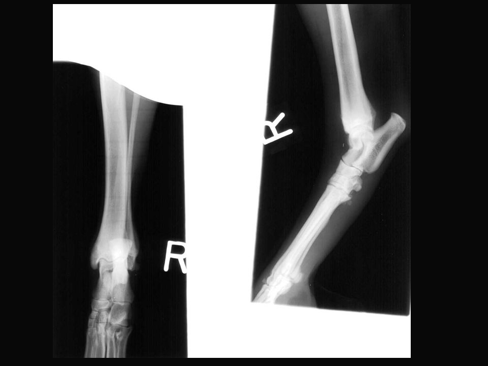
15
11-month old MN German Shepherd “Cisco” Hx: Intermittent lameness of right forelimb. Resists palpation of elbow.

16
11-month old MN German Shepherd “Cisco” RF –In the right elbow there is a defect in the medial humeral condyle. –In the left elbow a faint area of lucency is present in the medial humeral condyle. –Careful inspection of the left elbow reveals the presence of a tiny linear mineral opacity structure in the lucent area. This is the cartilage flap that has elevated from the bone and undergone mineralization. RD –Bilateral OCD of the medial humeral condyle

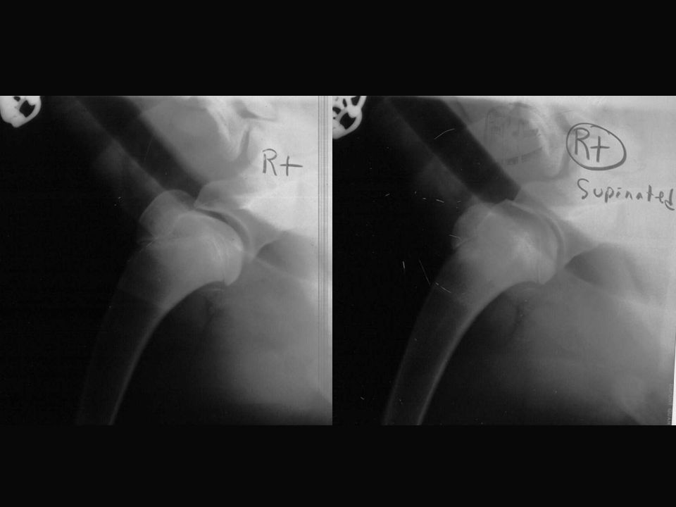
17
1-year old Rottweiler “Ruko” Hx: Left hind limb lameness. Mild swelling of the tarsus is present.

20
1-year old Rottweiler “Ruko” RF –There is mild soft tissue swelling surrounding the tarsus. –The subchondral bone of the medial ridge of the talus is abnormal. –The DP view of the right tarsus is normal. RD –Osteochondrosis of the medial ridge of the left talus

Similar presentations


 Shoulder Elbow Carpus Metacarpus Shoulder Elbow.>")


