Download presentation
Presentation is loading. Please wait.

2
Charachterized by: A nemia results when the rate of destruction exceeds the capacity of the m arrow to produce RBCs(The bone marro 8 times increased percentage of reticulocytes in the blood.

4
Etiologic classification of H. A. B – Extra corpuscular defects: acquired: 1. Immune hemolytic anemia: -- Isoimmune, as Rh & ABO hemolytic disease. -- Autoimmune. 2. Nonimmune hemolytic anemia: As occurs in severe burns, prosthetic heart valves, bacterial sepsis, malaria, venom & metallic poisoning as lead & copper A - Intracorpuscular defects; 1. R. C. membrane disorder as spherocytosis & ovalocytosis (elliptocytosis). 2. R. C. enzyme deficiencies: G6PD, pyrovate kinase deficiency. 3. Ineffective erythropoiesis :as Hemoglobinopathies as sickle cell disease & Hb C, D. E disease. thalassemia 4. Paroxysmal nocturnal hemoglobinuria.
. 2. R. C. enzyme deficiencies: G6PD, pyrovate kinase deficiency. 3. Ineffective erythropoiesis :as Hemoglobinopathies as sickle cell disease & Hb C, D. E disease. thalassemia 4. Paroxysmal nocturnal hemoglobinuria..")
5
Clinical manifestation: 1. Anemia: 2. Jaundice 3. Dark urine 4. Various degrees of splenomegaly. 5. Leg ulcers & gall stones are rare complications of some H.A. as S.C.A., thalassemia major & congenital spherocytosis

6
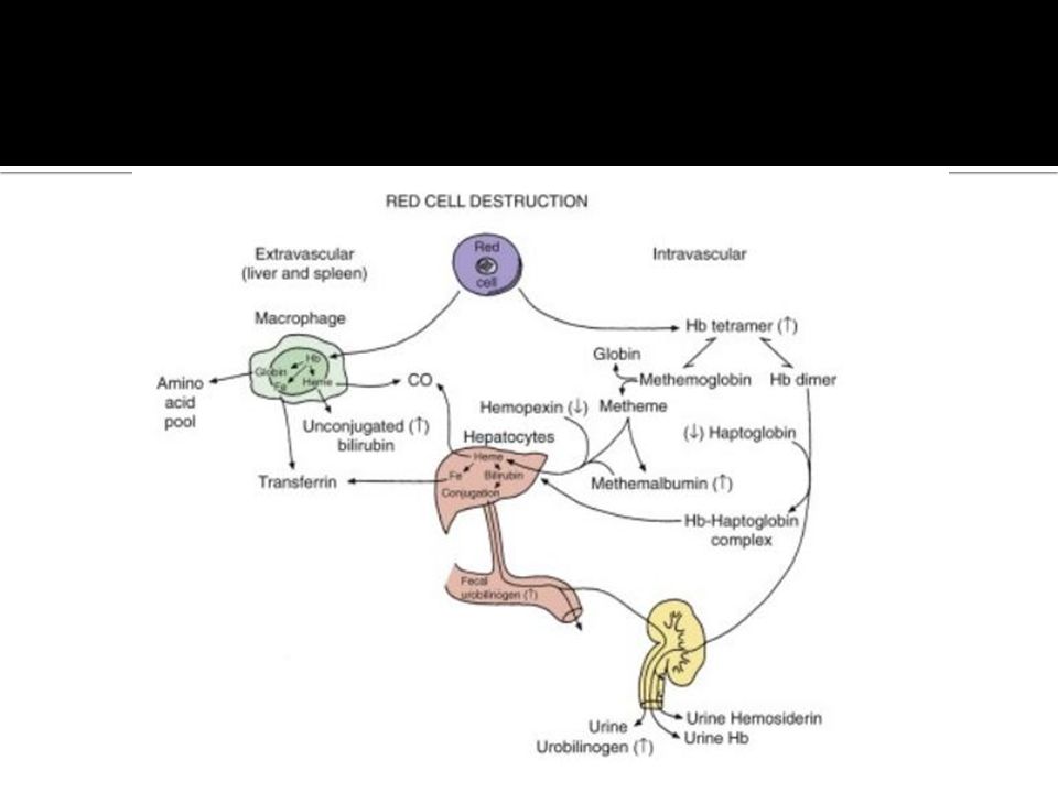
Laboratory: 1. Evidences of increased Hb breakdown: -- Hemoglobinemia (intravascular hemolysis) -- Raised serum bilirubin (mainly indirect bilirubin). -- Hemoglobinuria, released Hb ≥haptoglobin. -- Raised urine urobilinogen. -- Hemosiderinuria, 2. Evidences of increased erythropoiesis: -- Reticulocytosis, -- Increased normoblasts in the B. smear -- Bone marrow expansion, may produce, frontal bossing, mongoloid facies, bone pain & increased liability for fracture.
 -- Raised serum bilirubin (mainly indirect bilirubin). -- Hemoglobinuria, released Hb ≥haptoglobin. -- Raised urine urobilinogen. -- Hemosiderinuria, 2. Evidences of increased erythropoiesis: -- Reticulocytosis, -- Increased normoblasts in the B. smear -- Bone marrow expansion, may produce, frontal bossing, mongoloid facies, bone pain & increased liability for fracture..")
7
Laboratory: (cont.) 3. Morphological abnormalities of the R. C.: -- Spherocytes : spherocytosis& acquired H. A. -- Elliptocytosis congenital ovalocytosis & rarely in the other forms of H. A. -- Sickle cells in S. C. A. -- Fragmented RC in HUS -- Target cells, in thalassemia, HbC & S.C.A. -- Siderocytes :after splenectomy. (siderocytes are reticulocytes which contain iron granules, which are confirmed by “Prussian blue reaction”) 4. Evidences of shortened RC survival: 51Cr 5. Evidence of increased hemolysis; osmotic fragility test
 4. Evidences of shortened RC survival: 51Cr 5. Evidence of increased hemolysis; osmotic fragility test.")
8
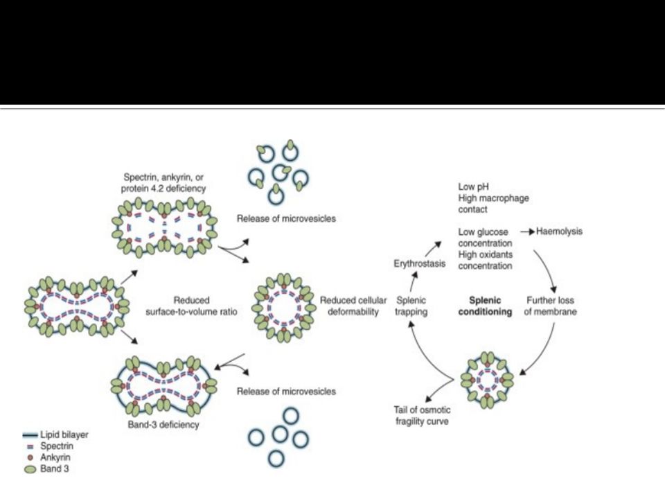
Definition:An A.D. inherited disorder characterized by R.C. stromal protein (spectrin) deficiency which makes the R.Cs. become spherical, rigid & more prone to lyse.
 deficiency which makes the R.Cs. become spherical, rigid & more prone to lyse..")
10
Clinical picture: Various degrees of anemia, jaundice splenomegaly hyperbilirubinemia (neonatal period) Aplastic crisis Hemolytic crisis is less common (leg ulcer and gall stone)
 Aplastic crisis Hemolytic crisis is less common (leg ulcer and gall stone)")
11
INV. Peripheral film shows microspherocytes MCH is normal but MCHC is increased. Increased reticulocyte count (except during aplastic crisis). Increased S. indirect bilirubin. Increased osmotic fragility, which becomes more exaggerated after R. C. incubation for 24 hour. Negative coombs test Hb electrophoresis shows normal Hb A. A, Hereditary spherocytosis. B, Hereditary elliptocytosis.
. Increased S. indirect bilirubin. Increased osmotic fragility, which becomes more exaggerated after R. C. incubation for 24 hour. Negative coombs test Hb electrophoresis shows normal Hb A. A, Hereditary spherocytosis. B, Hereditary elliptocytosis..")
12
Treatment: Splenectomy anemia & the accompanying symptoms. The crisis also disappears prevents gall stone formation (although spherocytosis & increased osmotic fragility however persist.) blood transfusion folic acid supplement are essential.
 blood transfusion folic acid supplement are essential..")
13
rare AD, Mild hereditary elliptocytosis produces no symptom s; more severe varieties can result in neonatal poikilocytosis (shape variation) and hemolysis In the rare instances when 2 abnormal alleles are inherited (HPP) severe hemolytic anemia abnormalities of α - and β-spectrin
 and hemolysis In the rare instances when 2 abnormal alleles are inherited (HPP) severe hemolytic anemia abnormalities of α - and β-spectrin")
14
Anemia, jaundice &splenomegaly are the main manifestations Reticulocyte count is increased osmotic fragility test & autohemolysis are normal. Splenectomy cures the symptomatic case Prognosis is good as longevity is not affected

15
Other rare R.C. shape defect which may cause H.A. -- Hereditary stomatocytosis; AR or AD in which RC with a slit- like central pallor predominates in the peripheral film. Some patients are symptomatic with anemia, jaundice & splenomegaly. Splenectomy may be beneficial. Hereditary acanthocytosis; an AR rare disorder in which there is marked irregularity of the RC surface. It’s seen in a syndrome called ”a- betalipoprotienemia): Steatorrhoea nervous system degeneration retinitis pigmentosa Symptoms are present since infancy & the condition is fatal during childhood. S. level of cholesterol is low
: Steatorrhoea nervous system degeneration retinitis pigmentosa Symptoms are present since infancy & the condition is fatal during childhood. S. level of cholesterol is low.")
17
This is an acquired type of H A which is caused by genetic deficiency of G6PD. is responsible for 2 clinical syndromes, episodic hemolytic anemia, chronic nonspherocytic hemolytic anemia. most com m on m anifestation : neonatal jaundice episodic acute hem olytic anem ia,

18
Hemolysis in the susceptible patients occurs after the administration of one of the following: Fava beans (ingestion or inhalation of its pollen), (Favism) Aspirin Sulphonamide as bactrim, & some food coloring agents which also contains sulfa. Furadantin & furazolidone Nalidixic acid (nigram) Paracetamol Antimalarials especially primaquine. Vit.K Phenacetin. Chloramphenicol. Naphthalene. Ciprofloxacin.
 Paracetamol Antimalarials especially primaquine. Vit.K Phenacetin. Chloramphenicol. Naphthalene. Ciprofloxacin..")
19
H.A. follows administration of the mentioned agents by 1-3 days jaundice, nausea, vomiting, epigastric pain & dark colored urine (hemoglobinurea). Types of epesodic H.A.: (G6PD A−) (chronic hemolysis) G6PD B− (G6PD Mediterranean).enz. Activity hemizygous males is <5% of norm al. (G6PD Canton) : common in chines
. Types of epesodic H.A.: (G6PD A−) (chronic hemolysis) G6PD B− (G6PD Mediterranean).enz. Activity hemizygous males is <5% of norm al. (G6PD Canton) : common in chines.")
20
Laboratory investigations: Rapid drop in Hb & R.C. counts Raised reticulocyte count Hemoglobinemia & hemoglobinurea Absent haptoglobin G6PD enzyme activity Treatment: Blood transfusion Sodium bicarbonate;

21
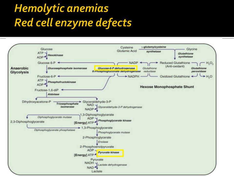
2- Pyrovate kinase deficiency; An A.R. disorder which may present in the neonatal period as jaundice &anemia. During infancy & childhood anemia, jaundice & splenomegaly are present. Osmotic fragility is normal. Reticulocytosis is present, with erythroid hyperplasia in the bone marrow. Diagnosis is confirmed by demonstration of reduced P.K. enzyme activity in the R.C.

22
Treatment: The severity of the disease decreases after childhood Exchange transfusion may be required in neonates. Blood transfusion on need Folic acid supplement Splenectomy

Similar presentations


. Introduction Hereditary spherocytosis is a class of hemolytic anemia. The disease occurs due to an intrinsic “membrane.>")


 Course code: MLHE-201 Supervisor: Prof. Dr Magda Sultan Outcome : The student will know : -The types of hemolytic anemias.>")


 A. Hereditary 1. Membrane defect (spherocytosis, elliptocytosis)>")


 Course code: MLHE-201 Supervisor: Prof>")