Download presentation
Presentation is loading. Please wait.

1
Otitis Media

2
Definition Inflammation of the middle ear. May also involve inflammation of mastoid, petrous apex, and peri-labyrinthine air cells

3
Classification Acute OM - rapid onset of signs & symptoms, < 3 wk course Sub-acute OM - 3 wks to 3 months Chronic OM - 3 months or longer

4
Acute Suppurative Otitis Media (ASOM)
Etiology Age: Common among children due to shorter Eustachian tube

5
Adenoiditis, Tonsillitis, Rhinitis, Sinusitis, Pharyngitis & infections secondary to cleft palate
Trauma to the TM Head injury Barotrauma

6
Pathology Catarrhal stage: is characterized by occlusion of Eustachian tube and congestion of middle ear. Stage of exudation: Exudate collects in the middle ear and ear drum is pushed laterally. Initially the exudate is mucoid, later it becomes purulent.

7
Pathology 3. Stage of suppuration: Pus in the middle ear collects under tension, stretches the drum & perforates it by pressure necrosis & the exudate starts escaping into external auditory canal 4. Stage of healing: The infection starts resolving from any of the stages mentioned & usually clears up completely without leaving any sequelae.

8
Pathology 5. Stage of complications: Infection may spread to the mastoid antrum. Initially it causes Catarrhal mastoiditis [congestion of the mastoid mucosa], stage of Coalescent mastoiditis & later empyeme of the mastoid

9
Clinical manifestations: ASOM
1. Catarrhal stage (stage of congestion) Fullness or heaviness in the ear Severe ear pain at night Deafness Tinnitus (ringing or buzzing in the ear) Autophony (spoken words of patient echo in his ears) TM (ear drum) gets retracted Cart wheel appearance of ear drum Absence of light reflex
 Fullness or heaviness in the ear. Severe ear pain at night. Deafness. Tinnitus (ringing or buzzing in the ear) Autophony (spoken words of patient echo in his ears) TM (ear drum) gets retracted. Cart wheel appearance of ear drum. Absence of light reflex.")
11
2. Stage of exudation All symptoms becomes more severe.

12
3. Stage of suppuration Perforation of Ear drum
Otorrhoea with mucoid purulent discharge Pulsatile discharge (ear discharge with each arterial dilation) [Lighthouse sign]
 [Lighthouse sign]")
13
4. Stage of healing Healing starts in this stage

14
5. Stage of complication Spread of infection to mastoid

15
Diagnosis Tuning fork test and audiometry Radiography
Bacteriological examination of the ear discharge Pneumatic otoscopy is gold standard

16
Treatment - AOM Systemic Local
Antibiotics: Tetracycline, erythromycin, ampicillin or penicillin for 6 days Systemic decongestants: Phenylephrine HCl Local Glycerine carbolic ear drops or warm olive oil reduces pain before perforation of TM. Antibiotic drops : Chloramphenicol, spirit boric drops is used after perforation of TM.

17
Surgery Myringotomy: The TM is incised to drain the middle ear cavity.
Myringo-puncture: Puncturing the ear drum with a long thick injection needle & aspirating the middle ear contents.

18
Chronic Otitis Media It is the chronic infection of middle ear cleft mucosa. Chronic Suppurative Otitis Media (CSOM): accompanied by continuous or intermittent otorrhoea Chronic Non-Suppurative Otitis Media: No otorrhoea Chronic Specific Otitis Media: Tb OM or syphilitic OM
: accompanied by continuous or intermittent otorrhoea. Chronic Non-Suppurative Otitis Media: No otorrhoea. Chronic Specific Otitis Media: Tb OM or syphilitic OM.")
19
CSOM Benign or tubotympanic type with central perforation of the ear drum: The disease is limited to the TM & the Eustachian tube. No complications occur as a rule. Dangerous or Attico-antral type with attic and marginal perforation: It is characterised by the presence of destructive cholesteatoma, which may spread beyond the ear cleft causing life threatening complications

21
Etiology:CSOM AOM which fails to heal. Acute necrotic OM
Traumatic large perforation Congenital cholesteatoma

22
Benign or tubotympanic type
Pathology Benign or tubotympanic type Etiological factors Necrosis of ear drum portion which has poor blood supply Necrosis of ossicular chain Sclerosis of mastoid bone Polyp formation

23
Dangerous / Attico-antral type
Cholesteatoma formation Polyps and granulation Perforation and retraction of ear drum Partial or complete damage of Ossicles

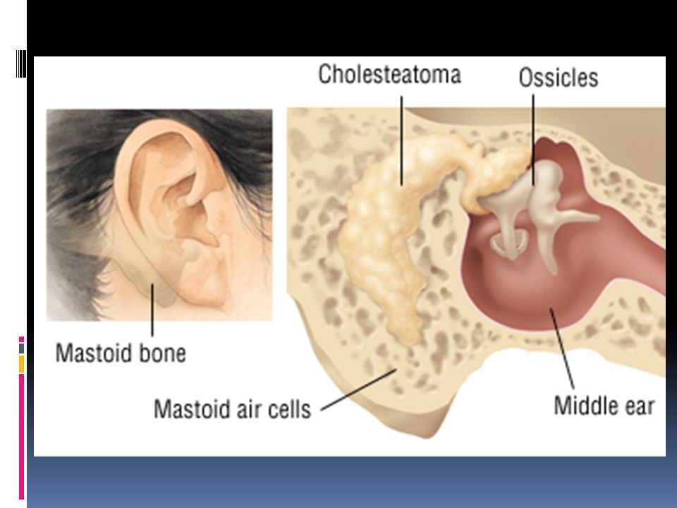
25
Cholesteatoma

26
Clinical stages Benign perforation Dangerous perforation
Active stage : discharge is actively flowing Quiescent stage : ear remains dry for up to 6 months Inactive stage : Ear remains dry for > 6 months Dangerous perforation Active stage Inactive stage

27
Diagnosis Examination of nose and pharynx to find any septic focus or an obstruction around the Eustachian tube Hearing test [voice test, tuning fork test, audiometry]: Conductive deafness up to 60 db hearing loss Radiology of the mastoid

28
Diagnosis Testing the patency of Eustachian tube:
Using ear drops Using Valsalva maneuver Otomicroscopy: perforation, cholesteatoma, polyps

29
Management of Benign Perforation
Adenoidectomy, tonsillectomy; treatment of sinusitis & DNS to remove the septic focci Antibiotic ear drops Chemical cautery using 50% trichloro acetic acid TT injection Tympanoplasty: Reconstruction of middle ear and ossicular chain after removing the active disease Myringoplasty : repair of defect in TM

30
Management of dangerous perforation
Suction and cleaning of cholesteatoma Excision of polyps and granulomas Mastoidectomy Atticotomy & atticoantrostomy tympanoplasty

31
Complications Mastoiditis Mastoid abscess Petrositis
Mastoid infection Extracranial complications Intracranial complications Mastoiditis Mastoid abscess Petrositis Facial nerve palsy Labyrinthitis Extradural abscess Subdural abscess Meningitis Sigmoid sinus thrombophlebitis Brain abscess Otitis hygrocephalus

32
Thank You

Similar presentations


.>")


. >")
 is inflammation.>")

>")

