Download presentation
Presentation is loading. Please wait.

1
Herniorrhaphy SUR 111

2
Hernias Definition Hernia orifice Hernia sac
Protrusion of the viscus (viscera) or abdominal organs through an opening in the wall of the cavity in which it is contained Hernia orifice Abdominal wall defect Hernia sac Out-pouching of peritoneum
 or abdominal organs through an opening in the wall of the cavity in which it is contained. Hernia orifice. Abdominal wall defect. Hernia sac. Out-pouching of peritoneum.")
3
Hernia pathology Congenital = Indirect hernia Acquired = Direct hernia
Both or combination = Pantaloon hernia

4
Hernia pathology Congenital = Indirect hernia
Follows the congenital defects that dilate the internal inguinal ring and pass though the deep inguinal ring into the scrotum. An indirect inguinal hernia is an inguinal hernia which results from the failure of embryonic closure of the internal inguinal ring after the testicle has passed through it. Mostly seen in males, due to testicular decent.

5
Indirect hernia

6
Hernia pathology Acquired = Direct hernia
Hernia sac passes within Hasselbach’s Triangle; breaches posterior inguinal wall (bulges “directly” through abdominal wall); passes medial to inferior epigastric artery; Goes through external inguinal ring only.
; passes medial to inferior epigastric artery; Goes through external inguinal ring only.")
7
Hesselbach’s Triangle
Hesselbach’s Triangle defined by Medially – lateral border of rectus abdominis Laterally – inferior epigastric vessels Base – inguinal ligament

8
May also be described as…
Reducible Contents easily put back Irreducible/Incarcerated - Contents cannot be put back Strangulated - Contents are stuck, and there is constriction of the tissues at the neck of the hernia, leading to reduced venous drainage and arterial occlusion

9
Hernias Based on Anatomical Location
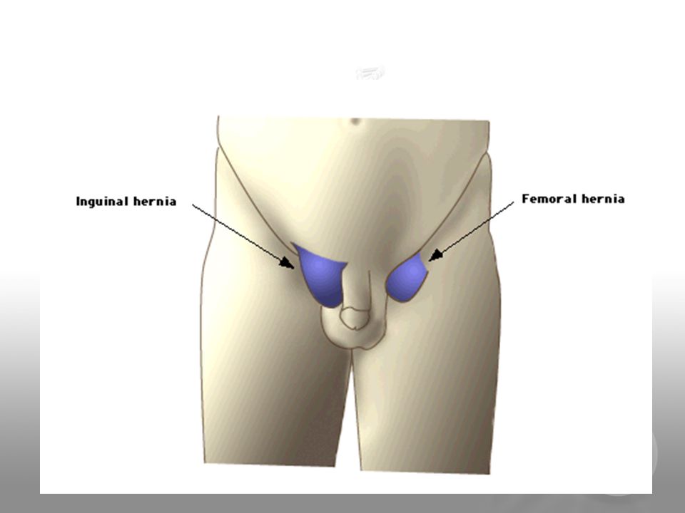
Groin Inguinal - 75% are this type. 50% are Indirect Inguinal hernias. 25% of males will have an inguinal hernia vs. 2% of females. More frequent on right than left side of groin. Femoral Ventral Incisional Diaphragmatic Esophageal hernia or hiatal hernia

10
incisional

12
2 Main Types of Inguinal Hernias

13
Which Type of Inguinal Hernia?

16
Symptoms of direct and indirect hernias
Bulge that enlarges when stand or strain, but can be asymptomatic Pain or dull sensation in groin Patients can present with complication (obstruction or strangulation-->10-20% of pts with inguinal hernia present with strangulation)
")
17
Hernia Repair Goal Identify hernia Reduce hernia Repair defect
27 min hernia video - good intro

18
Surgical Procedures Inguinal Hernias
Open repair of indirect inguinal hernia Anatomy of the suprapubic and inguinal region The inguinal canal begins as a defect or split in the transversalis fascia at the deep inguinal ring and continues to the superficial inguinal ring. In the male, this space is larger than in the female; this corresponds to the higher incidence of inguinal hernias in males. The spermatic cord, which follows the inguinal canal, contains the following structures: Spermatic fascia Cremaster muscle Genitofemoral nerve Ductus deferens Lymph vessels Pampiniform veins, which form the testicular vein Testicular artery Incising aponeurosis of external oblique muscle Blunt dissection of the hernia sac from the spermatic cord (Colorized from Moody FG: Atlas of ambulatory surgery, St Louis, 1999, Mosby.)
")
19
Inguinal Hernias, continued
Open repair of indirect inguinal hernia When tissue from within the abdomen pushes out through the defect, it may emerge between the layers. This is referred to as a hernia. Tissue that becomes entrapped in the defect is referred to as incarcerated hernia. If pressure on the incarcerated tissue is not relieved by reducing the hernia (replacing tissue to its correct anatomical location), its vascular supply can be cut off. This is referred to as a strangulated hernia. When tissue is strangulated, a tourniquet effect occurs at the site of the protrusion. This can lead to tissue necrosis. Strangulated bowel tissue can result in bowel obstruction—a surgical emergency An indirect inguinal hernia is a protrusion of abdominal viscera that traverses the inguinal canal from the deep inguinal ring and may extend through the superficial inguinal ring into the scrotum (or labia majorus of the female). In the male, the hernia extends along the length of the membrane covering the spermatic cord and can emerge though the superficial ring, causing a bulge under the skin. Opening the hernia sac Placing the purse-string suture at the neck of the hernia sac (Colorized from Moody FG: Atlas of ambulatory surgery, St Louis, 1999, Mosby.)
, its vascular supply can be cut off. This is referred to as a strangulated hernia. When tissue is strangulated, a tourniquet effect occurs at the site of the protrusion. This can lead to tissue necrosis. Strangulated bowel tissue can result in bowel obstruction—a surgical emergency. An indirect inguinal hernia is a protrusion of abdominal viscera that traverses the inguinal canal from the deep inguinal ring and may extend through the superficial inguinal ring into the scrotum (or labia majorus of the female). In the male, the hernia extends along the length of the membrane covering the spermatic cord and can emerge though the superficial ring, causing a bulge under the skin. Opening the hernia sac. Placing the purse-string suture at the neck of the hernia sac. (Colorized from Moody FG: Atlas of ambulatory surgery, St Louis, 1999, Mosby.)")
20
Open repair of indirect inguinal hernia
Open Inguinal Hernia with Patch Graft Open repair of indirect inguinal hernia Technique: A right or left inguinal incision is made. Layers of the abdominal wall are incised and the edges retracted. The spermatic cord is dissected away from preperitoneal fat and other surrounding tissues. The spermatic cord is retracted laterally with a small penrose drain. The indirect hernia sac is dissected from the cord and opened, and its content is pushed back into the abdomen. The hernia sac is ligated with ties or a purse-string suture. A synthetic mesh patch or plug is sutured in place over the defect. Abdominal wall is closed in layers. Suturing the mesh graft (Colorized from Moody FG: Atlas of ambulatory surgery, St Louis, 1999, Mosby.)
")
21
Laparoscopic Inguinal Hernia Repair
Laparoscopic direct hernia repair Surgical exposure during the laparoscopic approach is through transabdominal preperitoneal (TAPP) laparoscopy or by total extraperitoneal (TEP) surgery. In the TAPP approach, a pneumoperitoneum is performed and the inguinal canal entered from the abdominal cavity. The TEP approach avoids a pneumoperitoneum by inflating and entering the preperitoneal space with a balloon dissector, which functions as a tissue expander. Technique: Pneumoperitoneum is established and ports inserted in the abdomen. A small transverse incision is made above the direct hernia space (see Figure 21–7B). The weakened area in the pelvic floor is covered with a synthetic patch or other mesh system. The mesh is secured without tension using endoscopic staples or sutures. The peritoneum is closed. The pneumoperitoneum is released and port incisions are closed. Incising the peritoneum (Colorized from Ballantyne GH: Atlas of laparoscopic surgery, Philadelphia, 2000, Saunders.)
 laparoscopy or by total extraperitoneal (TEP) surgery. In the TAPP approach, a pneumoperitoneum is performed and the inguinal canal entered from the abdominal cavity. The TEP approach avoids a pneumoperitoneum by inflating and entering the preperitoneal space with a balloon dissector, which functions as a tissue expander. Technique: Pneumoperitoneum is established and ports inserted in the abdomen. A small transverse incision is made above the direct hernia space (see Figure 21–7B). The weakened area in the pelvic floor is covered with a synthetic patch or other mesh system. The mesh is secured without tension using endoscopic staples or sutures. The peritoneum is closed. The pneumoperitoneum is released and port incisions are closed. Incising the peritoneum. (Colorized from Ballantyne GH: Atlas of laparoscopic surgery, Philadelphia, 2000, Saunders.)")
22
Trocar Placement in Laparoscopic Hernia Repair
Laparoscopic direct hernia repair To create a pneumoperitoneum, trocars are placed in the abdomen. A primary port is placed in the umbilicus and secondary ports lateral to the rectus muscles (see Figure 21–7A). Placement of ports
. Placement of ports.")
24
Transabdominal Extraperitoneal (TEP) Inguinal Hernia Repair
A balloon expander inserted into the incision and inflated with air or normal saline Technique: A small periumbilical incision is made through the rectus sheath. Tissues are manually dissected and then retracted. A balloon tissue expander is introduced. The preperitoneal space is inflated and the expander removed. A balloon trocar may be inserted to seal the space and hold the trocar. Two additional 5 mm ports are created. The direct or indirect hernia is reduced, and polypropylene mesh is secured over the defect. The wounds are closed as for TAPP.

25
Femoral Hernia Surgical Goal
An open repair of a femoral hernia is performed to restore strength to the inguinal floor and prevent abdominal tissue from protruding into the inguinal canal The femoral hernia occurs most commonly in the female. The abdominal wall defect allows abdominal tissue to descend into the femoral canal inferior to the inguinal ligament. The femoral hernia is more likely to become strangulated than the inguinal hernia. It is apparent as a bulge below the crease line of the leg and inguinal ligament. Technique: The groin is incised on the affected side. The hernia sac is identified and opened. The sac is ligated and removed. Synthetic mesh is secured over the defect. The wound is closed in layers as for inguinal hernia.

26
Incisional or Ventral Hernia
To remodel a previous abdominal wall scar and provide sufficient strength to prevent a recurring hernia 58 min Ventral Hernia Video Short version: 4 min Lap Ventral Hernia repair A ventral or incisional hernia usually, but not always, occurs at the incisional site of a previous surgery. Weakness in the tissue layers can develop from a number of different causes: Previous or concurrent surgical site infection Extensive strain on the incision due to obesity (morbid obesity in particular) Poor tissue healing related to metabolic disease such as diabetes or alcoholism or infection Repeated surgeries in the same location Instrumentation for this procedure will be determined by the size of the patient as well as the size of the hernia. Examples: A five-year-old pediatric patient vs. a 300-pound adult patient
 Poor tissue healing related to metabolic disease such as diabetes or alcoholism or infection. Repeated surgeries in the same location. Instrumentation for this procedure will be determined by the size of the patient as well as the size of the hernia. Examples: A five-year-old pediatric patient vs. a 300-pound adult patient.")
27
Umbilical Hernia Repair
Completed to Repair Weakening of Abdominal Wall Around or Under Umbilicus Lap repair - 9 min Lap Umbicial Hernia repair Umbilical hernias are common in children and frequently disappear spontaneously by age two. In adults, these types of hernias appear more frequently in obese people. Umbilical hernias are potentially dangerous because they have small necks and frequently incarcerate. Surgical intervention is indicated in all adult asymptomatic umbilical hernias.

28
Spigelian Hernia Repair
Completed to Reduce Protrusion of Abdominal Viscera in “Spigelian Zone” SPIGELIAN hernias are ventral hernias occurring through the spigelian fascia along the Spieghels semilunar line and lie under the external oblique aponeurosis just outside the outer border of the Rectus or "six-pack" muscles. This procedure is completed to reduce a protrusion of a peritoneal sac, preperitoneal fat, or other abdominal viscera through the congenital or acquired defect in the muscle wall referred to as the “spigelian zone.” The “spigelian zone” is defined as the area of aponeurosis that lies between the linea semilunaris of the transverse abdominis muscle and the lateral edge of the rectus muscle.

29
Spigelian Hernia Repair

30
Anticipating Potential Problems With Abdominal Surgery
Any time you are cutting into the body there is a risk of something being cut that was not meant to be cut Depending on what structure is accidentally cut into, will determine what is needed by the surgical technologist Vascular structures will require sutures that are non-absorbable such as silk, Prolene or Ticron on a taper needle Other structures can often be repaired with chromic, Vicryl or Dexon on a taper needle

Similar presentations