Download presentation
Presentation is loading. Please wait.

1
The microscopic study of tissue. It is important to know the structure and function of tissues In order to understand how individual cells are organized And how tissues work to form organs and organ systems.

2
Learner Objectives To analyze the relationships between the structures and functions of tissues. To evaluate the cause and effect of disease and trauma on the structure and function of tissues. To research embryological development of tissues.

3
Epithelial Tissues Overall classification is based on the fact that they have very little extra-cellular matrix between the cells. These tissues cover organs, form structures, and has a “free surface.” They all have basement membrane – opposite the free surface; binds cells to underlying tissues. Blood supply does not penetrate the basement membrane. Diffusion of all gasses and nutrients; cells close to basement membrane are active, and those further away will die in stratified tissue.

4
Classification of Epithelium – by number of cell layers and the shape of cells. Simple- made of a single layer Stratified-made of two or more layers Pseudostratified-simple tissue appears to be layered Squamous-squished cells flattened and shape irregular Cuboidal- looks like a cube Columnar- rectangular shape columns Transitional- change between cuboidal and squamous

5
Cell Surfaces 1)Free surface – faces away from underlying tissue 2)Lateral - surfaces which face other cells 3)Basal – surface facing the basement membrane If the free surface is smooth: If the free surface has microvilli: If the free surface has cilia:
Free surface – faces away from underlying tissue 2)Lateral - surfaces which face other cells 3)Basal – surface facing the basement membrane If the free surface is smooth: If the free surface has microvilli: If the free surface has cilia:")
6
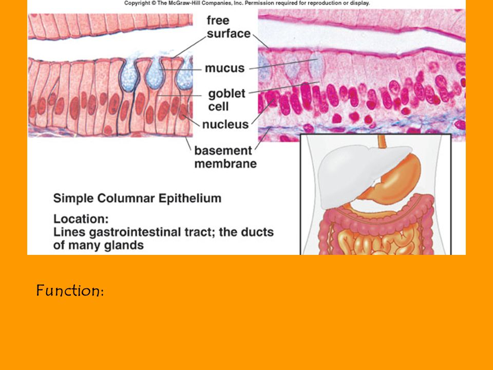
Function:

10
Connective Tissue

11
The essential Characteristic that distinguishes Connective tissue from the other 3 types of tissue is The non-living extra Cellular matrix. The specialized cells of the various Connective tissues are –Blasts, -Cytes, and -Clasts. The structure of the matrix gives Connective tissue types most of their Functional characteristics, Such as the Ability of bones and cartilage to bear weight, Of tendons and ligaments to withstand Tension, and the Dermis to withstand abrasions.

12
The Matrix is composed of: 1)Protein Fibers (Collagen, reticular, And Elastin). 2)Ground Substance – the shapeless Background against which the fibers Are seen. Hyaluronic acid is a long, unbranched, Polysaccharide chain composed of Repeating disaccharide units. It gives a very slippery quality to the Fluids that contain it. Proteoglycans are formed from proteins And polysaccharides – it is a molecule That can trap large quanities of water, Which gives them the capacity to return To their original shape when compressed.
Ground Substance – the shapeless Background against which the fibers Are seen. Hyaluronic acid is a long, unbranched, Polysaccharide chain composed of Repeating disaccharide units. It gives a very slippery quality to the Fluids that contain it. Proteoglycans are formed from proteins And polysaccharides – it is a molecule That can trap large quanities of water, Which gives them the capacity to return To their original shape when compressed..")
13
Sub-Classifications (Based on Matrix) 1.Fibrous A. Dense - Irregular (dermis of skin) - Regular (ligaments & tendons) 2.Fluid A. Blood 3.Solid A. Bone B. Cartilage
 - Regular (ligaments & tendons) 2.Fluid A. Blood 3.Solid A. Bone B. Cartilage.")
14
4. Special Connective Tissue Adipose, reticular and hemopoietic Adipose consists of adipocytes and comes In yellow and brown forms. Reticular tissue forms the framework of Lymphatic tissue, bone marrow, and the Liver. Hemopoietic is blood forming tissue – it is Mostly found in the marrow – Red marrow.

15
Fig. 4.6

16
Loose (Fibrous) Connective Tissue Areolar The protein fibers form a lacy network with numerous Fluid filled spaces. (Think of packing peanuts- hold organs in their places) Loose packing material in organs and attachment to the Skin. It contains collagen and elastin fibers and a variety of Cells, but mostly fibroblasts.
 Loose packing material in organs and attachment to the Skin. It contains collagen and elastin fibers and a variety of Cells, but mostly fibroblasts..")
17
Fig. 4.5

18
Dense (Fibrous) Connective Tissue Regular Protein fibers packed and fill almost all of the extra Cellular space. The fibers are oriented predominately in one direction. (Tendon or Ligaments) Irregular Protein fibers packed and fill almost all of the extra Cellular space. The fibers are oriented in a random meshwork. (Dermis of the skin)
 Irregular Protein fibers packed and fill almost all of the extra Cellular space. The fibers are oriented in a random meshwork. (Dermis of the skin).")
19
Fig. 4.7

20
Solid Connective Tissue Matrix with both protein fibers and ground substance (50/50) Cartilage – composed of chondrocytes located in spaces within the crystalized matrix called lacunae (little gap where cell lives); protein is within the matrix (collagen and elastin) plus proteoglycan aggregate (hyaluronic acid and proteoglycan). * Lack of blood supply = slow healing 3 types: Hyaline – large amounts of collagen and proteoglycans; fine collagen fibers evenly spaced in the ground substance (rib cage and trachea and joints). Fibrocartilage – more collagen than proteoglycans; thicker bundles of protein; slightly compressible and very tough (jaw, between vertebrae) Elastic cartilage – elastic fibers (ears)
. Fibrocartilage – more collagen than proteoglycans; thicker bundles of protein; slightly compressible and very tough (jaw, between vertebrae) Elastic cartilage – elastic fibers (ears).")
21
Fig. 4.8

22
Solid Connective Tissue Matrix with both protein fibers and ground substance (50/50) Bone – Hydroxyapatite (mineral inorganic portion- calcium phosphate crystals of bone); osteocytes (bone cells in the lacunae). 2 types: Cancellous – spongy; has spaces between trabeculae (bony bars & plates). Designed for strength. Compact – compact, has no spaces between the lamina (thin layers of bone that wrap in a circular pattern.) Provides reinforcement of body structure (elasticity And strength).
. Designed for strength. Compact – compact, has no spaces between the lamina (thin layers of bone that wrap in a circular pattern.) Provides reinforcement of body structure (elasticity And strength)..")
23
Fig. 4.9

24
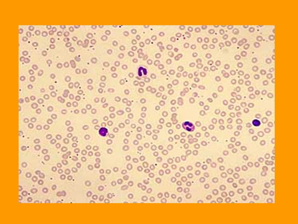
Predominantly Fluid Matrix Blood – composed of formed elements and plasma. Plasma is the liquid matrix with dissolved ions and nutrients. The formed elements are the cells and cell fragments: Erythrocytes – red blood cells Leukocytes – White blood cells Thrombocytes – platelets (fragments of giant cells found in bone marrow) The fluid nature of blood allows movement of cells, Allowing transportation of oxygen, nutrients, wastes, etc.
 The fluid nature of blood allows movement of cells, Allowing transportation of oxygen, nutrients, wastes, etc..")
26
Muscle Tissue Muscle tissue is contractile, and through contractions is designed to move the body, pump blood, and decrease the size of hollow organs. 3 types are classified by structure and function: Skeletal Cardiac Smooth

27
Table 4.3

28
Fig. 4.11

29
Fig. 4.12

30
Fig. 4.13

31
Nervous Tissue Conducts electrical signals called “action potentials” Neurons – nerve cells – composed of 3 parts: cell body, dendrites, and axons. Types of neurons: Multipolar Bipolar Unipolar Support cells – support cells of the brain, spinal cord, and peripheral nerves. Also form myelin sheaths around axons and produce cerebrospinal fluid. Schwann cells neuroglia

32
Fig. 4.14

33
End of all tissues structure and function

Similar presentations


 Connective.>")


