Download presentation
Presentation is loading. Please wait.

1
The Salivary Glands diseases

2
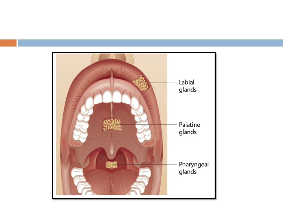
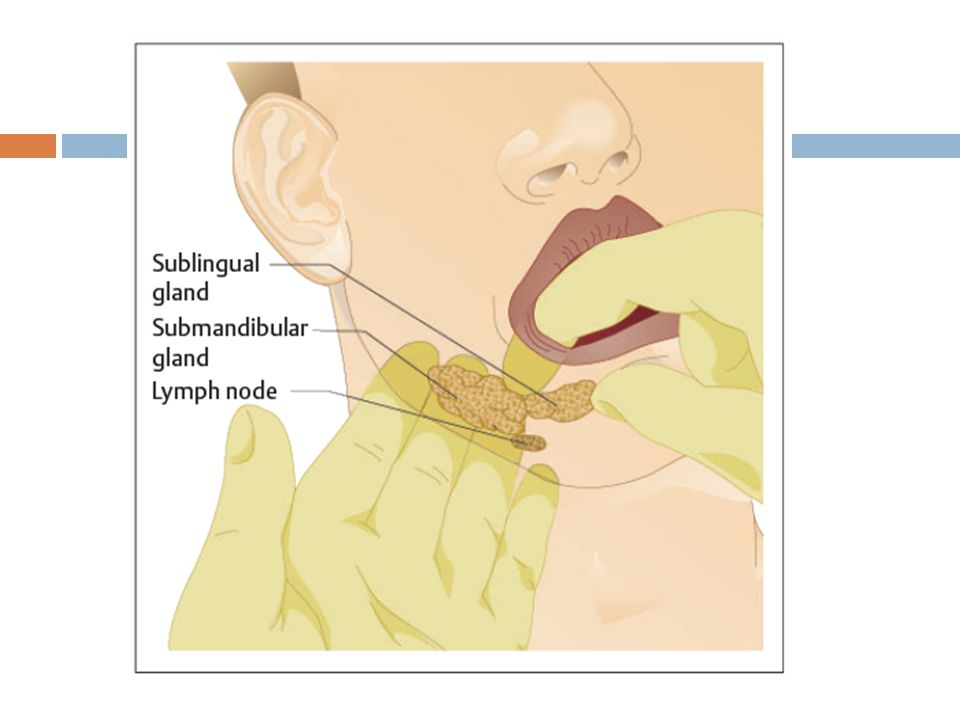
Clinical Anatomy of the Salivary Glands

7
Reactive lesions Mucocele is a clinical term that includes mucus extravasation phenomenon and mucus retention cyst.

9
Mucocele Etiology Extravasation type
Physical-traumatic injury to minor gland excretory duct Mucus extravasation into periductal soft tissue produces a local inflammatory response and granulation tissue “encapsulation.”

10
Clinical Presentation
Lower lip most common site; also buccal mucosa, anterior ventral tongue Painless bluish hue when mucin is near surface Often waxes and wanes in size Microscopic Findings Mucus pool surrounded by granulation tissue Macrophage and neutrophil response to free mucin Focal chronic sialadenitis

12
Differential Diagnosis
Presentation Microscopic findings Differential Diagnosis Hemangioma/ varix Pyogenic granuloma Salivary neoplasm Connective tissue neoplasm

13
Treatment Excision with associated local minor salivary glands Prognosis Occasional recurrence

14
Mucus Retention Cyst Etiology
Represents dilatation of salivary excretory duct due to obstruction Duct obstruction may be due to a mucous plug or sialolith formation

15
Clinical Presentation
Major or minor salivary glands affected in adulthood Asymptomatic, soft mucosal swelling Can occur at any intraoral minor salivary gland site, especially upper lip

18
Differential Diagnosis
Microscopic Findings Thin, dilated, epithelial-lined salivary excretory duct Lining is cuboidal to columnar with occasional mucus-producing cells present Adjacent salivary gland lobules minimally altered but may show obstructive inflammatory changes Diagnosis Microscopic findings Differential Diagnosis Extravasational mucocele Salivary gland neoplasm, especially mucoepidermoid carcinoma

19
Treatment Prognosis Excision of cyst with adjacent gland(s)
Recurrence is rare.

20
Necrotizing Sialometaplasia
Etiology Local ischemic injury of salivary gland lobules May be preceded by trauma or local anesthetic injury, or it may appear spontaneously

21
Clinical Presentation
Both major and minor salivary glands can be affected. Hard palate most common site, usually unilateral Initially a painful to dysesthetic submucosal swelling Ultimately, a central necrotic crater develops. May extend to and involve deep soft tissue and palatal bone

23
Microscopic Findings Salivary gland inflammation and lobular necrosis (necrosis is not always demonstrable on biopsy) Ductal squamous metaplasia (bland cytology) Lobular architecture of salivary glands persists
 Lobular architecture of salivary glands persists.")
24
Differential Diagnosis
Microscopic findings Differential Diagnosis Salivary gland neoplasm Squamous cell carcinoma Granulomatous disease

25
Treatment Follow-up only Prognosis Excellent

26
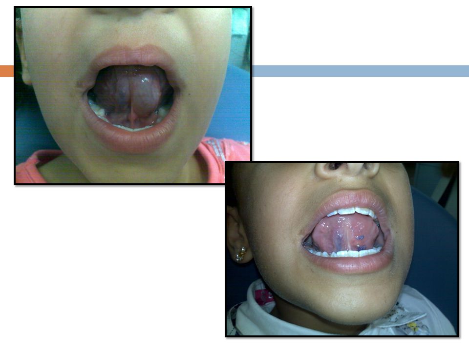
Ranula Etiology Obstruction of the sublingual (usually) or submandibular salivary gland by a sialolith or by trauma Secondary to obstruction, extravasation of saliva into the soft tissue of the floor of the mouth

27
Clinical Presentation
Unilateral, fluctuant, soft tissue mass on the floor of the mouth Usually has a bluish, slightly translucent quality When above the mylohyoid muscle, presentation is intraoral. If extravasation extends below the mylohyoid muscle, a plunging ranula forms. Occlusal radiographs may demonstrate a suspected sialolith.

29
An unusual clinical variant, the plunging or cervical ranula, occurs when the spilled mucin dissects through the mylohyoid muscle and produces swelling within the neck.

31
Diagnosis Demonstration of sialolith
Soft tissue imaging (T2-weighted magnetic resonance image) Aspiration of mucinous salivary fluid Excised tissue with granulation tissue lining around mucin pool
 Aspiration of mucinous salivary fluid. Excised tissue with granulation tissue lining around mucin pool.")
32
Differential Diagnosis
Dermoid cyst Salivary gland tumor Soft tissue tumor Cystic hygroma Thymic cyst

33
Treatment Prognosis Marsupialization as an initial procedure
Excision of the involved gland (extravasation type) Sialolithectomy (in obstructive type) Prognosis No recurrence with sialadenectomy Recurrence risk with sialolithectomy secondary to duct scarring or reformation of stone
 Sialolithectomy (in obstructive type) Prognosis. No recurrence with sialadenectomy. Recurrence risk with sialolithectomy secondary to duct scarring or reformation of stone.")
34
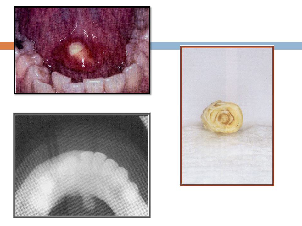
Sialolithiasis Etiology
Sialoliths are calcified structures that develop within the salivary ductal system. They are believed to arise from deposition of calcium salts around a nidus of debris within the duct lumen. This debris may include inspissated mucus, bacteria, ductal epithelial cells, or foreign bodies. The cause of sialoliths is unclear, but their formation can be promoted by chronic sialadenitis and partial obstruction. Their development is not related to any systemic derangement in calcium and phosphorus metabolism.

35
Salivary obstruction from stones, mucous plugs, and mucous extravasation phenomenon.

36
Clinical Presentation
Sialolithiasis can form in all of the salivary glands, including minor salivary glands, but the gland that most commonly produces such stones is the submandibular gland. The so‐called stones that form in the parotid duct system are rarely calcified and are actually mucous plugs that do not appear on radiographs. Stones that form in the submandibular duct system are almost always radiopaque because they are composed of calcium carbonate and calcium phosphate.

37
They can occur along any part of the duct and are most frequent at anatomic bends.
In the submandibular duct, stones are often found at the duct's bend around the posterior edge of the mylohyoid muscle. When sialoliths form, they will obstruct the duct either partially or completely. Therefore, individuals present with a painful swelling of the gland and usually with signs of secondary infection, including a suppurative exudate from the duct, fever, and mild to moderate leukocytosis . Individuals will report an increase in pain and swelling upon eating. The gland will be palpably firm and anywhere from mildly tender to very painful.

40
Diagnosis CT scan with 2‐ to 3‐mm cuts to identify the location of the stone. Most obstructed submandibular glands will have both inflammation and fibrosis in a homogeneous pattern throughout the gland.

41
Differential Diagnosis
Calcified lymph nodes from previously resolved tuberculosis Phleboliths (particularly if an old cavernous hemangioma were present) Tonsoliths Calcifications of the carotid bifurcation.
 Tonsoliths. Calcifications of the carotid bifurcation.")
42
Treatment Stones that are accessible in the floor of the mouth are removed via a direct approach, and the damaged duct is sutured to the mucosa of the floor of the mouth (sialodochoplasty). Parotid stones usually do not produce a long‐term clinical problem. Most are passed with parotid flow, and a few require removal from the duct with either a repair or duct transposition.
. Parotid stones usually do not produce a long‐term clinical problem. Most are passed with parotid flow, and a few require removal from the duct with either a repair or duct transposition.")
43
Prognosis If the sialolith has been present for a short time, the gland may recover after sialolith removal. If the sialolith is of long standing, the gland may harbor irreversible inflammation and fibrosis, so that it cannot recover even if the sialolith is removed.

44
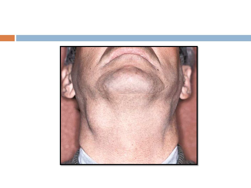
Sialorrhea (Sialosis)
Etiology Varied; may include idiopathic paroxysmal sialorrhea, parkinsonism, stomatitis (acute), newly inserted oral appliances, expectorants, neostigmine, and others
, newly inserted oral appliances, expectorants, neostigmine, and others.")
45
Clinical Presentation
Excess saliva resulting in drooling Angular cheilosis Diffuse parotid/submandibular salivary gland enlargement

47
Diagnosis Direct observation and analysis of history
Flow-rate measurement

48
Treatment Scopolamine If related to medication use, an alternate medication should be chosen, if possible. Prognosis Guarded/indeterminate

49
Adenomatoid hyperplasia
Etiology It is a nonneoplastic enlargement of the minor salivary glands of the hard palate. The cause is unknown, although there is some evidence to suggest that trauma plays a role.

50
Clinical Presentation
The palate is the chief site of involvement of this salivary gland hyperplasia. There is a male predominance, and age ranges from 24 to 63 years. The clinical presentation is a unilateral swelling of the hard and/or soft palate. This lesion is asymptomatic, broad based, and covered with intact mucosa of normal color and quality.

51
Differential Diagnosis
Salivary neoplasms Lymphoma Extension of nasopharyngeal or sinonasal disease into the oral cavity.

52
Treatment Subsequent to identification by means of an incisional biopsy, no treatment is necessary, given the purely benign nature of this process. Prognosis There is no neoplastic potential.

53
Sjögren’s Syndrome Etiology
An autoimmune disease resulting in exocrine gland dysfunction secondary to mononuclear cell infiltration Increased prevalence of human leukocyte antigen DR/DQ alleles Autoantibody production against nuclear antigens SS-A and SS-B No specific agent identified; postulations include the following: Potential role for viruses/retroviruses as cofactors Possible role of cytokine and hormonal influence on signal transduction and secretion

54
Clinical Presentation
Decrease in exocrine gland function Xerostomia Xerophthalmia/keratoconjunctivitis sicca Salivary and lacrimal gland enlargement (one-third of cases) Secondary effects of exocrine dysfunction are as follows: Dental caries Oral candidiasis Ocular/corneal discomfort Primary form: exocrine dysfunction dominates Secondary form: exocrine dysfunction; other associated autoimmune conditions—usually rheumatoid arthritis, less often lupus erythematosus
 Secondary effects of exocrine dysfunction are as follows: Dental caries. Oral candidiasis. Ocular/corneal discomfort. Primary form: exocrine dysfunction dominates. Secondary form: exocrine dysfunction; other associated autoimmune conditions—usually rheumatoid arthritis, less often lupus erythematosus.")
58
Diagnosis Demonstration of objective xerostomia and xerophthalmia
Serologic demonstration of associated SS-A or SS- B antibodies Correlation of clinical and serologic findings with labial salivary gland biopsy; demonstration of presence of periductal lymphocytic sialadenitis

59
Differential Diagnosis (Xerostomia/Parotid Gland Swelling)
• Sarcoidosis • Depression • HIV- associated • Autonomic neuropathy exocrinopathy • Graft-versus-host disease • Drug side effects • Alcoholism • Lymphoma • Diabetes mellitus • Bulimia

60
Treatment Directed at associated connective tissue or autoimmune disease Systemic corticosteroids if acute symptoms arise Usually symptomatic and preventative therapies are used, including the following: Reduction of oral dryness Pilocarpine Cemiveline Oral moisturizing agents (saliva substitutes) Gustatory stimulation Ocular moisture replacement Saline Synthetic glycoprotein solutions Carboxymethylcellulose sodium Ocular punctual occlusion Frequent dental/ophthalmic examinations
 Gustatory stimulation. Ocular moisture replacement. Saline. Synthetic glycoprotein solutions. Carboxymethylcellulose sodium. Ocular punctual occlusion. Frequent dental/ophthalmic examinations.")
61
Prognosis Guarded High risk of lymphoma compared with risk in those without autoimmune disease

Similar presentations


 Pathogenesis` (Mechanisms:inflammation) Clinical Features (Signs and Symptoms) Fever,>")


