Download presentation
Presentation is loading. Please wait.

1
PHYSIOLOGIC PHARMACOKINETIC MODEL
Pharmaceutical Technology & Biopharmaceutics Guided By : Presented By : Dr. Tejal A. Mehta PANKAJ LADDHA M.Pharm (Sem-3)
")
2
Introduction The model is drawn on the basis of known anatomic and physiological data and thus presents a more realistic picture of drug disposition in various organ and tissues. The number of compartments to be included in the model depends upon the disposition characteristics of the drug.

3
Advantages over conventional compartment modeling
1) Mathematical treatment is straight forward 2)The model gives exact description of drug concentration-time profile in organ or tissue. 3) Correlation of data in several animal species is possible and with some drugs, can be extrapolated to human.
 Mathematical treatment is straight forward. 2)The model gives exact description of drug concentration-time profile in organ or tissue. 3) Correlation of data in several animal species is possible and with some drugs, can be extrapolated to human.")
4
Disadvantage These comprehencive models is obtaining experimental data which is very exhaustive.

5
Type of model Blood Flow Limited Model.
Physiologic Pharmacokinetic Model with Binding.

6
Blood flow limited model
Drugs are carried by blood flow from the administration (input) site to various body organs. Physiologic pharmacokinetic models are mathematical models describing drug movement and disposition in the body based on organ blood flow and the organ spaces penetrated by the drug. In its simplest form, a physiologic pharmacokinetic model considers the drug to be blood flow limited
 site to various body organs. Physiologic pharmacokinetic models are mathematical models describing drug movement and disposition in the body based on organ blood flow and the organ spaces penetrated by the drug. In its simplest form, a physiologic pharmacokinetic model considers the drug to be blood flow limited.")
7
Drugs are carried to organs by arterial blood and leave organs by venous blood.
In such a model, transmembrane movement of drug is rapid, and the capillary membrane does not offer any resistance to drug permeation. Tissue compartment Blood C ven Cart,Qt

8
Uptake of drug into the tissues is rapid, and a constant ratio of drug concentrations between the organ and the venous blood is quickly established. This ratio is the tissue/blood partition coefficient: Ptissue= C tissue / C blood where P is the partition coefficient.

9
The rate of blood flow to the organ
The magnitude of the partition coefficient can vary depending on the drug and on the type of tissue. for example Adipose tissue has a high partition for lipophilic drugs. The rate of drug carried to a tissue organ Depend on The rate of blood flow to the organ

10
The tissue/blood partition coefficient
Tissue drug uptake The tissue/blood partition coefficient Depend on The rate of blood flow to the tissue is expressed as Qt (mL/min), and the rate of change in the drug concentration with respect to time within a given tissue/ organ is expressed as d(Vtissue Ctissue )/dt = Qt (Cin-Cout) d(Vtissue Ctissue )/dt = Qt (Cart-Cven)
, and the rate of change in the drug concentration with respect to time within a given tissue/ organ is expressed as. d(Vtissue Ctissue )/dt = Qt (Cin-Cout) d(Vtissue Ctissue )/dt = Qt (Cart-Cven)")
11
Where C art is the arterial blood drug concentration and C ven is the venous blood drug concentration. If drug uptake occurs in the tissue, the incoming concentration c art, is higher than the outgoing venous concentration, c ven. The rate of change in the tissue drug concentration is equal to the rate of blood flow multiplied by the difference between the blood drug concentrations entering and leaving the tissue/ organ.

12
In the blood flow-limited model, drug concentration in the blood leaving the tissue and the drug concentration within the tissue are in equilibrium, and C ven may be estimated from the tissue/blood partition coefficient that is C ven = C tissue/P tissue so d(Vtissue Ctissue )/dt = Qt (Cart-Ctissue/Ptissue) This equation describes drug distribution in a noneliminating organ or tissue group.
/dt = Qt (Cart-Ctissue/Ptissue) This equation describes drug distribution in a noneliminating organ or tissue group.")
13
For example, drug distribution to muscle, adipose tissue, and skin is represented in a similar manner by following equation

14
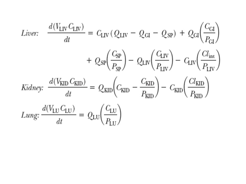
Typical eliminating tissue organ
For tissue organs in which drug is eliminated , parmeters representing drug elimination from the liver (k LIV) and kidney (k KID) are added to account for drug removal through metabolism or excretion. Tissue compartment Blood C ven Cart,Qt Drug eliminated Typical eliminating tissue organ
 and kidney (k KID) are added to account for drug removal through metabolism or excretion. Tissue compartment. Blood. C ven. Cart,Qt. Drug eliminated. Typical eliminating tissue organ.")
16
The mass balance for the rate of change in drug concentration in the blood pool is

17
Blood flow to organ in physiologic pharmacokinetic model
heart kidney adipose lung brain muscle bone skin liver spleen GI VENOUS BLOOD ARTERIAL BLOOD QBR QLU QH QMUS QSP QGI QBO QA QSK QLIV Urine Blood flow to organ in physiologic pharmacokinetic model

18
Lung perfusion is unique because the pulmonary artery returns venous blood flow to the lung, where carbon dioxide is exchanged for oxygen and the blood becomes oxygenated. The blood from the lungs flows back to the heart (into the left atrium) through the pulmonary vein, and the quantity of blood that perfuse the pulmonary system ultimately passes through the remainder of the body. In describing drug clearance through the lung, perfusion from the heart (right ventricle) to the lung is considered as venous blood.
 through the pulmonary vein, and the quantity of blood that perfuse the pulmonary system ultimately passes through the remainder of the body. In describing drug clearance through the lung, perfusion from the heart (right ventricle) to the lung is considered as venous blood.")
19
Therefore, the term describing lung perfusion are reversed compared to those for the perfusion of other tissues. With some drugs, the lung is a clearing organ besides serving as a merging pool for venous blood. In those case, a lung clearance term could be included in the general model

20
The system of differential equations used to describe the blood flow-limited model is usually solved through computer programs. The Runge–Kutta method is often used in computer methods for series of differential equations.

21
Physiologic Pharmacokinetic Model with Binding
The physiologic pharmacokinetic model assumes flow-limited drug distribution without drug binding to either plasma or tissues. In reality, many drugs are bound to a variable extent in either plasma or tissues. With most physiologic models, drug binding is assumed to be linear (not saturable or concentration dependent).
.")
22
Moreover, bound and free drug in both tissue and plasma are in equilibrium.
Further, the free drug in the plasma and in the tissue equilibrates rapidly. Therefore, the free drug concentration in the tissue and the free drug concentration in the emerging blood are equal:

23
Where f b is the blood free drug fraction, f t is the tissue free drug fraction, C t is the total drug concentration in tissue, and C b is the total drug concentration in blood. Therefore, the partition ratio, p t, of the tissue drug concentration to that of the plasma drug concentration is

24
By assuming linear drug binding and rapid drug equilibration, the free drug fraction in tissue and blood may be incorporated into the partition ratio and the differential equations. These equations are similar to those above except that free drug concentrations are substituted for c b. Drug clearance in the liver is assumed to occur only with the free drug.

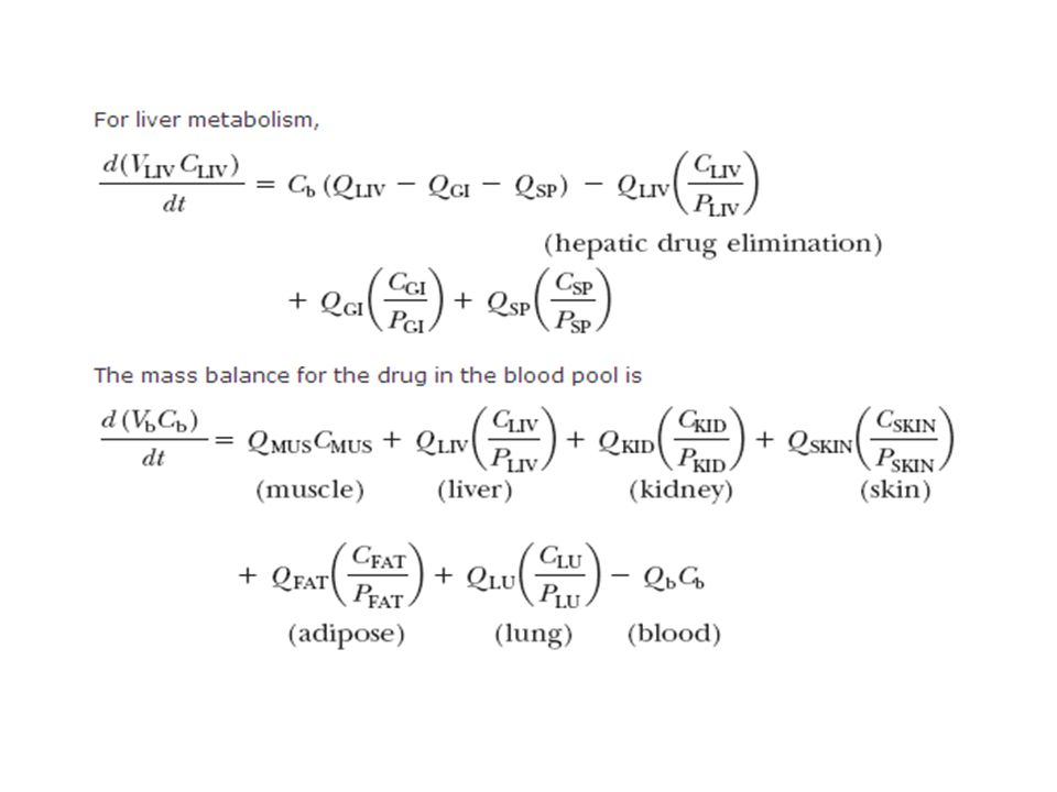
25
The inherent capacity for drug metabolism (and elimination) is described by the term Cl int
General mass balance of various tissues is described by Equation

27
The influence of binding on drug distribution is an important factor in interspecies differences in pharmacokinetics. In some instances, animal data may predict drug distribution in humans by taking into account the differences in drug binding. For the most part, extrapolations from animals to humans or between species are rough estimates only, and there are many instances in which species differences are not entirely attributable to drug binding and metabolism.

28
Mean Residence Time MRT describes the average time for all the drug molecules to reside in the body. The residence time for the drug molecules in the dose may be sorted into groups i (i = 1, 2, 3, , m) according to their residing time. The total residence time is the summation of the number of molecules in each group i multiplied by the residence time, t I , for each group. Total residence time for all the drug molecule in body Total number of drug molecule MRT =
 according to their residing time. The total residence time is the summation of the number of molecules in each group i multiplied by the residence time, t I , for each group. Total residence time for all the drug molecule in body. Total number of drug molecule. MRT =")
29
The summation of n i (number of molecules in each group) is the total number of molecules, N.
The drug dose (mg) may be converted to the number of molecules by dividing the dose (mg) by 1000 and the molecular weight of the drug to obtain the number of moles of drug, and then multiplying the number of moles of drug by x 1023 (Avogadro's number) to obtain the number of drug molecules.
 may be converted to the number of molecules by dividing the dose (mg) by 1000 and the molecular weight of the drug to obtain the number of moles of drug, and then multiplying the number of moles of drug by x 1023 (Avogadro s number) to obtain the number of drug molecules.")
30
For convenience, above equation may be written in terms of milligrams (instead of molecules) by substitution of n i with Dei x f, where Dei is the number of drug molecules (as mg) leaving the body with residence time t i (i = 1, 2, 3, , m). The f is a conversion factor.
. The f is a conversion factor..")
31
Mean Residence Time—IV Bolus Dose
The drug concentration in the body after an IV bolus injection for a drug that follows the pharmacokinetics of a one-compartment model is given by where V D is the apparent volume of distribution, k is the first-order elimination rate constant, and t is the time after the injection of the drug. (1)
")
32
The rate of change in the amount of drug in the body with respect to time (dD p/dt) reflects the rate at which the drug molecules leave the body at any time t. Although all drug molecules enter the body at the same time, the exit time, or the residing time, for each molecule is different. Equation (2) is obtained by taking the derivative of Equation(1), with all the drug molecules exiting the body from t = 0 to ∞. (2)
 is obtained by taking the derivative of Equation(1), with all the drug molecules exiting the body from t = 0 to ∞. (2)")
33
Alternatively, the rate of drug molecules exiting at any time t is given by
At any time t, dDe molecules exit. Therefore, multiplying Equation (3) by t on both sides yields the residence for each molecule exiting with a residence time t. Summation of the residence time for each drug molecule, and division by the total number of molecules, estimates the mean residence time. (3)
 by t on both sides yields the residence for each molecule exiting with a residence time t. Summation of the residence time for each drug molecule, and division by the total number of molecules, estimates the mean residence time. (3)")
34
As shown in Equation (4), the MRT is related to the product of the elimination rate constant k and the function describing drug elimination in the body. MRT is the integrated normalized form of the differential function representing drug amount (or concentration) in the body. The term differential probability is used to reflect that the function is a probability density function (PDF), which represents the residence time probability of a molecule in the population. (4)
 in the body. The term differential probability is used to reflect that the function is a probability density function (PDF), which represents the residence time probability of a molecule in the population. (4)")
35
The mean residence time is the normalized (divided by D 0) differential of the function governing drug elimination in the body. When a function is normalized, it becomes dimensionless, without units. Equation (4)was derived in terms of amount of drug. Because D 0 = C 0pVD, substituting for D 0 with C 0pVD into the right side of Equation (4)yields
was derived in terms of amount of drug. Because D 0 = C 0pVD, substituting for D 0 with C 0pVD into the right side of Equation (4)yields.")
36
Above equation may be used to determine MRT directly or may be rearranged to following Equation by dividing the numerator and denominator by k to yield a moment equation. The plasma concentration equation [function f(t)] multiplied by time and integrated from 0 = ∞ gives a term called the first moment of the plasma drug curve.
] multiplied by time and integrated from 0 = ∞ gives a term called the first moment of the plasma drug curve.")
37
The denominator is the area under the curve, AUC∞0
The denominator is the area under the curve, AUC∞0. The AUC∞ 0 is equal to ∫∞0C pdt or D 0/V Dk. Because C 0 p = D 0/V D, the denominator of Equation (4) is the AUC∞ 0 of the time–concentration curve (AUC∞ 0 = D 0/kV D) Where AUMC is the area under the (first) moment-versus-time curve from t = 0 to infinity. AUC is the area under plasma time-versus-concentration curve from t = 0 to infinity. AUC is also known as the zero moment curve. MRT = AUMC AUC
 is the AUC∞ 0 of the time–concentration curve (AUC∞ 0 = D 0/kV D) Where AUMC is the area under the (first) moment-versus-time curve from t = 0 to infinity. AUC is the area under plasma time-versus-concentration curve from t = 0 to infinity. AUC is also known as the zero moment curve. MRT = AUMC. AUC.")
38
Detrmination of MRT log Cp Cp*t AUMC CUM AUMC AUC cum AUC 100 2 89.687
Time Cp log Cp Cp*t AUMC CUM AUMC AUC cum AUC 100 2 89.687 1 79.374 39.687 71.188 63.002 3 50.007 44.85 4 39.693 5 31.506 157.53 6 25.007 22.428 7 19.849 17.802 8 15.755 126.04 9 12.506 11.216 10 9.926 99.26 42 0.006 0.252 0.0055 43 0.005 0.215 0.2335 0.0045 44 0.004 0.176 0.1955 0.0035 45 0.003 0.135 0.1555 0.0025 550.59 46 0.002 0.092 0.1135 2841.8 47 0.094 0.093 48 0.096 0.095 0.0015 49 0.001 -3 0.049 0.0725

39
Ke = 2.303* = 0.231 Tail AUMC=Cp*t/k+ Cp/k2 = Total AUMC = Tail AUC=C*/K = Total AUC = MRT=AUMC/AUC =

40
Statistical Moment Theory
Statistical moment theory provides a unique way to study time-related changes in macroscopic events. A macroscopic event is considered as the overall event brought about by the constitutive elements involved. For example, in chemical processing, a dose of tracer molecules may be injected into a reactor tank to track the transit time (residence time) of materials that stay in the tank. The constitutive elements in this example are the tracer molecules, and the macroscopic events are the residence times shared by groups of tracer molecules.
 of materials that stay in the tank. The constitutive elements in this example are the tracer molecules, and the macroscopic events are the residence times shared by groups of tracer molecules.")
41
Each tracer molecule is well mixed and distributes noninteractively and randomly in the tank.
In the case of all the molecules (∫D0 0dDe = D 0) that exit from the tank, the rate of exit of tracer molecules (–dDe/dt) divided by D 0 yields the probability of a molecule having a given residence time t A mathematical formula describing the probability of a tracer molecule exited at any time is a probability density function. MRT is merely the expected value or mean of the distribution.
 that exit from the tank, the rate of exit of tracer molecules (–dDe/dt) divided by D 0 yields the probability of a molecule having a given residence time t. A mathematical formula describing the probability of a tracer molecule exited at any time is a probability density function. MRT is merely the expected value or mean of the distribution.")
42
The moment curve shows the characteristics of the distribution.
The moment theory facilitates calculation of MRT and related parameters from the kinetic function, as discussed below. A probability density function f(t) multiplied by tm and integrated over time yields the moment curve (Eq.5). The moment curve shows the characteristics of the distribution. where f(t) is the probability density function, t is time, and m is the mth moment. (5)
 multiplied by tm and integrated over time yields the moment curve (Eq.5). The moment curve shows the characteristics of the distribution. where f(t) is the probability density function, t is time, and m is the mth moment. (5)")
43
For example, when m = 0, substituting for m = 0 yields Equation 6, called the zero moment,µ 0
If the distribution is a true probability function, the area under the zero moment curve is 1. Substituting into Equation 5 with m = 1, Equation 7 gives the first moment µ 1 The area under the curve f(t) times t is called the AUMC, or the area under the first moment curve. The first moment, µ1, defines the mean of the distribution. (6) (7)
 times t is called the AUMC, or the area under the first moment curve. The first moment, µ1, defines the mean of the distribution. (6) (7)")
44
Similarly, when m = 2, Equation 5 becomes the second moment, µ2:
where µ2 defines the variance of the distribution. Higher moments, such as µ3 or µ4, represent skewness and kurtosis of the distribution. Equation 5 is therefore useful in characterizing family of moment curves of a distribution.

45
The principal use of the moment curve is the calculation of the MRT of a drug in the body.
The elements of the distribution curve describe the distribution of drug molecules after administration and the residence time of the drug molecules in the body. The plasma equation, f(t) = c0pe–kt , was converted to a pdf [ie, f(t) = ke –kt ].
 = c0pe–kt , was converted to a pdf [ie, f(t) = ke –kt ].")
46
It can be shown that µ0 for this function = µ1 (total probability adds up to 1 by summing zero moment), and the mean of the function is the area under the first moment curve (the mean is the MRT).
, and the mean of the function is the area under the first moment curve (the mean is the MRT).")
47
REFERENCES Shargel L., Susanna Wu-Pong, Andrew B.C. Yu , “Applied Biopharmaceautics &Phamacokinetics”,5th Edn.,2005,p.no Kar Ashutosh, “ Essentials Biopharmaceutics & pharmacokinetics” , 1 st edn, 2010,Elsevier publication ,Page no

Similar presentations


>")


