Download presentation
Presentation is loading. Please wait.

1
Cytoskeleton and Cell Motility
Nancy Freitag

2
Objectives What regulates the shape and assembly of the cell cytoskeleton? How do pathogens exploit host cell actin assembly?

3
Overview The cytoskeleton and cell motility
The dynamics of actin assembly The cell cytoskeleton as a target for pathogens Actin elongation-based propulsion: bacterial motility and cell movement (PAPER)
")
4
The actin cytoskeleton and cell motility
Cytoplasmic system of fibers crucial to cell motility Plays a structural role Undergoes rearrangement which can produce movement

6
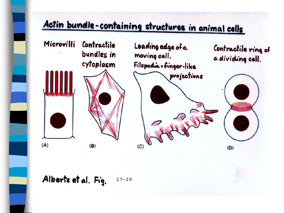
Actin provides framework & determines cell shape

7
Filaments are organized into bundles & networks held together by cross-linking proteins

8
Actin filaments give shape to microvilli

9
Cell locomotion Cell moves forward by extending filipodia & lamellipodia Focal adhesions are formed Cell is pulled forward

10
Actin monomers and filaments
Actin is the most abundant intracellular protein. Highly conserved. G-actin = actin monomer. F-actin = filamentous polymer. Each actin monomer contains Mg2+ complexed with either ATP or ADP

11
G-actin has two lobes separated by a deep cleft where ATP binds.

12
G-actin can assemble into F-actin in vitro under the right ionic conditions; no other proteins are required to produce filaments.

13
Actin filaments in solution

15
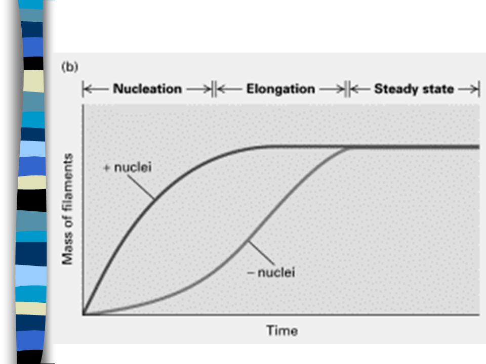
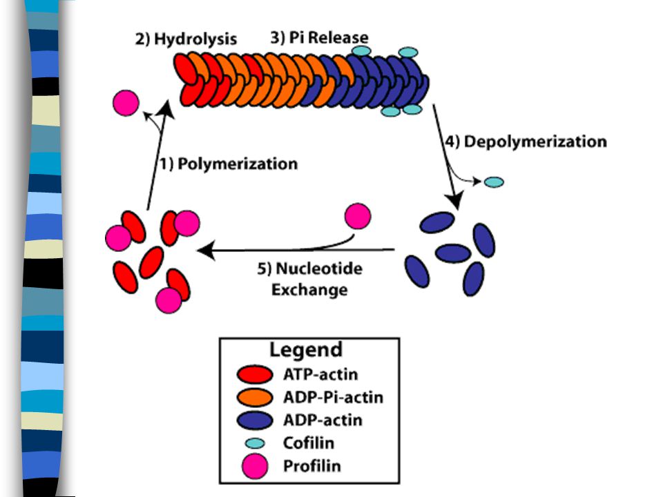
Dynamics of actin assembly in vitro: a brief overview
Lag phase: G-actin aggregates into short, unstable oligomers. An oligomer of 3 or 4 subunits acts as a nucleus for further polymerization

16
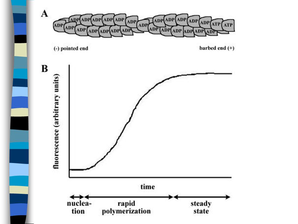
Elongation: addition of monomer
to both ends Steady state: G-actin monomers exchange with subunits at both ends w/no change in total mass

18
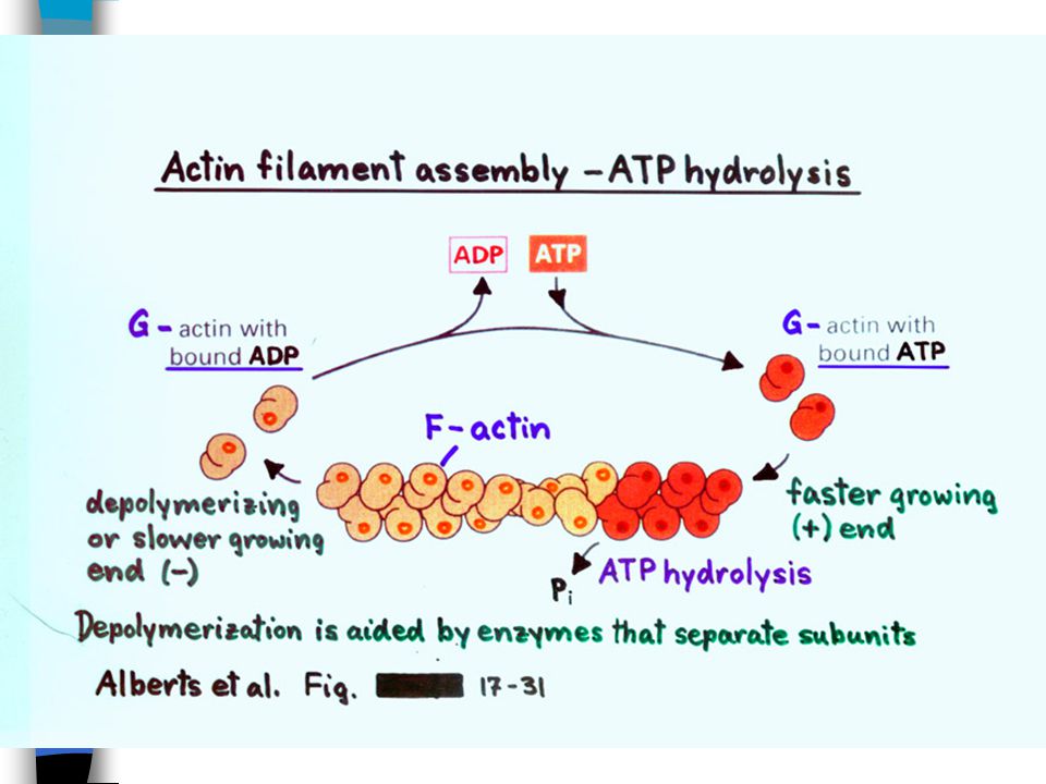
Each actin monomer is bound to a molecule
of ATP. Following addition of monomer, ATP is hydrolyzed to ADP.

20
Critical concentration (Cc)
The equilibrium concentration of a pool of unassembled actin The measure of the ability of a solution of G-actin to polymerize Above Cc a solution of actin will polymerize Below Cc F-actin will depolymerize

21
Cc

22
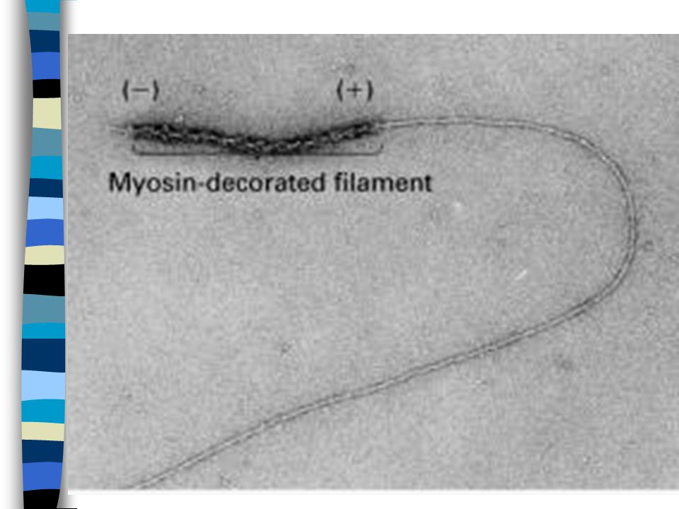
Actin filaments grow faster at one end than at the other
The barbed end, or (+) end, elongates 5 to 10 times faster than the pointed, or (-) end. The difference in elongation reflects the difference in Cc values at the two ends.
 end, elongates. 5 to 10 times faster than the. pointed, or (-) end. The difference in elongation reflects the. difference in Cc values at the two ends.")
24
Actin filaments grow faster at one end than at the other
Below Cc (+) end: no filament growth occurs Between Cc (+) and Cc (-): growth occurs at the (+) end (treadmilling) Above Cc (-): growth occurs at both ends
 end: no filament growth occurs. Between Cc (+) and Cc (-): growth occurs at the (+) end (treadmilling) Above Cc (-): growth occurs at both ends.")
25
Treadmilling

26
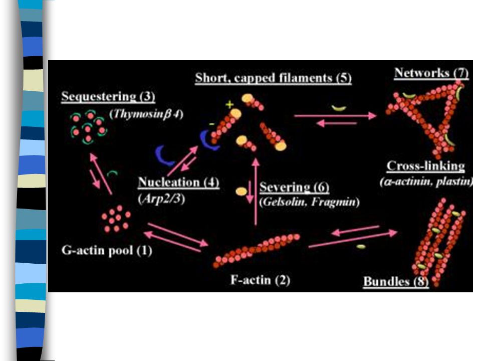
Actin polymerization is regulated by proteins that bind G-actin
Cc for a cell is ~ 0.2 uM. Concentration of G-actin is 50 uM to 200 uM. Pool of G-actin is maintained by proteins that sequester G-actin

27
Examples of proteins that sequester G-actin
Thymosin b4: sequesters free ATP-G-actin. Acts as a buffer. Profilin: sequesters actin, and promotes the exchange of ATP for ADP-G-actin.

28
Actin filament length is controlled by proteins that cap or sever filaments

29
Actin filament length is controlled by proteins that cap or sever filaments
Gelsolin and cofilin: break actin network into shorter fragments. Alter conformation of actin subunit, causing breakage, & then remain bound. Bound protein prevents addition of new monomers, an activity called capping.

32
Assays for actin polymerization
Pyrene actin assays: spectrofluorometric assay. Fluorescently tagged actin gives a wavelength-specific signal when polymerized. Cytoplasmic extracts: can add or deplete factors.

34
Pathogen-mediated cytoskeletal rearrangements
Prevention of uptake: inhibition of phagocytosis or pedestal formation Invasion: induced uptake Actin-based motility: intracellular motility and intercellular spread

36
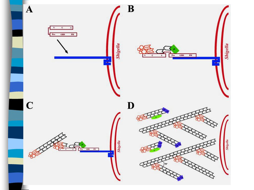
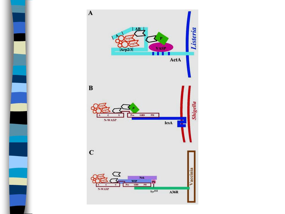
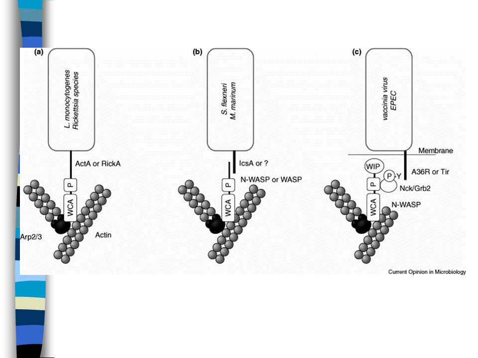
Pathogens that exploit actin-based intracellular motility
Listeria monocytogenes Shigella flexneri Mycobacteria Burkholderia Rickettsia Vaccinia virus

37
L. monocytogenes as a tool for defining actin assembly
From Tilney and Portnoy, J. Cell Biology 1989

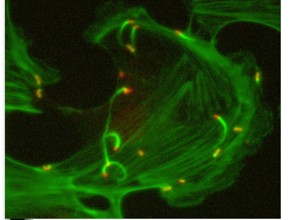
39
What is required for actin-based motility within the cytosol?
Immunofluorescence studies indicated the presence of a variety of proteins associated with actin tails… But which ones are required for movement? Which ones are simply binding actin?

40
Identification of ActA
Search for bacterial mutants unable to spread within cells led to identification of the actA gene product wild type actA mutant

41
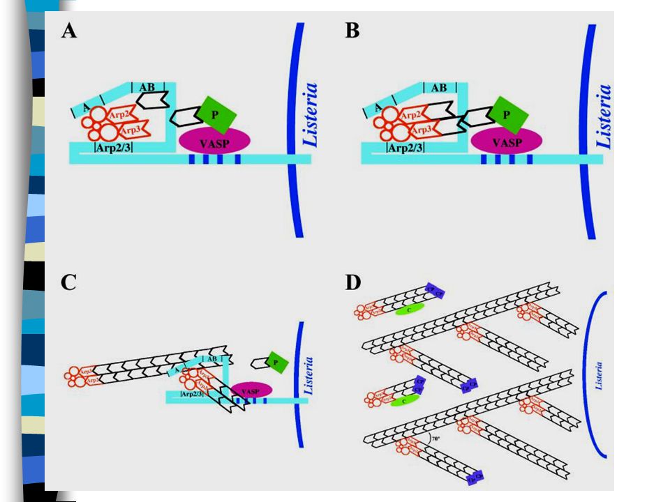
ActA and a host protein complex, Arp 2/3,
were found to co-localize at the base of L. monocytogenes actin tails within the cytosol

42
Domains of ActA SP = signal peptide WH2 & Arp 2/3 = bind Arp 2/3
AB region = monomeric actin binding

43
ActA + Arp 2/3 function as a
highly efficient nucleation site actin, actin + ActA Arp 2/3 Arp 2/3 + ActA ActA

44
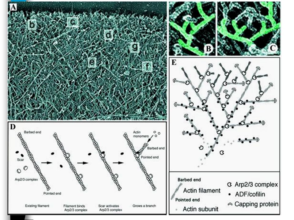
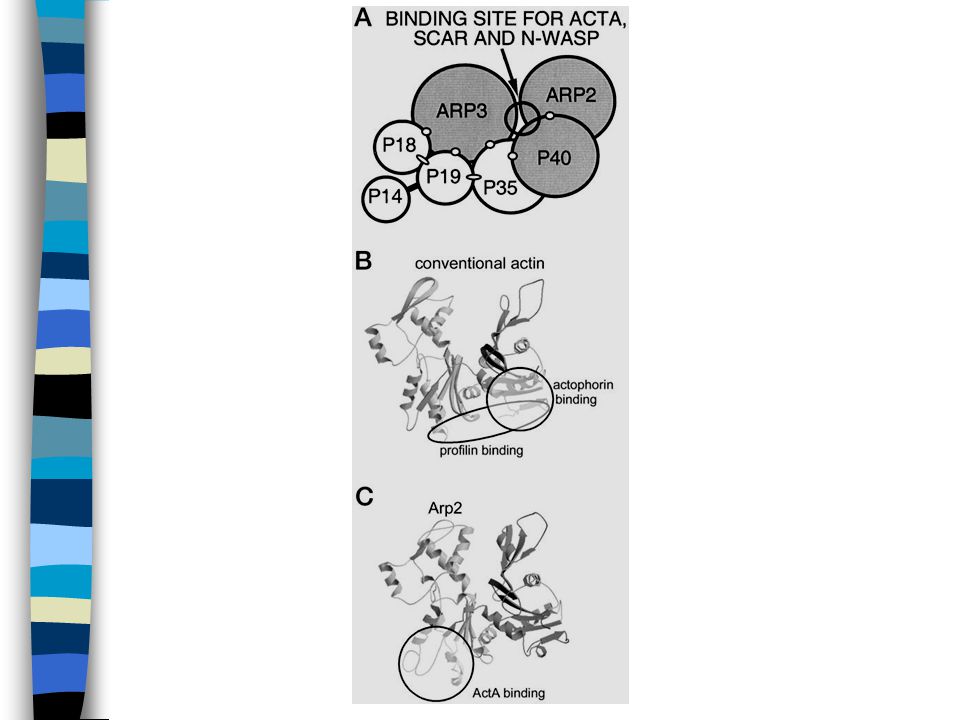
Arp 2/3 complex Complex of 7 polypeptides Present in all eukaryotes
Only known factor that stimulates nucleation of actin at barbed ends Can bind to the sides of filaments and stimulate polymerization Requires activation

46
Wiskott-Aldrich protein family (WASP)
Activate Arp 2/3 Contain WH2 domains, acidic domains, and proline rich regions WASP, N-WASP, Scar

53
Fascin-mediated propulsion of Listeria monocytogenes independent of frequent nucleation by the Arp2/3 comple J. Cell Biology 165: 2004

54
Figure 1

55
Figure 2

56
Figure 3

57
Figure 4

58
Figure 5

59
Figure 6

60
Figure 7

61
Figure 8

62
Figure 9

63
Figure 10

64
Fig. 5. The transition from asters to stars
Haviv, Lior et al. (2006) Proc. Natl. Acad. Sci. USA 103, Copyright ©2006 by the National Academy of Sciences
 Proc. Natl. Acad. Sci. USA 103, Copyright ©2006 by the National Academy of Sciences.")
65
Additional references
Actin-based motility of intracelllular microbial pathogens. Micro Mol Biol Rev. (2001) 65: Interaction of human Arp 2/3 complex and the Listeria monocytogenes ActA protein in actin filament nucleation. Science (1998) 281:
 65: Interaction of human Arp 2/3 complex and the Listeria monocytogenes ActA protein in actin filament nucleation. Science (1998) 281:")
Similar presentations


, intermediate.>")


 Macrophage cytoskeleton Cytoskeleton of a lung cell in mitosis.>")

 Actin-binding proteins affect the localized assembly or disassembly of the actin.>")
 Actin filaments>")


, microtubules and intermediate filaments. Not surprisingly, the actin skeleton is dynamic, not.>")


