Download presentation
Presentation is loading. Please wait.

1
Introduction Histology is a science which studies the normal microstructure of the human body and the relationship between the body’s structure and function. cells are the smallest structural and functional unit of human body

2
Key points of this class
Methods employed in Histology

3
1. Histology 2. Embryology Histo=Tissue Logy=Study Logy= study
Embryo= unborn or unhatched offspring Logy= study

4
What we will learn in this course
Tissue: 4 basic tissues Organ system

5
Why it is important to learn histology?
To recognize normal tissue and cells To acquire basic skill which you will use throughout your career!!!

6
What you need to do Look Think Compare Remember Idealized images

7
2. Basic methods of histology
Observation of histological slides Procedure: Specimen Fixation Embedding Sectioning Staining Observation

8
Fixation solution (fixative)
LM--- 4% formaldehyde – Fixative and antibacterial agent EM--- glutaraldehyde followed by osmium tetroxide

9
Embedding Ethanol----dehydration Xylene----clearing
Paraffin----embedding

10
Sectioning Microtome thickness 2~10 μm Units M,mm,μm,nm
1nm=0.001μm=10-6mm=10-9m

11
Microtome

12
Staining Basophilia , Acidophilia, Neutrophilia, metachromasia
Basic dyes: toluidine blue, methylene blue, hematoxylin (blue) Acid dyes: orange G, eosin, acid fuchsin (pink) common staining: Hematoxylin & Eosin Specific staining: silver
 Acid dyes: orange G, eosin, acid fuchsin (pink) common staining: Hematoxylin & Eosin. Specific staining: silver.")
13
Hematoxylin & Eosin (H&E)
H&E is the most commonly method in histological study Hematoxylin Blue Eosin Pink

14
Hematoxylin Eosin H&E

15
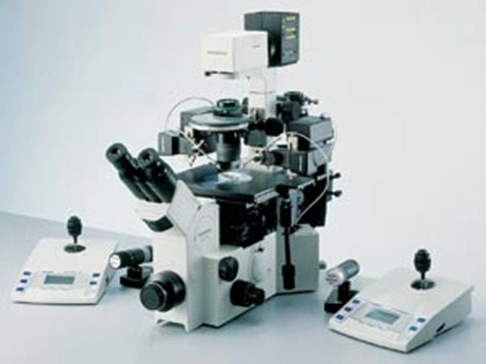
3. Microscopes Light microscope Specific microscope
(1). Phase-contrast microscope (2). Differential interference microscope (3). Fluorescence microscope (4). Confocal microscope
. Phase-contrast microscope. (2). Differential interference microscope. (3). Fluorescence microscope. (4). Confocal microscope.")
17
Electron microscope EM Transmission - plain
Scanning – platinum or gold layers, 3 dimensional

18
Photograph of the transmission electron microscope.

19
Schematic view of a transmission electron microscope with its lenses and the pathway of the electrons. CCD, charged coupled device.

20
Schematic view of a scanning electron microscope.

21
4. Other methods A. Autoradiography
B. Histochemistry and cytochemistry PAS (periodic acid Schiff reaction) Feulgen reaction C. Immunocytochemistry Antigen and antibody reaction D. In situ hybridization DAN or RNA single strand complementary
 Feulgen reaction. C. Immunocytochemistry. Antigen and antibody reaction. D. In situ hybridization. DAN or RNA single strand complementary.")
22
Other methods (continue)
E. Cell culture —stem cells F. Transgenic animal G. Micromanipulation and cloning

24
Summary 1. Histology is the study of the tissues of the body and of how these tissues are arranged to constitute organs. 2. Histology=Microscopic anatomy 3. Section preparation and HE staining 4. Other methods

Similar presentations


 iris diaphragm.>")


 directly in fixed.>")


 or 1000000000.>")


