Download presentation
Presentation is loading. Please wait.

1
Coregistration and Normalisation By Lieke de Boer & Julie Guerin

2
Presentation Overview Recap: Steps of Preprocessing Coregistration Normalisation Smoothing Summary SPM Guidelines

3
Steps of Preprocessing MNI str fct str MNI fct str 2 MNI 3 1 4 4 is a blurry version of 3 1.Realign & Unwarp 2.Coregister 3.Normalise 4.Smooth Raw data (fct) Raw data (str) MNI fct
 Raw data (str) MNI fct")
4
CoregistrationNormalisation & Smoothing

5
Recap: 1. Realignment Aligning all fMRI scans to reference scan –Linear transformations (translations and rotations) fct str fct MNI fct Translation Rotation
 fct str fct MNI fct Translation Rotation.")
6
Recap: 1. Unwarping Non-linear transformations Adjusts for deformations in magnetic field

7
2. Coregistration Cross modal (structural/functional) realignment to the same native space. Within-subjects Structural (high resolution) Functional (low resolution) MNI fct str
 Functional (low resolution) MNI fct str.")
8
Coregistration: Step 1 Registration –Similar to realignment –Fitting source image (T1 structural) to reference image (T2 functional) Translation Rotation
 to reference image (T2 functional) Translation Rotation")
9
Coregistration: Step 2 Reslicing/Interpolation Estimating what value of intensity each voxel represents in a functional image. Structural (high resolution; 1mm 3 ) ≠ Functional (low resolution; 3mm 3 ) –Reslicing estimates the intensity of surrounding voxels in functional scan so that functional voxels correspond with structural voxels.
 ≠ Functional (low resolution; 3mm 3 ) –Reslicing estimates the intensity of surrounding voxels in functional scan so that functional voxels correspond with structural voxels..")
10
Coregistration: Step 2 (Continued) Methods of Interpolation: Nearest Neighbour Linear Interpolation B-Spline Interpolation (Higher-Order Interpolation) zczc ?
 Methods of Interpolation: Nearest Neighbour Linear Interpolation B-Spline Interpolation (Higher-Order Interpolation) zczc")
11
intensity # voxels intensity Coregistration: Forming a Joint Histogram T2* (functional) histogramT1 (structural) histogram Registered Joint Histogram UN-Registered Joint Histogram
 histogramT1 (structural) histogram Registered Joint Histogram UN-Registered Joint Histogram")
13
3. Normalisation Coregistered images Standardised space fctstr MNI Talairach AtlasMNI Template

14
Why Normalise? Statistical power Group analyses Generalise findings/Representative Cross-study comparisons (standard coordinate system) ≠
 ≠.")
15
Normalisation Aligning and warping to standardised space Template fitting: –Right/Left –Anterior/Posterior –Superior/Inferior GOAL: voxel to voxel correspondence between brains of many subjects

16
Normalisation: Optimisation Difficulties Aim: to find an identical fit between the subjects’ brain and the template brain. Reality: difficult to match brains when size/shape are so different –not structurally or functionally homologous –maximise similarity within reasonable limits/expectations.

17
Normalisation: Affine Transformation Linear Transformations Translations across axes Rotations around axes Scaling and zooming axes Shearing or skewing, i.e. angle changes between pairs of axes

18
Normalisation: Refine with Warping Applies non-linear warping to images to match template. Apply deformations/displacements to move voxels from original location to template location in multiple dimensions Affine RegistrationNon-linear Registration

19
Normalisation: Risk of Overfitting Warping is completely flexible and can therefore introduce unrealistic deformations = overfitting. Non-linear registration using regularisation. Non-linear registration without regularisation.

20
Normalisation: Regularisation Rather have less good match: compromise between reasonably good match & realistic deformation –Uses Bayesian framework: probability function of determining appropriate warp amounts. Regularisation sets limits to warp parameters, and ensures voxels stay close to their neighbours.

21
Normalisation: Segmentation Different scanners, noise, artifacts, magnetic field properties, etc. prevent data from being uniformly and predictably adjusted to template.

22
Segmentation Tissue Probability Maps (TPM) from standard space help predict tissue differentiation in subjects. Segmentation helps correctly identify which tissue type the voxels of interest belong to.

23
Normalisation: Generative Model Each tissue type has Gaussian probability density function for intensity. Goal is to get the best estimate of tissue probabilities. Generative Model (Bayesian) – fitting a Gaussian Mixture Model (GMM) to the joint histogram
 – fitting a Gaussian Mixture Model (GMM) to the joint histogram.")
24
4. Gaussian Smoothing Smoothing: adjusts for any residual differences and alignment errors. –Reduces signal to noise ratio. –Better spatial overlap –Better normally distributes data; enables statistical analyses How: Convolution calculates a weighted average of neighbouring voxels -- each voxel gets replaced by a weighted average of itself and its neighbours.

25
Keep In Mind: fMRI analyses are not precise and are based on multiple data adjustments that ultimately alter the raw data substantially. However, relatively reliable and robust inferences can be made when sample sizes are large enough, and when appropriate statistical analyses are performed.

26
Summary MNI str fct str MNI fct str MNI 4 4 is a blurry version of 3 fct 1. Realign & Unwarp 2. Coregister 3. Normalise & 4. Smooth

27
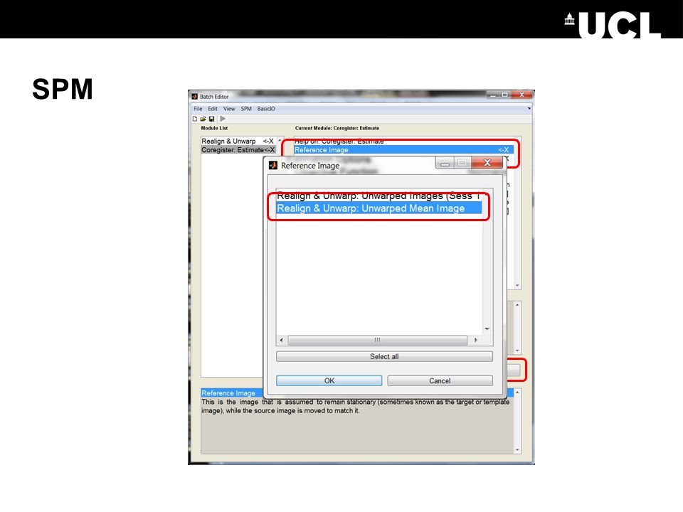
SPM

31
Tissue probability maps = 3 files: white matter, grey matter, CSF (Default) Masking image = exclude regions from spatial normalization (e.g. lesion) Data = Structural file (batched, for all subjects) Parameter File = Click ‘Dependency’ (bottom right of same window) Images to Write = Co- registered functionals (same as in previous slide)
 Data = Structural file (batched, for all subjects) Parameter File = Click ‘Dependency’ (bottom right of same window) Images to Write = Co- registered functionals (same as in previous slide).")
32
SPM

34
References http://www.ucl.ac.uk/stream/media/swatch?v=1d42446d1c34 (Ged Ridgway’s preprocessing lecture)http://www.ucl.ac.uk/stream/media/swatch?v=1d42446d1c34 SPM videos: http://www.fil.ion.ucl.ac.uk/spm/course/video/ SPM Homepage: http://www.fil.ion.ucl.ac.uk/spm/ Suz Prejawa MfD Resources 2011-2012
 v=1d42446d1c34 SPM videos: SPM Homepage: Suz Prejawa MfD Resources")
35
And thanks to Ged!

Similar presentations


>")


