Download presentation
Presentation is loading. Please wait.

1
Radiology Case Presentation By Matt Cole

2
Clinical Information Clinical history: 60 year old white female who presented with a 1 week history of abdominal pain, worse in the RLQ, with recent increased intensity. Some intermittent N/V, no changes in bowel habits, no decreased appetite, no fever/chills.

3
Clinical Information Physical Exam: Afebrile, Vital Signs within normal limits, Abdominal exam revealed tenderness in the RLQ, positive rebound tenderness with pain radiating to the RLQ, minimal rigidity or guarding. Lab tests showed a normal white count, normal U/A, normal LFTs, normal lipase

4
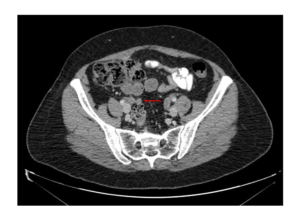
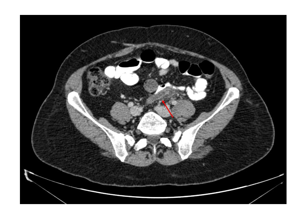
Imaging The following imaging studies were obtained: –Chest x-ray –Abdominal series –Abdomen/Pelvis CT Both the CXR and the Abdominal series were within normal limits. The CT showed the following…

10
CT Findings Abnormally enlarged appendix (9.5 mm) with mild adjacent inflammatory stranding, compatible with appendicitis. Appendix lies in the mid and left pelvis and not the RLQ.

11
Hospital Course Based on history, physical exam, and CT findings, she was felt to have appendicitis. She was taken to the OR where a laparoscopic appendectomy was performed. The appendix was noted to be inflamed but not perforated. Final pathology reported the diagnosis to be “acute appendicitis”.

12
Radiographic features of appendicitis Plain Abdominal Radiograph –The presence of a calcified appendiceal fecalith occurs in fewer than 10% of cases. –Radiographic signs suggesting appendicitis include convex lumbar scoliosis, obliteration of right psoas margin, right lower quadrant air-fluid levels, air in the appendix, or localized ileus. –In rare cases, a perforated appendix may produce pneumoperitoneum. Ultrasound –Especially useful for pediatric appendicitis. –The finding of a noncompressible dilated appendix is a strong indicator of nonperforated appendicitis. –After perforation, ultrasound can identify a periappendiceal phlegmon or abscess formation. –Additional findings that can support the diagnosis of appendicitis include the presence of appendicoliths, fluid in the appendiceal lumen, focal tenderness over the inflamed appendix, and a transverse diameter of 6 mm or more.

13
CT diagnosis of Appendicitis The most useful features to diagnose appendicitis on CT include enlarged appendix (> 6cm), appendiceal wall thickening, periappendiceal fat stranding, and appendiceal wall enhancement. Other features can include appendicolith, appendiceal intraluminal air, intramural air, and abscess.

14
ACR CODE ACR CODE: 75.29

15
References Grainger & Allison's Diagnostic Radiology: A Textbook of Medical Imaging, 4th Ed., Copyright © 2001 Churchill Livingstone, Inc. www.emedicine.com

Similar presentations