Download presentation
Presentation is loading. Please wait.

1
Examination of the Knee Thursday SM Conference August 30, 2007

2
Exam Settings 1.Sideline Exam (on the field triage) 2.Training room (post game eval) 3.Office/clinic Exam (delayed + detailed)
 2.Training room (post game eval) 3.Office/clinic Exam (delayed + detailed)")
3
Sideline Exam Purpose: determine disposition 1.Transfer (severe injury) 2.Hold out(mild – moderate) 1.Observe + re-examine 2.Provide first aide 3.Return to action (mild ?– no injury?)
 2.Hold out(mild – moderate) 1.Observe + re-examine 2.Provide first aide 3.Return to action (mild – no injury )")
4
Sideline Exam Routine Determine mechanism Point of maximum tenderness Maneuver producing most pain Determine severity of damage

5
Case Presentation 22 year old collegiate wrestler Contact injury to left knee Medial-sided knee pain

6
Mechanism ? –Foot planted –Outside force –Pain + “pop”

7
Sideline Exam Pain “on inside”

8
Medial Pain (Differential Diagnoses) Medial Collateral Ligament sprain Hamstring strain Gastroc strain Medial Meniscus tear
 Medial Collateral Ligament sprain Hamstring strain Gastroc strain Medial Meniscus tear")
9
First… find Joint line

10
Maximum Tenderness? Pain “on inside” Tender over MCL

11
Most Painful maneuver? Straight Valgus? –Straight –30 degrees External rotation?

12
Sprain Severity? Classify by laxity Best exam technique –One-handed –Two handed

13
Check both sides! Laxity normal ??? –“Plastic man” –Post exercise

14
Anterior Drawer at 20-30 degrees “Modified Lachman’s”

15
Exam Settings 1.Sideline Exam (on the field triage) 2.Training room (post game eval) 3.Office/clinic Exam
 2.Training room (post game eval) 3.Office/clinic Exam")
16
Two handed technique Trap ankle on Iliac crest Both hands on joint line –Palpate both joint lines

17
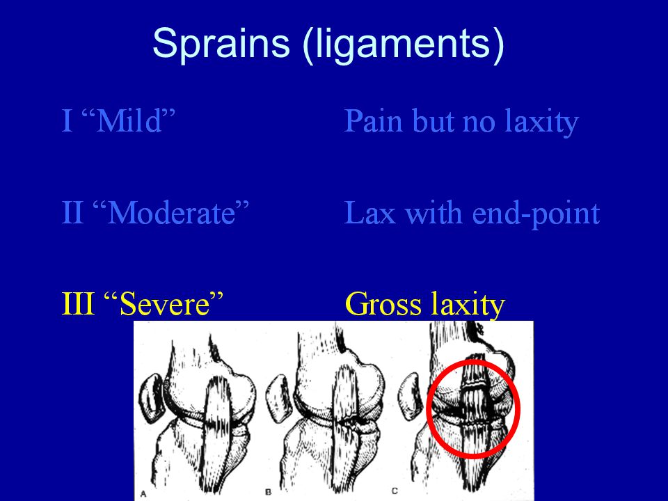
Sprains (ligaments)
")
20
Hughston Laxity Classification Grade I – 1-4 mm laxity Grade II – 5-9 mm laxity Grade III – >10 mm laxity (soft endpoint) »Hughston JC, Andrews JR, Cross MJ, Moschi A: Classification of knee ligament instabilities. Part I. The medial compartment and cruciate ligaments. »J Bone Joint Surg Am 58:159-172, 197

21
Two handed technique Trap ankle on Iliac crest Both hands on joint line –Palpate both joint lines

22
Two handed technique

23
Collegiate football Severity vs. Return Grade I – 10.6 days Grade II – 19.5 days Derscheid, G.L. and J.G. Garrick. MCL injuries in football: Non-operative management of grade I and grade II sprains. Am J Sports Med, 1981. 9(6): p. 365-8.
: p")
24
Sideline estimate ( Crowley-Albright 30 consecutive FB cases) 1 mm = 1 week 2 mm = 2 weeks 3 mm = 3 weeks 6 mm = 6 weeks
 1 mm = 1 week 2 mm = 2 weeks 3 mm = 3 weeks 6 mm = 6 weeks")
25
Time Loss From Sport Severity of injury Compliance??

26
Exam Settings 1.Sideline Exam (on the field triage) 2.Training room (post game eval) 3.Office/clinic Exam
 2.Training room (post game eval) 3.Office/clinic Exam")
27
Office Exam What is important about the MCL exam? –Knee stability in full extension

28
Knee Hemarthrosis Differential Diagnosis ACL70% Meniscus50% Fracture20% Patellar dislocation PCL

29
Value of MRI? When should an MRI be done? –When knowledge of location of injury might influence treatment –When additional injury is suspected Instability at full extension should increase suspicion of cruciate injury »Mazzocca, A.D., et al., Valgus medial collateral ligament rupture causes concomitant loading and damage of the anterior cruciate ligament. »J Knee Surg, 2003. 16(3): p. 148-51.
: p")
30
Location MCL Tissue damage Proximal ruptures heal more quickly than distal but have more stiffness Complete ruptures can displace into the joint Damage over entire ligament associated with persistent laxity after non-operative treatment –Nakamura, N., S. Horibe, et al. (2003). "Acute grade III MCL injury of the knee associated with ACL tear. usefulness of MRI in determining treatment regimen." –Am J Sports Med 31(2): 261-7.
. Acute grade III MCL injury of the knee associated with ACL tear. usefulness of MRI in determining treatment regimen. –Am J Sports Med 31(2):")
31
Grade III – Gross instability Laxity at full ext (no endpoint) Indicates complete rupture of MCL –Evaluate posteromedial capsule –Evaluate for cruciate injury ACL PCL –Evaluate for Patellar Dislocation
 Indicates complete rupture of MCL –Evaluate posteromedial capsule –Evaluate for cruciate injury ACL PCL –Evaluate for Patellar Dislocation")
32
Pivot shift techniques Re-entry tests –MacIntosh –Hughston Jerk Exit tests –Losee (5 tests) –Slocum –Low profile
 –Slocum –Low profile")
33
The “Pivot-Shift” “Low Profile” Technique “exit” type pivot (in-to-out of place) No valgus Limit arc to last 20 degrees
 No valgus Limit arc to last 20 degrees")
34
Losee Tests See video

35
Active Quad Self Induction of Pivot Shift

36
LCL??? or MCL???

37
Reverse pivot Ask patient “In or out?”

38
Meniscus tears Joint line tenderness –Most sensitive but least specific (Fu) Squat and duck walk test McMurray' s test –Modified McMurray' s test –Most sensitive but least sensitive Appley’s test –Modified Appley’s test Full Extension?? –Pain anterior joint line

39
Modified McMurray' s test

40
Displaced Meniscus? (bucket-handle tear) Lacks full extension –No screw home –Pain anterior joint line –Rotation affects degree of pain Lacks full flexion –Rotation affects degree of pain Rotation OK in mid range flexion
 Lacks full extension –No screw home –Pain anterior joint line –Rotation affects degree of pain Lacks full flexion –Rotation affects degree of pain Rotation OK in mid range flexion.")
41
Anterior knee pain Osgood Schlatter' s Jumper’s knee P-F Chondromalacia Synovitis (Plica?) P-F instability
 P-F instability")
Similar presentations


>")


 Tear. Development of LCL Tear A varus force to the medial aspect of the knee while bearing weight can put enough stress.>")