Download presentation
Presentation is loading. Please wait.

1
Gastrointestinal Bleeding Dr.Mirzaei

2
Bleeding: oropharynx => Anus
Acute: rapid loss of blood even shock Chronic: anemia, fatigue Maybe the first symptom of GI disease Self limited or need for intervention

3
Hematemesis , coffee-ground
Melena (50 – 60 cc) Hemato chezia Occult blood in stool (10 cc)
 Hemato chezia. Occult blood in stool (10 cc)")
4
Upper G I Bleeding Lower G I Bleeding Obscure G I Bleeding

5
UPPER GI BLEEDING

6
Causes of Upper GI Bleeding
PUD 40% Oesophagitis 10% Varices 5% Mallory – Weiss Syndrome (longitudinal tear in the mucosa of the GE junction) 5% Erosive Disease 6% Neoplasm 4% Other 6% No Obvious Cause 24%
 5% Erosive Disease 6% Neoplasm 4% Other 6% No Obvious Cause 24%")
7
Massive Upper GI Bleeding
Acute Bleeding Proximal to the ligament of treitz Requires blood transfusion

8
Massive Upper GI Bleeding
PUD Gastritis Mallory weiss Syndrome Esophagogastric Varices

9
Massive Upper GI Bleeding ( Less Common Causes)
Neoplasm (malignant – benign) Angiodysplasia Dieulafoy’s Lesion (Congenital arteriovenous malformation) Arterioenteric Fistula (Aortic Graft-Repair of visceral artery aneurysm)
 Angiodysplasia. Dieulafoy’s Lesion (Congenital arteriovenous malformation) Arterioenteric Fistula (Aortic Graft-Repair of visceral artery aneurysm)")
10
History P. U. D-Heart burn – reflux
Drugs (NSAID- stroid- anticoagulant) Alcohol Cirrhosis
 Alcohol. Cirrhosis.")
11
Peptic ulcer disease Bleeding may be the first symptom DU: GU = 4 : 1

12
Upper GI Bleeding Most common complication of PUD
Most peptic ulcer related death Typically Present with melena and/or hematemesis

13
Management Resuscitation Continuous IV PPI
Large-bore IV access (2 IV line) Foley catheterization NGT + irrigation with normal saline (room temperature) Continuous IV PPI
 Foley catheterization. NGT + irrigation with normal saline (room temperature) Continuous IV PPI.")
14
Managment Lab test CBC, Hb, HCT, Platelet BUN - Cr – Na – K PT, PTT
L.F.T ABG + E.C.G

15
Upper GI Bleeding due to peptic ulcer
Acid suppression + NPO ¾ will stop ¼ will continue to bleed or will rebleed All mortalities & operations occur in this group

16
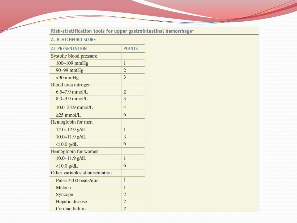
Risk Stratification Magnitude of the Hemorrhage - Shock - Hematemesis
- Transfusion > 4 units in 24 h - Hypotension - Tachycardia - Oliguria - Low Hct - Pallor - Altered Mentation

17
Risk Stratification Comorbidities - Lung - Liver - Kidney - Heart Age
Anticoagulated or immunosuppressed

18
Risk Stratification Endoscopic Findings Bleeding from varices
Active bleeding or Visible vessel

21
High Risk Patients (25%) Type & Crossmatch Admit to ICU
Consult Surgeon Consult gastroenterologist Start continuous infusion of PPI

22
High Risk Group (25%) Endoscopy within 12 hours after correction of coagulopathy (Diagnosis the cause – Assess the need for hemostatic therapy) Endoscpic hemostasis Arteriography (occasionally) Operation
 Operation.")
23
Endoscopic Therapy Injection with epinephrine Electrocautery
Clip (exposed vessel)
")
26
Indications of Operation
Massive Bleeding unresponsive to Endoscopic Therapy Transfusion requirement of > 4-6 Unit Persistent bleeding or rebleeding after one or more endoscopic therapy Lack of availability of a therapeutic endoscopist Lack of availability of blood for transfusion Repeat hospitalization for bleeding ulcer Concurrent indication: Perforation – Obstruction

27
Indications of Early Elective Operation
After initially successful endoscopic treatment Elderly Patients Multiple comorbidity (don’t tolerate another episode of Hemorrhage) Deep ulcer overlying a large vessel :posterior duodenal bulb(Gastroduodenal Artery) or lesser gastric curve (left gastric artery)
 Deep ulcer overlying a large vessel :posterior duodenal bulb(Gastroduodenal Artery) or lesser gastric curve (left gastric artery)")
28
LOWER GI BLEEDING

29
Symptoms Unexplained Iron – Deficiency Anemia (Occult Blood)
Hematochezia Dark or Clot Rectal Bleeding Massive Shock

30
Causes Hemorrhoids Fissure SRU IBD Malignancy Polyps

31
Causes - Angiodysplasia
Usually in cecum & R.T Side colon Non congenital or Neoplastic but Degenerative No relation with other skin & visceral vascular lesions with age Usually small < 5 mm

32
Causes - Angiodysplasia
Colonoscopy or Angiography for diagnosis 80 % self limited 50 % Recurrence during 3 years Treatment options: laser, electrocoagulation ,surgery

33
Causes - Diverticulosis
Left sided colon Cause of > 50% massive lower GI Bleeding

34
Causes Meckel’s Diverticulum Infectious Colitis A-V malformation
Ischemic colitis Mesenteric Thrombosis

35
History Weight loss Abdominal Pain / Cramp Recent Bowel Habit Change
+ Ve Family hx of colorectal CA Drug History

36
Management Resuscitation (2 IV Line)
Correction of coagulopathy, thrombocytopenia Lab test CBC, Hb, HCT, Platelet BUN - Cr – Na – K PT, PTT L.F.T ABG + E.C.G

37
Identify the Source NGT:
- Return of Bile => Source of Bleeding is distal to the ligament of treitz - Blood => Upper GI Bleeding

38
Proctoscopy + DRE Rectal Tumors Hemorrhoids SRU Proctitis
Rectal Polyps Varices

39
Colonoscopy Stable Patients Rapid Bowel Prep 4-6 h Therapeutic
- Cautery - Injection of Epinephrine

40
99 mTC RBC Scintigraphy Massive Bleeding Responsive to conservative treatment (Stable Patients) Extremely Sensitive Detection of 0.1 ml/min bleeding Localization is imprecise Intermittent bleeding (can repeat till 30 h)
")
41
Positive TC => Angiogaphy
To localize bleeding (the most definite for localization) Detection of 0.5 cc/min Infusion of vasopressin or angioembolization (Therapeutic) Catheter can left for laparotomy
 Detection of 0.5 cc/min. Infusion of vasopressin or angioembolization (Therapeutic) Catheter can left for laparotomy.")
42
Barium Enema Double contrast
Difficult, poor prep, unsuccessful colonoscopy

44
Obscure GI Bleeding

45
90% lesions for GI Bleeding are within the reach EGD and colon
<10 % GI Bleeding, No source by endoscopic studies Overt 80 % : Hematemesis, Melena, Hematochezia Occult 20% : Iron-Deficiency Anemia, Positive Guaiac Most lesions in small intestine Angiodysplasia 75 % Neoplasms 10 % Meckel’s diverticulum: most common in children

46
Crohn’s Infectious enteritis NSAID induced ulcers & erosions Vasculitis Ischemia Varices Diverticula Intussusception

47
Enteroscopy Push => 60 cm Jejunum (+ therapeutic)
Sonde => % of the small intestinal mucosa can be examined (No Biopsy or therapy) Wireless Capsule => Success rate 90% Radiotelemetry, portable, detectors attached to the patient’s body, stable patient but continues to bleed, success rate 90 %
 Wireless Capsule => Success rate 90% Radiotelemetry, portable, detectors attached to the patient’s body, stable patient but continues to bleed, success rate 90 %")
48
Enteroscopy Intraoperative Enteroscopy Oral Cecum Enterotomy
Exam during insertion rather than withdrawal

49
Enteroclysis Small Bowel follow – through MR Enterography Angiography (angiodysplasia, vascular tumors) 99 mTC – labeled RBC Scan (Meckel’s Diverticulum)
")
Similar presentations


 682-3793; (p) 413-3222.>")

