Download presentation
Presentation is loading. Please wait.

1
Bio& 242: Unit 2 / Lecture 1

2
Major Functions of the Kidneys and the Urinary System
Regulation of blood ionic composition Maintenance of blood osmolarity Regulation of blood volume Regulation of blood pressure Regulation of blood pH

3
Major Functions of the Kidneys and the Urinary System
Release of hormones calcitriol – active form of Vitamin D, helps control calcium homeostasis. erythropoietin – stimulates RBC production Regulation of blood glucose levels via gluconeogenesis

4
Major Functions of the Kidneys and the Urinary System
8. Excretion of wastes and foreign substances

5
Location of the kidney The Kidney is Retroperitoneal:
In a pocket of the parietal Peritoneum against the dorsal wall of the abdomen. Three layers of tissue surround each kidney: 1. renal fascia (outermost layer) 2. adipose capsule (middle layer) 3. renal capsule (innermost layer)
 2. adipose capsule (middle layer) 3. renal capsule (innermost layer)")
6
The Male Urethra Specializations of the male urethra:
Prostatic urethra Membranous urethra Penile urethra Urology: The branch of Medicine related to health care of the male and female Urinary system (Bladder and urethra) and the male reproductive system is called.
 and the male reproductive. system is called.")
7
Nephron: The Functional Unit of the Kidneys
Nephrology: The specialized branch of medicine that deals with structure, function of the Kidney in urine formation. Cortical Nephrons: 80 to 85% of nephrons. Have short Loops of Henle that lay mainly in the cortex Juxtamedullary Nephrons: 15 to 20% of nephrons. Have long Loops of Henle that extend into the deepest regions of the medulla. Produce the most concentrated urine.

8
The Anatomy of a Nephron
Subdivision of a Nephron: Renal Corpuscle Proximal Convoluted tubule Descending Loop of Henle Ascending Loop of Henle Distal Convoluted tubule Collecting duct Papillary duct

9
Urine Drainage through the Kidney and body
From papillary duct Minor Calyx Major Calyx Renal pelvis Ureter Urinary Bladder Urethra: prostatic membranous penile

10
Blood flow through the Kidney

11
Basic Functions of a Nephron
Nephrons perform three basic functions: 1. glomerular filtration 2. tubular reabsorption 3. tubular secretion

13
The Glomerular Filtration Membrane
The filtration membrane is the filtering unit of a nephron. This endothelial-capsular membrane consists of: 1) the glomerular endothelium 2) the glomerular basement membrane 3) slit membranes between pedicels of podocytes
 the glomerular endothelium. 2) the glomerular basement membrane. 3) slit membranes between pedicels of podocytes.")
14
The Glomerular Filtration Membrane

15
Filtration Pressures and Glomerular Filtration Rate
Filtration Pressure is the force that drives the fluid and its dissolved substances through the glomerular filter Net Filtration pressure NPF (or Net Hydrostatic Pressure NHP) is the difference between three pressures: 1. Glomerular (blood) hydrostatic pressure GHP or GBHP 2. Capsular Hydrostatic Pressure (CHP) 3. (Blood) Colloid Osmotic Pressure (BCOP) The relationship can be expressed by NPF = GBHP – (CHP + BCOP) Glomerular Filtration Rate: amount of filtrate the kidneys produce each minute. (about 125 ml per minute) Determined by a creatinine clearance test
 is the difference between three pressures: 1. Glomerular (blood) hydrostatic pressure GHP or GBHP. 2. Capsular Hydrostatic Pressure (CHP) 3. (Blood) Colloid Osmotic Pressure (BCOP) The relationship can be expressed by. NPF = GBHP – (CHP + BCOP) Glomerular Filtration Rate: amount of filtrate the kidneys produce each minute. (about 125 ml per minute) Determined by a creatinine clearance test.")
16
Factors affecting filtration rate in the kidney

17
Regulation of Glomerular Filtration Rate Renal Auto-regulation
Major Stimulus Mechanism Effect on GFR Myogenic Stretching of afferent arteriole walls due to increased systematic BP Contraction of smooth muscles in afferent arteriole wall Decrease GFR by constricting the lumen Decline in glomerular blood pressure Dilation of AA and G. capillaries Constriction of EA Increases GFR

18
Regulation of Glomerular Filtration Rate Neural Regulation
Major Stimulus Mechanism Effect on GFR Tubuloglomerular feedback Rapid increase in Na+ and Cl- In lumen at the macula densa due to increased BP Decreased release of Nitric Oxide by JGA causing AA constriction Decrease GFR and filtrate volume

19
Regulation of Glomerular Filtration Rate Neural Regulation
Major Stimulus Mechanism Effect on GFR Sympathetic Nerves (Autonomic) Acute fall in systematic blood pressure. Release of norepinephrine Constriction of afferent arterioles Decrease GFR and filtrate volume to maintain blood volume
 Acute fall in systematic blood pressure. Release of norepinephrine. Constriction of afferent arterioles. Decrease GFR and filtrate volume to maintain blood volume.")
20
Regulation of Glomerular Filtration Rate Hormonal Regulation
Major Stimulus Mechanism Effect on GFR Angiotensin II Decreased blood volume or decreased blood pressure Constriction of both afferent and efferent arterioles Decreases GFR Atrial natriuretic peptide Stretching of the arterial walls due to increased blood volume Relaxation of the mesangial cells increasing filtration surface Increases GFR

21
Regulation of Glomerular Filtration Rate Hormonal Regulation
Major Stimulus Mechanism Effect on GFR Antidiuretic hormone ADH Increased Angiotensin II or decreased volume of extracellular fluid Stimulate insertion of aquaporin-2 (water channels) In apical membrane or principal cells Increases blood volume to return GFR to normal Aldosterone Secreted from adrenal cortex because of increased Angiotensin II levels Increases reabsorption of Na+ and water by principal cells of the DCT collecting duct
 In apical membrane or principal cells. Increases blood volume to return GFR to normal. Aldosterone. Secreted from adrenal cortex because of increased Angiotensin II levels. Increases reabsorption of Na+ and water by principal cells of the DCT collecting duct.")
22
juxtaglomerular apparatus (JGA)
Consist of the juxtaglomerular cells of an afferent or efferent arteriole and the macula densa cells of the distal convoluted tubule. The JGA helps regulate blood pressure and the rate of blood filtration by the kidneys.

23
Angiotensin II Pathway
Renin is released to the blood by JGA cells due to decreased renal blood flow or perfusion. Renin converts a plasma protein (angiotensinogen) into angiotensin I Angiotensin-Converting Enzyme (ACE) in the lungs convertes Angiotensin I into Angiotensin II
 into angiotensin I. Angiotensin-Converting Enzyme (ACE) in the lungs convertes Angiotensin I into Angiotensin II.")
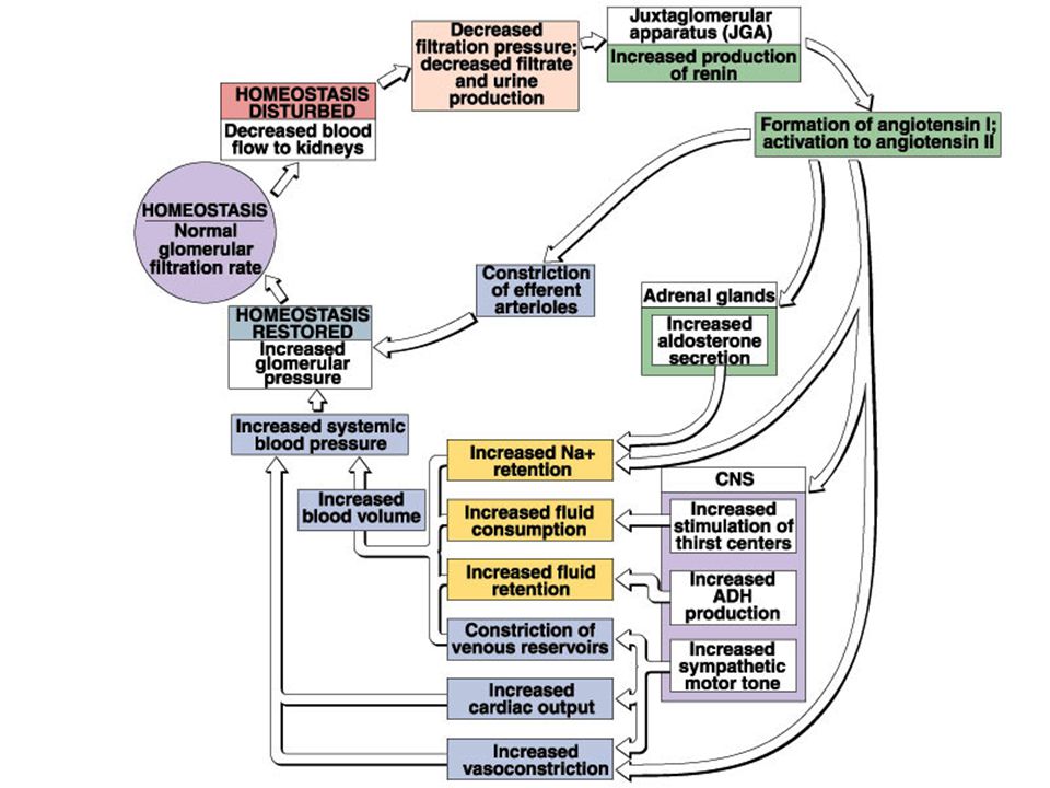
24
Renin – Angiotensin - Aldosterone System

26
Urine Concentration via Countercurrent Multiplication
Thin descending limb of Henle is permeable to water but not solutes Thick ascending limb of Henle is impermeable to water and solutes. Contains active transport mechanisms for sodium and chloride.

27
Urine Concentration via Countercurrent Multiplication
Sodium and Chloride are reabsorbed by thick ascending limb into the peritubular fluid These ions elevate the medulla osmotic pressure This increases osmotic flow of water out of the thin descending limb Increased osmotic potential of tubular filtrate increases active transport in the TAL

28
Urine Concentration via Countercurrent Multiplication

29
Roles of the Different Nephron Regions in Urine Formation
Proximal Convoluted tubule Reabsorption: 60%-70% of water (108 to 116 L/D) (obligatory water reabsorption) 100% of glucose and other sugars, amino acids, and some vitamins 60%-70% sodium and chloride, along with calcium, magnesium, phosphate, and bicarbonate Secretion: Hydrogen ions, ammonium ions, creatinine, drugs, toxins
 (obligatory water reabsorption) 100% of glucose and other sugars, amino acids, and some vitamins. 60%-70% sodium and chloride, along with calcium, magnesium, phosphate, and bicarbonate. Secretion: Hydrogen ions, ammonium ions, creatinine, drugs, toxins.")
30
Roles of the Different Nephron Regions in Urine Formation
Loop of Henle Reabsorption: Descending limb 25% of the water (obligatory water reabsorption) Thick Ascending limb 20-25% of the sodium and chloride to help maintain the countercurrent system
 Thick Ascending limb % of the sodium and chloride to help maintain the countercurrent system.")
31
Roles of the Different Nephron Regions in Urine Formation
Distal Convoluted Tubule Reabsorption: Up to 5% of water under ADH control (principle cells) (Facultative water reabsorption) Variable amounts of sodium and chloride under Aldosterone control (principle cells) Variable amounts of bicarbonate (intercalated cells) Variable amounts of Calcium controlled by parathyroid hormone Secretion: Hydrogen ions, ammonium ions, Creatinine, drugs , toxins
 (Facultative water reabsorption) Variable amounts of sodium and chloride under Aldosterone control (principle cells) Variable amounts of bicarbonate. (intercalated cells) Variable amounts of Calcium. controlled by parathyroid hormone. Secretion: Hydrogen ions, ammonium ions, Creatinine, drugs , toxins.")
32
Roles of the Different Nephron Regions in Urine Formation
Collecting Duct Reabsorption: Variable amounts of water under ADH control (principle cells) (Facultative water reabsorption) Variable amounts of sodium and chloride under Aldosterone control (principle cells) Variable amounts of bicarbonate (intercalated cells) Secretion: Potassium and hydrogen ions
 (Facultative water reabsorption) Variable amounts of sodium and chloride under Aldosterone control (principle cells) Variable amounts of bicarbonate. (intercalated cells) Secretion: Potassium and hydrogen ions.")
33
Summary of the roles of the different nephron regions in urine formation

Similar presentations


 System>")
