Download presentation
Presentation is loading. Please wait.

1
The patient and her problem:

3
Aneurysms can be treated by applying a clip to the “neck” Aneurysm

4
Aneurysms can be treated by applying a clip to the “neck”

6
However, if they get too big: 1.Danger of the clip occluding the parent artery 2.Can’t see around the aneurysm to clip it safely Our patient failed a “test balloon occlusion” of her ICA. Therefore, cannot afford to lose the artery, or to have it clamped off for prolonged perionds of time.

7
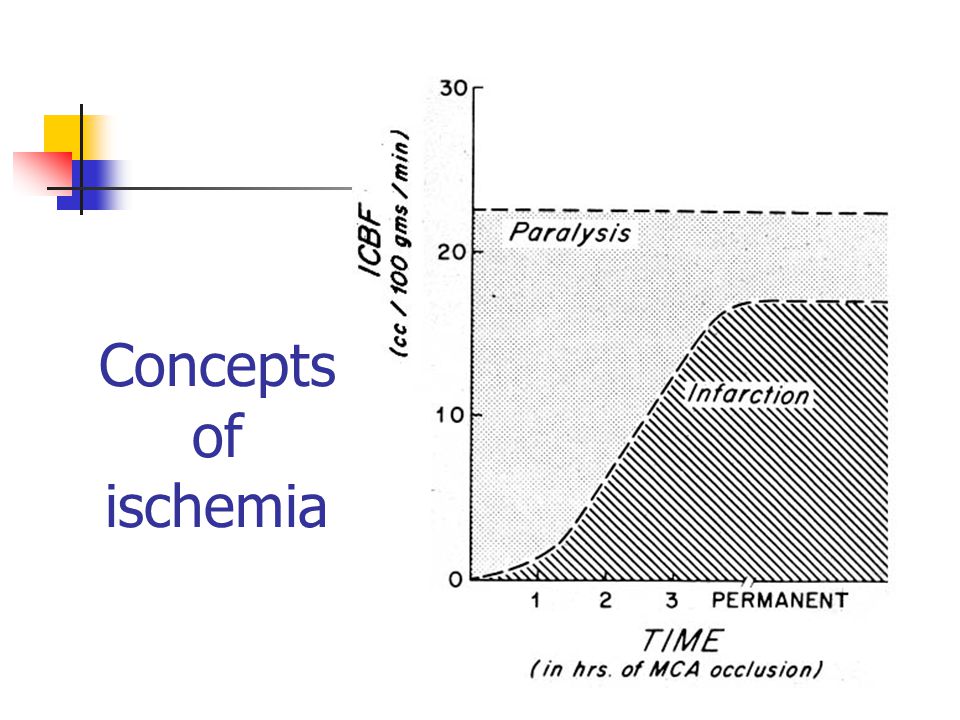
Need to devise a strategy to protect the brain from the lack of blood flow (ischemia) during the surgery.
 during the surgery.")
8
Concepts of ischemia

10
John Olney & The Excitotoxicity Theory: Glutamate as a neurotoxin

11
Glutamate receptors & Ischemia

12
Glutamate receptors

13
Glutamate evokes a 2- component EPSP AMPA (rapid) NMDA (slow)
 NMDA (slow)")
14
Ionotropic glutamate receptors Are associated with an ion channel N-methyl-D-aspartate (NMDA): composed of 4 or 5 subunits (NR1 and NR2a,b,c,d) -amino-3-hydroxy-5-methyl-4-isoxazole propionic acid (AMPA): composed of 4 or 5 subunits (GluR1-4) Kainate receptors: GluR5-7, KA1-2 These receptors are presumed to have different physiological functions.
: composed of 4 or 5 subunits (NR1 and NR2a,b,c,d) -amino-3-hydroxy-5-methyl-4-isoxazole propionic acid (AMPA): composed of 4 or 5 subunits (GluR1-4) Kainate receptors: GluR5-7, KA1-2 These receptors are presumed to have different physiological functions.")
15
Glutamate also activates metabotropic receptors These are glutamate receptors that do not contain an ion channel. These receptors, when activated, act by initiating 2 nd messenger cascades that result in the mobilization of intracellular Ca 2+ stores. The role of these receptors is less clear than ionotropic receptors (NMDA and AMPA), but is felt to be importante in neuronal function and dysfunction.
, but is felt to be importante in neuronal function and dysfunction..")
16
Glutamate receptors are strategically localized in the synapse

17
NMDA receptors on a cortical neuron

19
AMPA receptors on a cortical neuron

21
Molecular Model of the Postsynaptic Density at a Central Excitatory Synapse

22
Organization of the PSD Hypothetical organization of presynaptic (NT, nerve terminal) and postsynaptic (SP, spine) structures. Synaptic vesicles (orange spheres) release glutamate into the synaptic cleft, which in turn stimulates NMDA (blue rectangle), AMPA (red, yellow rectangle), and metabotropic (brown membrane protein) glutamate receptors. In the spine, actin cables (vertical pink filaments) are linked to brain spectrin (red, horizontal molecules). Also present in the spine are endoplasmic reticulum (blue membranous structure) and calmodulin (green ovals). Numerous kinases and proteases are not shown because their localization is not established.
 release glutamate into the synaptic cleft, which in turn stimulates NMDA (blue rectangle), AMPA (red, yellow rectangle), and metabotropic (brown membrane protein) glutamate receptors. In the spine, actin cables (vertical pink filaments) are linked to brain spectrin (red, horizontal molecules). Also present in the spine are endoplasmic reticulum (blue membranous structure) and calmodulin (green ovals). Numerous kinases and proteases are not shown because their localization is not established..")
23
PSD details: TrkB responds to BDNF (pink receptor; blue ligand). The neuroligan (green rectangle) cytoplasmic domain binds PSD- 95 (blue, green, yellow ovals). PSD-95 binds GKAP/ SAPAP/ DAP protein (red) and the NMDA receptor (blue rectangle), which in turn binds -actinin (orange) and actin (pink). Two AMPA receptors (red, yellow rectangle) are shown. Each binds GRIP, which has seven PDZ domains (red circles). Dimerization of GRIP via N termini is hypothetical.
 cytoplasmic domain binds PSD- 95 (blue, green, yellow ovals). PSD-95 binds GKAP/ SAPAP/ DAP protein (red) and the NMDA receptor (blue rectangle), which in turn binds -actinin (orange) and actin (pink). Two AMPA receptors (red, yellow rectangle) are shown. Each binds GRIP, which has seven PDZ domains (red circles). Dimerization of GRIP via N termini is hypothetical..")
24
Effect of calcium: Ca2+ that enters the spine through the NMDA receptor in response to receptor binding of glutamate (yellow circle) is proposed to activate calmodulin (green oval), which displaces -actinin and actin (orange oval, pink chain) from the NMDA receptor NR1 subunit C terminus.
 is proposed to activate calmodulin (green oval), which displaces -actinin and actin (orange oval, pink chain) from the NMDA receptor NR1 subunit C terminus.")
25
Why Glutamate Receptors are Important in Neurology: Glutamate is present in millimolar quantities in most cells, including neurons and glia Glutamate is the main excitatory neurotransmitter in the mammalian CNS Glutamate is released in large quantities during Stroke Trauma Epilepsy Possibly in chronic neurological disorders

26
Why Glutamate Receptors are Important in Neurology: Excess glutamate is released at the synapse through Synaptic activity Reverse operation of glutamate transporters Reduced re-uptake (due to reduced ATP levels) Glutamate levels may rise at the synapse to hundreds of micromolar, which is enough to cause excitotoxicity
 Glutamate levels may rise at the synapse to hundreds of micromolar, which is enough to cause excitotoxicity")
27
What happens to neurons with excess glutamate? Normal Neuron

28
What happens to neurons with excess glutamate? Cell Swelling Dendritic Beading Axons: no change (?) Glutamate
 Glutamate.")
29
Excess glutamate kills neurons through Ca 2+ overload

30
Ms. M.O., 47 yrs old. Admitted to hospital 7 days after a sudden, severe, headache, with Rt sided paralysis

31
Ms M.O. Vs Mrs S.N. Concept of vasospasm after SAH

32
Ms. M.O., 47 yrs old. Admitted to hospital 7 days after a sudden, severe, headache, with Rt sided paralysis

33
Ms. M.O., 47 yrs old. Admitted to hospital 7 days after a sudden, severe, headache, with Rt sided paralysis

34
Ms. M.O., 47 yrs old.

35
Progressive infarction Progressive Brain Swelling

36
Ms. M.O., 47 yrs old. Decompressive craniotomy

37
Last 2 lectures: Continue on why glutamate toxicity is important in neurological diseases Learn about Calcium Homeostasis in cells and why it is important Devise, based on what we have learned, strategies to protect the brain neurons and axons against stroke Find out what we did to prevent stroke for our patient with the giant aneurysm Show a short video of how an aneurysm gets fixed Cover the brain’s other cells, and how they might be important.

Similar presentations


 - made.>")


 + zF E axon myelin sheath dendrites nerve endings nt release nt receptors Cell body synapse.>")


