Download presentation
Presentation is loading. Please wait.

1
Blood Physiology Professor A.M.A Abdel Gader MD, PhD, FRCP (Lond., Edin), FRSH (London) Professor of Physiology, College of Medicine & King Khalid University Hospital Riyadh
, FRSH (London) Professor of Physiology, College of Medicine & King Khalid University Hospital Riyadh")
3
Lecture # 4 & 5 Leucocytes White Blood Cells (WBCs) Granulocytes, The Monocyte-Macrophage System
 Granulocytes, The Monocyte-Macrophage System")
4
Leucocytes (WBCs) General Characteristics & types of WBCs Genesis (Production) of WBCs Life Span of WBCs Defense properties of neutrophils & macrophages –Chemotaxis –Diapedesis –Amaeboid Motion –Phagocytosis
 General Characteristics & types of WBCs Genesis (Production) of WBCs Life Span of WBCs Defense properties of neutrophils & macrophages –Chemotaxis –Diapedesis –Amaeboid Motion –Phagocytosis")
5
Blood Film

7
Hematopoiesis

8
Leucocytes (WBCs) – cont. General Characteristics & types of WBCs Types of WBC 1.Granular (polymorphnuclear): Neutrophil 62%. –10-16um, nucleus 2-5 lobes, purple cytoplasmic granules Eosinophil 2.3%. –12-18um, 2 lobes nucleus, coarse red granules Basophil.4%. –10-14um, rarely segmented nucleus, nucleus hidden by large round bluish granules
: Neutrophil 62%. –10-16um, nucleus 2-5 lobes, purple cytoplasmic granules Eosinophil 2.3%. –12-18um, 2 lobes nucleus, coarse red granules Basophil.4%. –10-14um, rarely segmented nucleus, nucleus hidden by large round bluish granules.")
9
Leucocytes (WBCs) – cont. General Characteristics & types of WBCs Types of WBC 2.Agranular WBC –Monocytes 5.3% 15-20um, kidney shape nucleus –Lymphocyte 30% round nucleus –small (5-8um) –large (9-15um)
 –large (9-15um).")
10
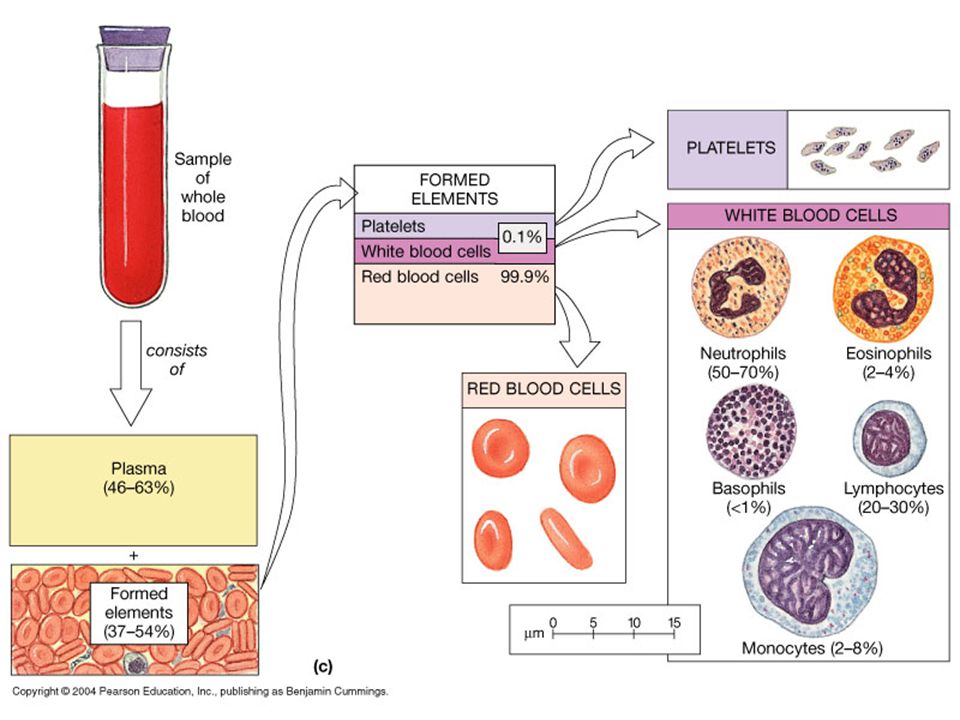
Formed Elements of Blood Red blood cells ( erythrocytes ) White blood cells ( leukocytes ) –granular leukocytes neutrophils eosinophils basophils –agranular leukocytes lymphocytes = T cells, B cells, and natural killer cells monocytes Platelets
 White blood cells ( leukocytes ) –granular leukocytes neutrophils eosinophils basophils –agranular leukocytes lymphocytes = T cells, B cells, and natural killer cells monocytes Platelets")
11
Leucocytes (WBCs) – cont. Genesis (Production) of WBCs Pluripotential stem cell Committed Stem cell RBCs WBCs Platelets MylocyticLymphocytic Linage
 of WBCs Pluripotential stem cell Committed Stem cell RBCs WBCs Platelets MylocyticLymphocytic Linage.")
12
Hematopoiesis

13
Leucocytes (WBCs) – cont. Genesis (Production) of WBCs- leucopoiesis) Sites of WBC formation Granulocytes (neutrophil, basophil, eosinophil): –bone marrow Agranulocytes –lymphocytes- bone marrow, thymus, lymphoid tissues –monocytes- bone marrow
 of WBCs- leucopoiesis) Sites of WBC formation Granulocytes (neutrophil, basophil, eosinophil): –bone marrow Agranulocytes –lymphocytes- bone marrow, thymus, lymphoid tissues –monocytes- bone marrow.")
14
Life Span of WBCs Granulocytes: 4 to 8 hrs (transit time ) in blood circulation 4 to 5 hrs in tissues –In infections life span a few hours Monocytes : –10 to 20 hrs in blood circulation –Leave capillaries to tissues, increase in size to become tissue macrphages which live for months
 in blood circulation 4 to 5 hrs in tissues –In infections life span a few hours Monocytes : –10 to 20 hrs in blood circulation –Leave capillaries to tissues, increase in size to become tissue macrphages which live for months")
15
Life Span of WBCs- cont. Lymphocytes: A few hrs in blood circulation >> tissues >> lymph >>> Blood (Recirculation) ?Life span: weeks to months
 Life span: weeks to months.")
16
Leucocytes (WBCs) General Characteristics & types of WBCs Genesis (Production) of WBCs Life Span of WBCs Defense properties of neutrophils & macrophages –Chemotaxis –Diapedesis –Amaeboid Motion –Phagocytosis
 General Characteristics & types of WBCs Genesis (Production) of WBCs Life Span of WBCs Defense properties of neutrophils & macrophages –Chemotaxis –Diapedesis –Amaeboid Motion –Phagocytosis")
17
Defense properties of neutrophils & macrophages Attack and destroy bacteria, viruses Sequence of events: –Chemotaxis –Diapedesis –Amaeboid Motion –Phagocytosis

19
Netrophils function-cont. Defense properties of neutrophils & macrophages- cont Phyagocytosis: Engulfing and killing of bacteria or any invading organism Steps: Chemotaxis: –Bacterial & viral toxins Products of damaged tissues : attract neutrophil to accumulate at infected site. –Opsonization: plasma substances (IgG) attached to the bacteria to make them easy to phagocyte
 attached to the bacteria to make them easy to phagocyte.")
20
Diapedesis

21
http://www.whfreeman.com/immunology/CH01/diapedesis.htm

22
Defense properties of neutrophils & macrophages Attack and destroy bacteria, viruses Sequence of events: –Chemotaxis –Diapedesis –Amaeboid Motion –Phagocytosis

24
Defense properties of neutrophils & macrophages Attack and destroy bacteria, viruses Sequence of events: –Chemotaxis –Diapedesis –Amaeboid Motion –Phagocytosis

25
Phagocytosis- cont. Phagocytosis is selective: Distinguish self from non-self…... How? –Normal tissues have smooth surface –Normal tissues have protective protein surface –Antibodies coating bacteria ( Opsonization)
.")
26
Phagocytosis

27
Phagocytosis by neutrophils- cont. Neutrophils attach to bacteria & encircled it with pseudopodia and take it into a vacuole (phagosome). One Neutrophil can engulf 3 to 20 bacteria One Macrophage can engulf up to 100 bacteria Microbial killing: fusion of neutrophil granules with vacuole, –Discharge of lysozyme, myeloperoxidase enzymes into the vacuole, killing and digesting the engulfed bacteria. –Release of Free radicals by oxidizing agents: superoxide, hydrogen peroxide to kill the bacteria
. One Neutrophil can engulf 3 to 20 bacteria One Macrophage can engulf up to 100 bacteria Microbial killing: fusion of neutrophil granules with vacuole, –Discharge of lysozyme, myeloperoxidase enzymes into the vacuole, killing and digesting the engulfed bacteria. –Release of Free radicals by oxidizing agents: superoxide, hydrogen peroxide to kill the bacteria.")
29
Leucocytes (WBCs) – cont. Types of WBC 1.Granular (polymorphnuclear): Neutrophil 62%. –10-16um, nucleus 2-5 lobes, purple cytoplasmic granules Eosinophil 2.3%. –12-18um, 2 lobes nucleus, coarse red granules Basophil.4%. –10-14um, rarely segmented nucleus, nucleus hidden by large round bluish granules

30
Blood Film

31
Eosinophils Function: Phagocytosis: Phagocytosis is same as neutrophil, but less efficient Chemotaxis: eosinophil attracted towards chronic inflammation/allergic tissue ( allergic disease of skin & lungs) By eosinophil chemotactic factor Phagocytose (& detoxify) antigen/antibody complexes
 By eosinophil chemotactic factor Phagocytose (& detoxify) antigen/antibody complexes")
32
Eosinophils cont, High eosinophil count: –Parasitic (hook worm, ascaris, bilharzia) –Allergic (asthma, rhinitis, drug reaction) –Allergic skin diseases
 –Allergic (asthma, rhinitis, drug reaction) –Allergic skin diseases")
33
Leucocytes (WBCs) – cont. Types of WBC 1.Granular (polymorphnuclear): Neutrophil 62%. –10-16um, nucleus 2-5 lobes, purple cytoplasmic granules Eosinophil 2.3%. –12-18um, 2 lobes nucleus, coarse red granules Basophil.4%. –10-14um, rarely segmented nucleus, nucleus hidden by large round bluish granules

34
Leucocytes (WBCs) – cont. Types of WBC 1.Granular (polymorphnuclear): Neutrophil 62%. –10-16um, nucleus 2-5 lobes, purple cytoplasmic granules Eosinophil 2.3%. –12-18um, 2 lobes nucleus, coarse red granules Basophil.4%. –10-14um, rarely segmented nucleus, nucleus hidden by large round bluish granules

35
Blood Film

36
Basophils Similar to tissue mast cells Non-phagocytic cells Granules: dark blue color. Granules contain: –Heparin –Histamine –Serotonin (5HT). Released during allergic reactions
. Released during allergic reactions.")
37
Hematopoiesis

38
Blood Film

40
Defensive Functions of the Monocytes Directly: –phygocytosis of bacteria, dead cells etc Indirectly: –Cooperates with lymphocytes by: Recognizing the foreign body Ingesting the foreign body Processing the foreign body Presenting it to lymphocytes

41
Monocyte-macrophage system Reticulo-endothelial System

42
Reticuloendothelial System- RES Blood Monocyte Tissue macrophage Attached (fixed) Mobile Function is phagocytosis of: Bacteria Viruses Dead tissues Foriegn particles Immune function
 Mobile Function is phagocytosis of: Bacteria Viruses Dead tissues Foriegn particles Immune function")
43
Reticuloendothelial System- RES RES is widespread in the body Cells of the RES: –Monocytes (blood macrophages) –Mobile and fixed tissue Macrophages –Specialiazed endothelial cells in bone marrow, ly mph nodes and spleen –Reticular cells of lymph nodes spleen & bone marrow.
 –Mobile and fixed tissue Macrophages –Specialiazed endothelial cells in bone marrow, ly mph nodes and spleen –Reticular cells of lymph nodes spleen & bone marrow.")
44
Reticuloendothelial System- RES Cells of the RES - Distribution: Tissue Macrophages in skin SC tissues Tisssue Macrophages of lymph nodes Tissue macrophages in lungs Macrphages (kupffer cells) in the liver Macrphages in the spleen and bone marrow
 in the liver Macrphages in the spleen and bone marrow")
45
Reticuloendothelial System- RES Blood Monocyte Tissue macrophage Attached (fixed) Mobile Function is phagocytosis of: Bacteria Viruses Dead tissues Foriegn particles Immune function
 Mobile Function is phagocytosis of: Bacteria Viruses Dead tissues Foriegn particles Immune function")
Similar presentations


 –Leukocytes (white blood.>")

>")


, FRSH (London) Professor of Physiology, College of Medicine & King Khalid University.>")

>")


