Download presentation
Presentation is loading. Please wait.

1
Transport Processes

2
Fig 36.2

3
A. Passive Transport I. Membrane Transport 1. Mechanisms 2. Requirements

4
Fig 36.7

5
B. Active Transport 1. Mechanisms 2. Requirements

6
1. Occur at virtually every level of biological organization. 2. Enzymes transport electrons, protons, acetyl groups. 3. Membranes transport material across themselves. 4. Cells transport material to and from other cells, and within themselves. 5. Whole organisms transport water, etc from one organ to another B. Why Transport?

7
A. Background 1. Plants adapted to the division of resources in the land environment (soil & air) by the differentiation of the plant body into roots and shoots. 2. But this created a new dilemma, the need to transport materials between roots and shoots. 3. Sometimes up to 100m away, in all kinds of environmental conditions. 4. What’s a poor plant to do? II. Transport in Plants
 by the differentiation of the plant body into roots and shoots. 2. But this created a new dilemma, the need to transport materials between roots and shoots. 3. Sometimes up to 100m away, in all kinds of environmental conditions. 4. What’s a poor plant to do. II. Transport in Plants.")
8
1. Cellular – uptake of H 2 O and solutes by individual cells. 2. Short-distance - between cells 3. Long-distance – throughout whole plant (xylem & phloem) B. Three levels of transport in plants: 4. Scenarios and Terms
 B. Three levels of transport in plants: 4. Scenarios and Terms.")
9
An artificial cell consisting of an aqueous solution enclosed in a selectively permeable membrane has just been immersed in a beaker containing a different solution. The membrane is permeable to water and to the simple sugars glucose and fructose but completely impermeable to the disaccharide sucrose.

10
a. sucrose b. glucose c. fructose Which solute(s) will exhibit a net diffusion into the cell?
 will exhibit a net diffusion into the cell")
11
Which solute(s) will exhibit a net diffusion out of the cell? a. sucrose b. glucose c. fructose
 will exhibit a net diffusion out of the cell a. sucrose b. glucose c. fructose")
12
Which solution is hypertonic to the other? a)the cell contents b)the environment
the cell contents b)the environment")
13
In which direction will there be a net osmotic movement of water? a)out of the cell b)into the cell c)neither
out of the cell b)into the cell c)neither.")
14
After the cell is placed in the beaker, which of the following changes will occur? a)The artificial cell will become more flaccid. b)The artificial cell will become more turgid.
The artificial cell will become more flaccid. b)The artificial cell will become more turgid..")
15
A. Definition Water potential refers to the free energy of water, its capacity to do work. By definition pure free water has a water potential of zero. B. Factors 1. Increased heating, pressure, or elevation 2. Decreased III. Water Potential

16
= s + p + m s = osmotic potential p = pressure potential m = matric potential C. Components of

17
a. Osmotic potential is a measure of the effect that solutes have on water potential. b. Pure water has an osmotic potential of zero. c. Adding solutes decreases the osmotic potential because water interacts with solutes. (more solutes = more negative) d. Thus s is always negative (if solute is present) s = osmotic potential
 d. Thus s is always negative (if solute is present) s = osmotic potential.")
18
a. A measure of the effect that pressure has on water potential. b. Pressure can be positive (when something is compressed). Pushing c. Pressure can be negative (when something is stretched or pulled). Called tension. d. Water can handle large amounts of tension because of cohesion – the tendency of water to stick to itself (H bonding) p = pressure potential
. Pushing c. Pressure can be negative (when something is stretched or pulled). Called tension. d. Water can handle large amounts of tension because of cohesion – the tendency of water to stick to itself (H bonding) p = pressure potential.")
19
Figure 3.1

20
a. Matric potential is a measure of water’s adhesion to non-dissolved but hydrophilic structures such as cell walls, membranes, soil particles etc. b. Adhesion can only decrease water’s free energy. c. So matric potential is always negative m = matric potential

21
Why would water move to where it is less free? –Water acts to dilute, hydrate, decrease tension –i.e. water acts to stabilize water potentials –If two Ψ’s are equal, no net movement of water Water moves from regions of high Ψ (less negative) to regions of lower Ψ (more negative).
 to regions of lower Ψ (more negative)..")
22
Ψ = -15MPa Ψ = -300MPa Examples Ψ = +46MPa Ψ = -22MPa

23
Fig 36.8

24
1. A flaccid cell ( p = 0). No pressure is being exerted against the inside of the cell wall. Cell is not firm. Why? Solute concentration within the cell is lower than surroundings. Water leaves the cell. 2. A turgid cell ( p = +) is filled with water, exerting pressure against its cell walls. Cell is firm. Why? Solute concentration within the cell is higher than surroundings. Water moves into the cell. D. Cells, Water , Terms
 is filled with water, exerting pressure against its cell walls. Cell is firm. Why. Solute concentration within the cell is higher than surroundings. Water moves into the cell. D. Cells, Water , Terms.")
25
1. Hypotonic solution – low solute concentration 2. Hypertonic solution – high solute concentration 3. A flaccid cell placed in a hypotonic solution will? 4. A turgid cell placed in a hypertonic solution will? This causes the cell membrane to shrink away from the cell wall (i.e. plasmolyze). E. Terms for cells and water movement
. E. Terms for cells and water movement.")
26
Fig 36.9a

27
Fig 36.9b

28
A. Three routes for lateral transport: 1. Trans-membrane – water & solutes move across the plasma membranes and cell walls of adjacent cells 2. Symplast – movement through a continuum of cytoplasm connected by the plasmodesmata of cell walls. 3. Apoplast – extracellular pathway; movement through the continuous matrix of cell walls IV. Cell to Cell Transport

29
Fig 36.8

30
a. Guard Cells i. Mechanism: Opening K + is pumped into GC by active transport. Proton pump creates membrane potential that drives K + in. Thus Ψ inside cell is lower than outside cell. Water diffuses into the guard cell. Sunlight, circadian rhythms, & low CO 2 concentration in leaf air spaces stimulate the proton pumps & thus stomatal opening 4. Examples of Short Distance Transport

31
ii. Mechanism: Closing Proton pumps no longer active (darkness) K + is lost from the GC, creating lower water potential outside cell. Water flows out of GC and cells become flaccid Stomatal closure during the day stimulated by water stress – not enough water to keep GCs turgid
 K + is lost from the GC, creating lower water potential outside cell. Water flows out of GC and cells become flaccid Stomatal closure during the day stimulated by water stress – not enough water to keep GCs turgid.")
32
Fig 36.15

33
i. Description Leaves of these plants can flex & fold in response to stimuli Motor cells are the “joints” where this flexing occurs. Accumulate or expel potassium to adjust their Ψ & thus turgidity. ii. Examples: Venus’ flytrap Oxalis – leaves fold in sunlight to minimize transpiration; open in shade Transpiration = loss of water vapor from the stomata b. Motor Cells

34
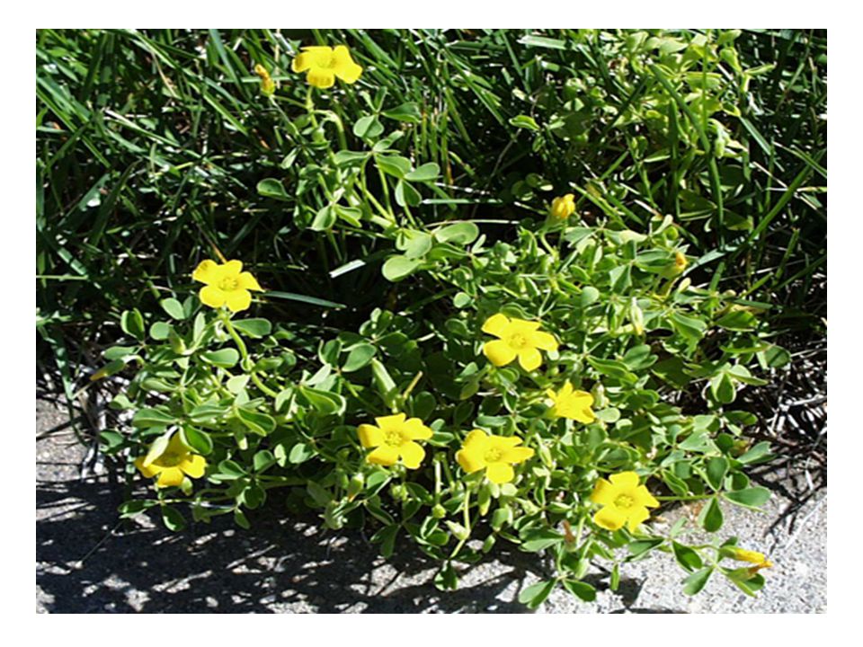
http://www.uccs.edu/~ppbotany/Colo_family/Oxalid/oxalis_stricta_P.htm

36
i. Description Cell walls have many finger–like projections on the inner surface. The plasma membrane is pressed firmly against these convolutions, creating an increase in surface area Greater surface area means more molecular pumps & thus high – volume solute transport Found in areas of rapid, high volume transport: salt- excreting glands, sugar loading into phloem c. Transfer Cells

37
i. Description Soil particles coated with water, minerals; adhere to hydrophyllic epidermal cells of root hair Soil solution moves freely through epidermal cells & cortex via symplast and apoplast pathways Endodermis – selective barrier to soil solution between cortex & stele. Sealed together by the waxy Casparian strip (Suberin) – forces soil solution in apoplast to pass through the selectively permeable membrane of the endodermis. Once through the endodermis, soil solution freely enters the xylem d. Root Cells
 – forces soil solution in apoplast to pass through the selectively permeable membrane of the endodermis. Once through the endodermis, soil solution freely enters the xylem d. Root Cells.")
38
Fig 36.9 Suberin

40
Xylem: Transpiration (evaporation from leaves) creates a tension which pulls sap up from the roots, in direction of lower Ψ. Phloem: Hydrostatic pressure at one end of the sieve tube forces sap to the other end of the tube (= bulk flow). V. Long Distance Transport
. V. Long Distance Transport.")
41
1. Forces for Xylem sap both pushed & pulled up the stem WHY? a. Root Pressure Stele has high concentration of minerals, decreasing Ψ. Water flows in, creating pushing pressure b. Guttation – exudation of water droplets by leaves during the night when transpiration is low. Caused by root pressure A. Xylem Transport = sap

43
i. Transpiration – cohesion – tension mechanism ii. Transpirational pull: Ψ of air typically << than Ψ of leaf, thus evaporation. iii. Water remaining in leaf is tightly adhered to cell walls in the mesophyll iv. This adhesion & surface tension of the water creates negative pressure – the pulling force c. Cohesion/Adhesion

44
Figure 36.14

45
d. Aided by: i. Cohesiveness of water – allows water to be pulled up in a continuous column without breaking ii. Adhesion of water to hydrophyllic cell walls of the xylem, creates tension (negative pressure/ pull) iii. Diameters of tracheids & vessel elements are small, so lots of surface area for adhesion iv. Since movement is by bulk flow (i.e. no membranes to pass through), Ψ s is not involved in the overall process
 iii. Diameters of tracheids & vessel elements are small, so lots of surface area for adhesion iv. Since movement is by bulk flow (i.e. no membranes to pass through), Ψ s is not involved in the overall process.")
46
Fig 36.15

47
ExamplesMax. Speed (cm/hr) Conifers120 Angiosperm Trees600 - 4400 Herbs6,000 Vines15,000 (0.6 mi/hr) 2. Speed of Xylem Sap Translocation
 Conifers120 Angiosperm Trees Herbs6,000 Vines15,000 (0.6 mi/hr) 2. Speed of Xylem Sap Translocation.")
48
3. Forces that act against transpirational pull: A. Gravity B. Hydraulic resistance Why is xylem transport in trees much slower than in herbs?

49
a. Guard cells! – balance two contrasting needs of the plant: i. Conserve water ii. CO 2 for photosynthesis b. Water Use Efficiency (WUE) = g H 2 O lost / g CO 2 assimilated by photosynthesis average = 600/1 4. Control of Transpiration
 = g H 2 O lost / g CO 2 assimilated by photosynthesis average = 600/1 4. Control of Transpiration.")
50
c. Adaptations by Desert plants to increase their WUE: i. Small thick leaves (less SA for water loss) ii. Thick cuticle iii. Stomata only on bottom of leaves iv. High-volume water storage (cacti) v. Crassulacean Acid Metabolism (CAM) – plants take in CO 2 only at night, so that stomata only have to be open at night.
 ii. Thick cuticle iii. Stomata only on bottom of leaves iv. High-volume water storage (cacti) v. Crassulacean Acid Metabolism (CAM) – plants take in CO 2 only at night, so that stomata only have to be open at night..")
51
a. Why? Transpirational pull is greater than the delivery of water by the xylem. Cells lose turgor pressure & stomata close 5. Wilting

52
Trans-stomatalTrans-cuticular Herbs: Coronilla varia1810190 Stachys recta1620180 Oxytropis pilosa1600100 Trees: Pinus sylvestris (pine)52713 Picea abies (spruce)46515 Fagus sylvaticus (birch)33090 b. Trans-stomatal and Trans-cuticular Transpiration (gH 2 0/dm 2 /hr)
.")
53
1. Direction and Material a. Phloem sap = sucrose, amino acids, hormones b. Sieve tubes carry sap from sugar source (e.g. leaves) to sugar sink (e.g. growing roots, shoot tips, stems, flowers, fruits) c. Thus not unidirectional e.g. tubers can be source in spring and sink in fall B. Phloem Transport
 to sugar sink (e.g. growing roots, shoot tips, stems, flowers, fruits) c. Thus not unidirectional e.g. tubers can be source in spring and sink in fall B. Phloem Transport.")
54
a. Sap moves into sieve tubes via companion cells by symplast and apoplast pathways b. Loading is by active transport only – why? Sap in sieve tubes is highly concentrated with solutes, thus phloem unloading is passive (source to sink) 2. Phloem loading
 2. Phloem loading.")
55
Fig 36.19

56
a. Pressure-flow Hypothesis: i. Phloem loading creates high sugar concentration at source, decreasing Ψ. ii. Thus water flows into sieve tubes, creating hydrostatic pressure (pushing pressure: positive). iii. Less pressure at sink end, where sugar is leaving sieve tube for consumption iv. Thus movement from source to sink 3. Mechanism
. iii. Less pressure at sink end, where sugar is leaving sieve tube for consumption iv. Thus movement from source to sink 3. Mechanism.")
57
Fig 36.18

58
ExamplesMaximum Speed (cm/hr) conifer stem13-48 angiosperm tree 48 vine 72 grass 168 sunflower 240 corn 660 4. Speed of Phloem Sap Translocation

59
We learn to move on, but we never stop learning..

Similar presentations