Download presentation
Presentation is loading. Please wait.

1
The eyes have it. Ophthalmologic emergencies Cecilia Guthrie, MD Emory University

2
Case- History CC : Blind in right eye 9y.o. HM hit in right eye with a rock 24 hours ago by same age boy at school No loss of consciousness and eye pain on being struck resolved quickly Some persistent blurriness of vision Mother noted red marks on white parts of eye, but no other injury

3
Case- History 2 hours to presentation, pt developed headache, dizziness, and loss of vision in right eye. Denied fever, pain, repeated trauma and vomiting. PMH - Asthma Meds - albuterol mdi Allergies - none FH - N/C ROS - o/w negative

4
Case - PE VS : 88 16 116/60 HEENT - no signs of trauma OD –mild ptosis of right upper eyelid. No echymosis of eyelid. –Sclera with multiple subconjunctival hemorrhages –globe intact w/o swelling –conjunctiva injected diffusely –fundus - unable to visualize the retina

5
Case - PE –Unable to visualize the pupil secondary to blood in anterior chamber. –Pt could distinguish light –EOMI OS –PERRLA –No erythema, normal sclera and conjunctiva –4mm--> 2mm Visual Acuity: OD none OS 20/40

7
Ophthalmology trauma Ocular trauma is leading cause of visual loss in the pediatric population Estimated approximately 1 million eye injuries occurring in children annually. M>F 3:1 Adolescent males at increased risk 1/2 of injuries occur secondary to sporting activities (baseball, basketball)
.")
8
Ophthamology trauma BB guns, paint ball guns,sticks, collision with fixed objects Visual system matures at 9 years of age Amblyopia may occur May be difficulty to obtain mechanism of injury, history and exam

9
You did what?? Mechanism of injury Time of injury Place of injury Caregiver at time of injury Initial intervention Possibility of retained foreign body Pertinent PMH and ocular hx Any vision changes

10
Open…Your…Eyes!! Non contact aspects of exam first Suspect ruptured globe, don’t touch eye Assess visual acuity in each eye separately Pupils Ocular motility Lids and orbits

11
Open… Your… Eyes!! Examine conjunctiva and sclera for lacs or foreign body Cornea for abrasion or laceration- flourescein Anterior chamber depth and clarity Assess red reflex

14
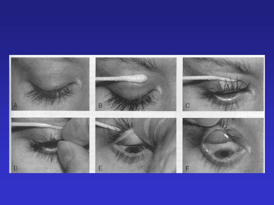
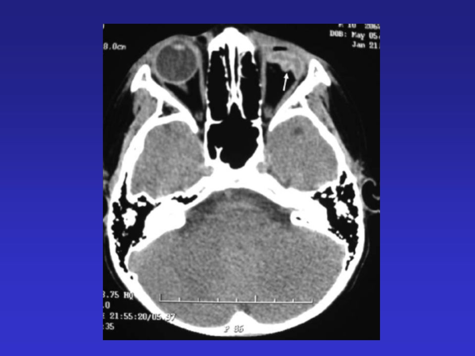
Trauma Foreign Body Foreign bodies can lodge underneath the upper eyelid or on the anterior surface Foreign body sensation Pain on blinking Watery Discharge Unilateral Photophobia

20
Trauma Foreign Body Extraocular vs intraocular Treatment –Topical anesthetic (tetracaine) –Eversion of the lid and flush with water –Remove foreign body –Question of retained foreign body after flush-call opthomology –Flourescein after flushing
 –Eversion of the lid and flush with water –Remove foreign body –Question of retained foreign body after flush-call opthomology –Flourescein after flushing")
23
Trauma Subconjunctival hemorrhage Unilateral Underlying sclera not visible Adjacent conjunctiva normal No discharge No pain Vision intact

24
Trauma Subconjunctival hemorrhage Etiology –Minor trauma –Bleeding disorders –Anticoagulation therapy –Hypertension –Coughing, vomiting Treatment –Resolves in 2-3 weeks

27
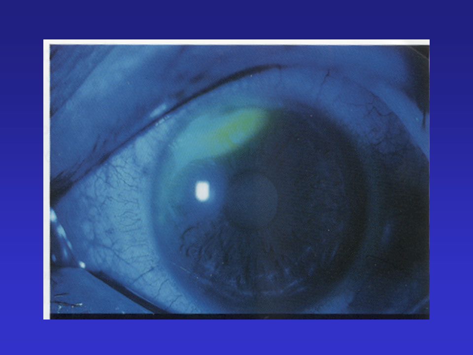
Trauma Corneal abrasion Cornea –Epithelium –Bowman membrane (protective layer) –corneal stroma (90% of thickness) –Descemet membrane –Endothelium Superficial to Bowman membrane If deeper to Bowman membrane - scar
 –corneal stroma (90% of thickness) –Descemet membrane –Endothelium Superficial to Bowman membrane If deeper to Bowman membrane - scar")
28
Trauma Corneal abrasion Moderate to severe pain Photophobia Conjunctival erythema Tearing Diagnosis –Better exam with topical anesthetic –Fluorescein

29
Trauma Corneal abrasion Treatment –Topical antibiotic therapy for 4-5 days –Patching vs No Patching –Cool compresses intermittently –Tylenol or ibuprofen –Cycloplegic agents for severe pain 5% homotropine 1% cyclopentolate (cyclogel) –If not healing in 48 hours, opthamology referral
 –If not healing in 48 hours, opthamology referral")
31
Trauma Eyelid lacerations Determine if laceration or injury to globe/conjunctiva underneath the eyelid laceration, especially with pointed objects Determine if a complete perforation of eyelid present Determine if involvement of tearducts

34
Trauma Eyelid lacerations Uncomplicated superficial eyelid lacerations may be sutured by ED physician –Shallow sutures used –Sedation may be needed

35
Trauma Eyelid lacerations Indications for opthamology consult –Full thickness perforation of lid –Ptosis –Involvement of the lid margin –Possible damage to tear drainage system –Tissue avulsion –Global injury

36
Trauma “Black eye” Can be associated with traumatic iritis, hyphema and cataracts. Dramatic ecchymosis and swelling may occur from mild trauma because of loose connections of eyelid skin and underlying tissues. Resolving midline forehead injuries/ hematomas can cause bilateral ecchymosis

38
Trauma Orbital fractures Most common-inferior and medial walls 50% of pediatric orbital fractures are associated with other ocular injuries Enopthalmia or proptosis Decreased extraocular muscle movement –hallmark of orbital fracture –entrapped muscle/tissue –orbital hemorrhage

39
Trauma Orbital fractures Inferior wall fx - infraorbital nerve injury Superior (roof) wall fx - pulsating proptosis Diploplia - eom entrapment May be subtle with normal rim CT of orbits with head CT (especially with possible superior wall fx)
 wall fx - pulsating proptosis Diploplia - eom entrapment May be subtle with normal rim CT of orbits with head CT (especially with possible superior wall fx)")
44
Trauma Orbital fractures Ophthalmology consult May also need “face” consult- If no entrapment, hemorrhage or global injury and fracture is nondepressed or displaced, may not require surgery Broad spectrum antibiotics Don’t blow nose

46
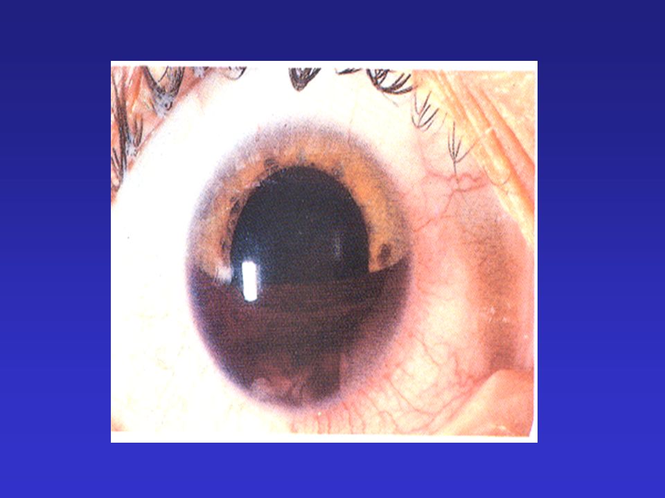
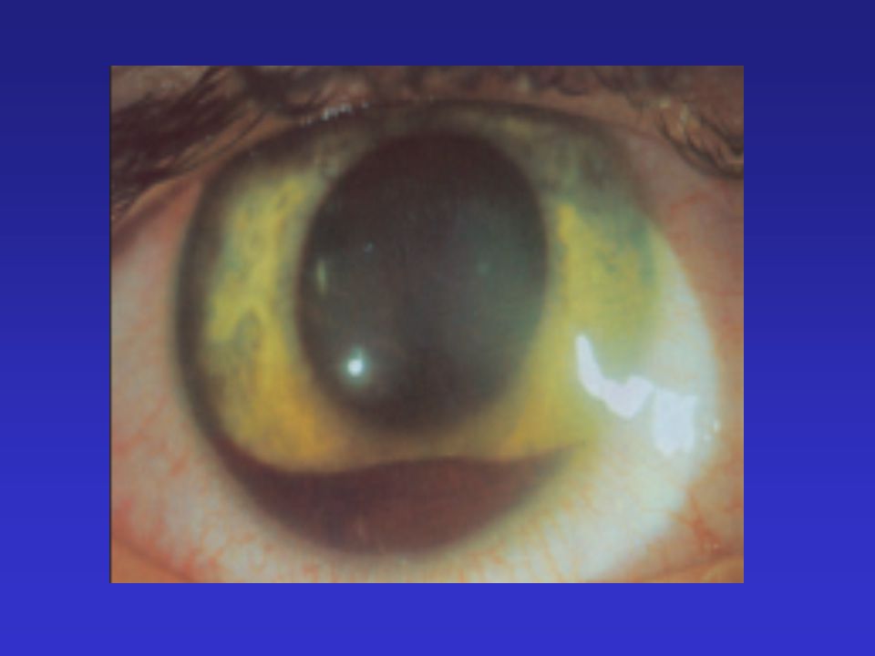
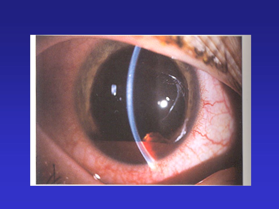
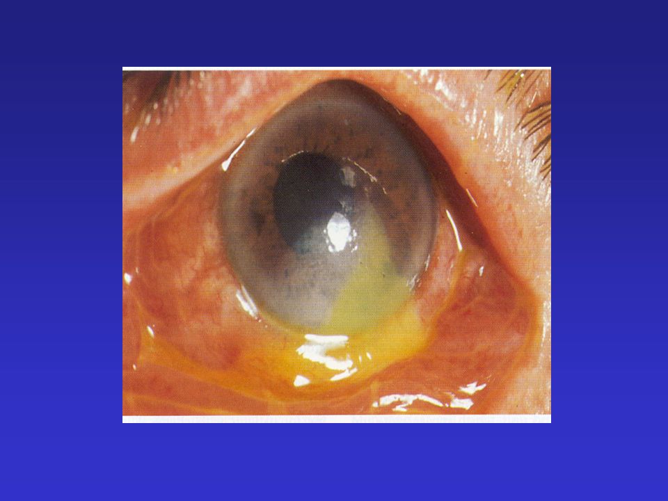
Trauma Hyphema Children and young adults M>F (4:1) After blunt trauma to the face/eye Trauma stretching of iris and ciliary body tear Blood in the anterior chamber Layering
 After blunt trauma to the face/eye Trauma stretching of iris and ciliary body tear Blood in the anterior chamber Layering")
48
Trauma Hyphema 3-5 days post injury, spontaneous rebleeding Rebleed complications –Corneal staining –Secondary glaucoma –Optic atrophy Sickle cell disease patients –Increased risk of rebleeding –~30% have increased intraocular pressure (10-20 normal) –Central artery occlusion and optic nerve damage with marginal increases in intraocular pressure
 –Central artery occlusion and optic nerve damage with marginal increases in intraocular pressure")
49
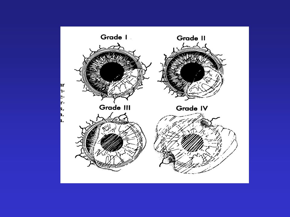
Trauma Hyphema DistributionSeverityDegree of Hyphema 0microscopic 58%1< 1/3 20%21/3- 1/2 14%3½-3/4 8%4¾-complete (8 ball)
")
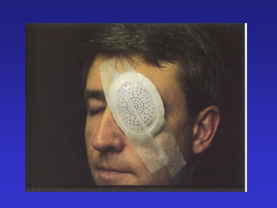
52
Trauma Hyphema Treatment –Eye shield –Strict bedrest with head elevated 45º –Ophthalmology consult –Long acting cycloplegic –Aminocaproic acid (antifibrinolytic) Initial: 200mg/kg/dose po (max 6 gm) Maintenance 50 - 100 mg/kg/dose q 6 hr. –Admission for 30% hyphema

55
Trauma Iritis Eye pain Photophobia Visual loss Ciliary flush Constricted pupil on affected side 24 - 72 hours after blunt injury to the eye ball

58
Trauma Iritis Treatment –Ophthalmology consult –Short acting cyclopegia –Topical antibiotic

60
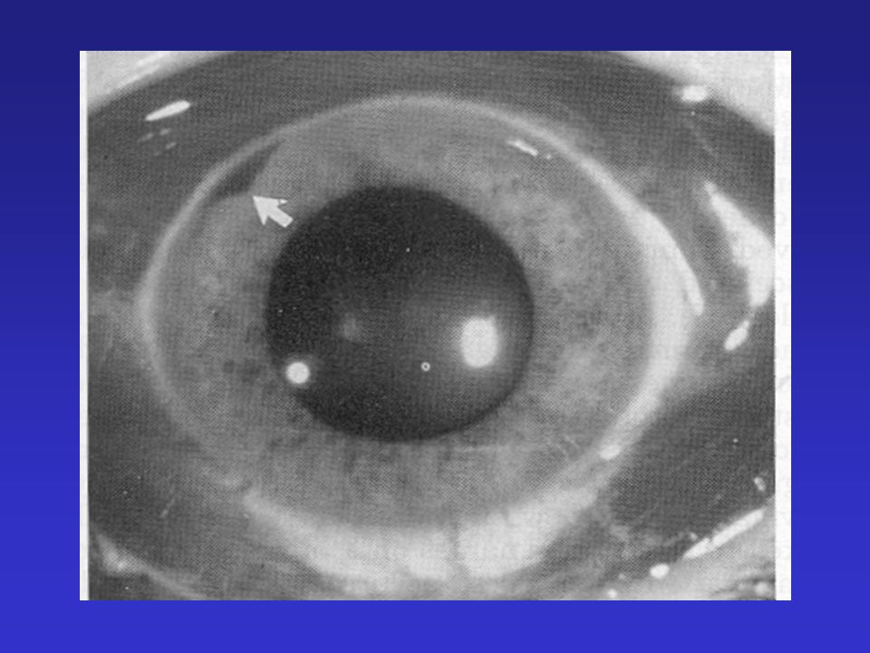
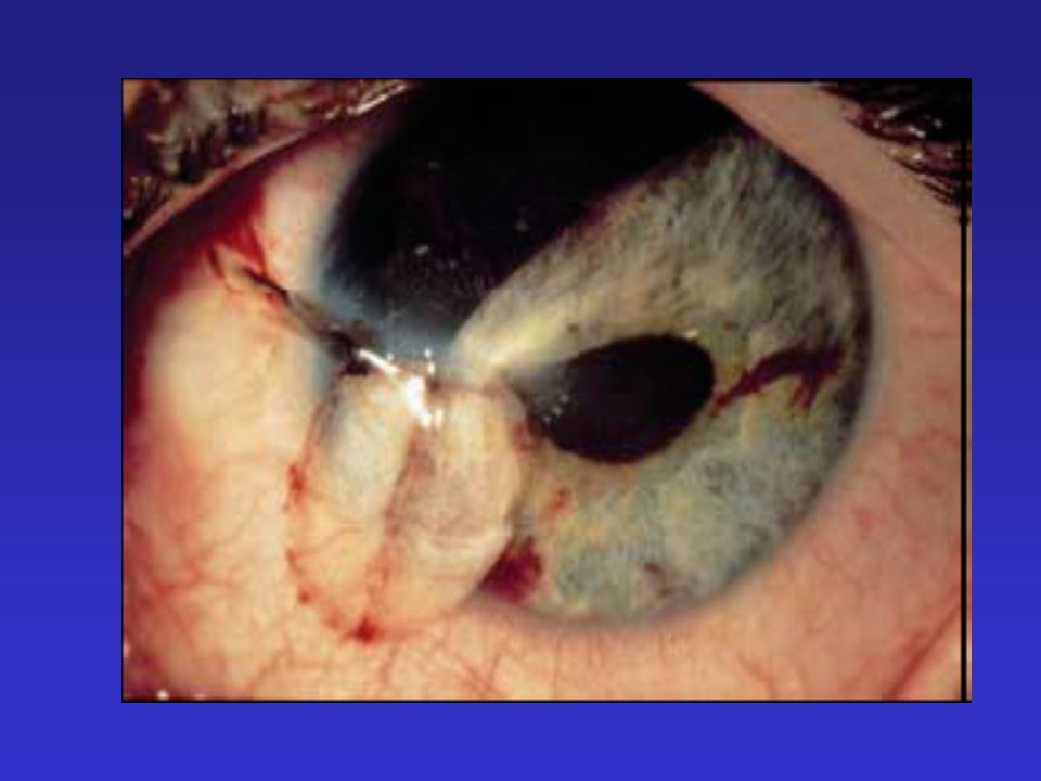
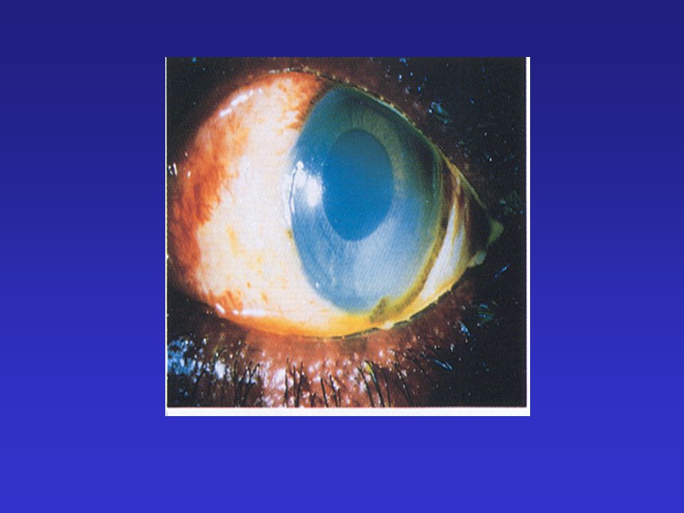
Trauma Ruptured globe S/P blunt trauma or projectile of sharp object –Guns (22%), sticks/tree branches (11%) M>F 6:1 Laceration or puncture of the sclera or cornea Iris or choroid plugs wound –tear drop pupil –brown,blue or black on scleral surface
, sticks/tree branches (11%) M>F 6:1 Laceration or puncture of the sclera or cornea Iris or choroid plugs wound –tear drop pupil –brown,blue or black on scleral surface")
64
Trauma Ruptured Globe If small may have a normal global appearance Limbus most susceptible area 360 degree subconjunctival hemorrhage-be suspicious

65
Trauma Ruptured Globe Management –Keep pt calm-may need sedation –Cover eye with hard shield –Ophthalmology consult-True emergency –CT of orbits –Tetanus –Antibiotics

67
Conjunctivitis Chemical injuries Chemical contact Most common cause are voluntary eye solutions –Neomycin, atropine, pilocarpine, idoxuridine, gentamycin Neonate Discontinue irritating agent

68
Chemical Burns Alkali Burns –Most serious burns –Hair straighteners, lye, ammonia –Penetrates cornea coagulative necrosis Acid Burns –Limiting burn –Protein precipitation in the corneal epithelium and stroma Limits acid penetration of the cornea Corneal opacifications Conjunctiva blanching

71
Chemical Burns Topical anesthetic Immediate irrigation-1 L NS Check pH of eye-conjunctiva Continue to irrigate until pH is normal Ophthalmology consult Red is better than white

72
Chemical Injuries Mace/Tear gas/Pepper spray –Superficial injury –Flush eyes well Super glue –Ophthalmic ointment –Check global movement beneath closed eyelid

74
Radiation Injury Prolonged exposure to ultraviolet light w/o proper eye protection Ultraviolet keratitis-corneal epithelium swells and dies Foreign body sensation Photophobia Pain Redness blepharospasm

75
Radiation Injury Fluorescein-diffuse punctate staining Treatment similar to corneal abrasion

76
References Albert, D and Jakobiec F Eds. Atlas of Clinical Ophthalmology. Philadelphia: W.B. Saunders, 1996 Arffa, R. Craven L ed. Grayson’s Diseases of the Cornea. St. Louis: Mosby, 1997 Levine, L “Pediatric ocular trauma and shaken infant syndrome” Peditr Clinic N Am. 2003;50 (1) Hatton MP, et al. “Orbital fractures in children” J Am Society of Opthalmic Plastic and Reconstructive Surgery. 2001;17(3), 174-9. Walton, W. et al. “Management of Traumatic Hyphema.” Survey of Opthalmology. 2002;47(4) 297-334. Fleisher and Ludwig. Textbook of Pediatric Emergency Medicine, 4th ed. Philadelphia: Lippinicott Williams and Wilkins, 200.
 Hatton MP, et al. Orbital fractures in children J Am Society of Opthalmic Plastic and Reconstructive Surgery. 2001;17(3), Walton, W. et al. Management of Traumatic Hyphema. Survey of Opthalmology. 2002;47(4) Fleisher and Ludwig. Textbook of Pediatric Emergency Medicine, 4th ed. Philadelphia: Lippinicott Williams and Wilkins,")
Similar presentations


 2-Perforating Injury 3-Perforating Injury & retained foreign body 4-Chemicals ( acid – alkaline ) & burns 5-Sonar.>")


