Download presentation
Presentation is loading. Please wait.

1
Prostate Gland, Seminal vesicle & Ejaculatory Ducts
Dr. Sama-ul-Haque

2
Objectives Discuss the site, size and relation of prostate.
Describe the fasciae covering and related to the prostate (true and false capsules). Describe its anatomical and surgical lobes. Discuss its blood, nerve supply and lymphatic drainage. Compare between the site of enlargement of the prostate in senile prostatic hyperplasia and cancer prostate.
. Describe its anatomical and surgical lobes. Discuss its blood, nerve supply and lymphatic drainage. Compare between the site of enlargement of the prostate in senile prostatic hyperplasia and cancer prostate.")
3
Objectives Define the seminal vesicle.
Discuss its site, relation, blood and nerve supply. Discuss its lymphatic drainage. Define the ejaculatory duct. Describe its site, beginning and termination.

4
Prostate Gland Fibromuscular glandular organ Site:
Glandular part : 2/3rd Fibromuscular part: 1/3rd Site: Lies between the neck of the urinary bladder above and the urogenital diaphragm below Size: 1.25 inches (3 cm)
")
5
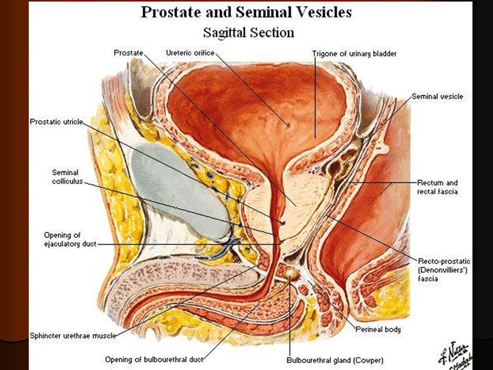
Prostate Gland Relations: Superiorly: Neck of the urinary bladder
Urethra enters the center of the base of the prostate Inferiorly: Urogenital Diaphragm Urethra leaves prostate just above its apex on the anterior surface

6
Location of Prostate Gland

7
Prostate Gland Relations: Anteriorly: Symphysis pubis
Puboprostatic ligaments (Pubic bones) Posteriorly: Rectal Ampulla Fascia of Denonvillier (Rectoprostatic fascia) Laterally: Levator Ani
 Posteriorly: Rectal Ampulla. Fascia of Denonvillier. (Rectoprostatic fascia) Laterally: Levator Ani.")
9
Capsule of the Prostate Gland
True capsule: Prostate is surrounded by a fibrous capsule which contains prostatic plexuses of nerves and veins False capsule: Visceral layer of pelvic fascia forms a fibrous prostatic sheath outside the true capsule

10
Prostate Gland Apex Base Anterior surface Posterior surface
Inferolateral surfaces

12
Anatomical lobes of Prostate Gland
1. Anterior lobe Lies in front of urethra Muscular Devoid of glandular tissue 2. Median or Middle lobe Lies between urethra and the ejaculatory ducts Glandular

13
Anatomical lobes of Prostate Gland
3. Posterior lobe Lies behind urethra and below ejaculatory ducts Glandular 4. Right lateral lobe Lies on the side of the urethra 5. Left lateral lobe

14
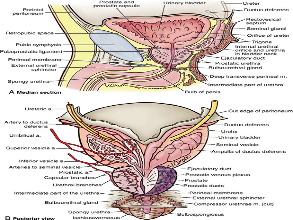
Blood supply of Prostate Gland
Arterial supply: Branches of Internal Iliac artery Branches from inferior vesical artery Branches from middle rectal artery Branches from Internal pudendal artery Venous drainage Veins form prostatic plexus Drain into internal iliac vein

17
Lymphatic drainage & Nerve supply of Prostate Gland
Internal iliac nodes Nerve supply Inferior hypogastric plexus

20
Benign Prostatic Hypertrophy (BPH)
Occurs in middle age Median or Middle lobe is involved Projects into urinary bladder Obstructs prostatic urethra and internal urethral orifice

21
Prostatic Cancer Occurs in old age (after 55 years usually)
Posterior and lateral lobes are involved Palpated during PR examination Can spread to iliac and sacral lymph nodes and can spread to vertebral column and brain through venous channels

22
Prostatectomy TURP (Transurethral resection of prostate)
")
23
Seminal vesicles Lobulated organ 2 inches (5 cm) long
Lies on the posterior surface of the urinary bladder Seminal vesicle joins the vas deferens to form ejaculatory ducts on each side

24
Blood supply of Seminal vesicles
Arterial supply Branches from inferior vesical artery Branches from middle rectal artery Venous drainage Drain into internal iliac vein Lymphatic drainage Internal iliac nodes

26
Ejaculatory ducts Less then 1 inches (2.5 cm) long
Formed by the union of duct of seminal vesicle and vas deferens Pierce posterior surface of the prostate Opens into the prostatic part of the urethra

28
Thank You

Similar presentations


 Middle rectal artery (anterior division of internal iliac) Inferior.>")


